radio final images
1/380
There's no tags or description
Looks like no tags are added yet.
Name | Mastery | Learn | Test | Matching | Spaced | Call with Kai |
|---|
No analytics yet
Send a link to your students to track their progress
381 Terms

dark blue
phleboliths

light blue
triticeous cartilage and thyroid cartilage

dark green
calcified lymph nodes
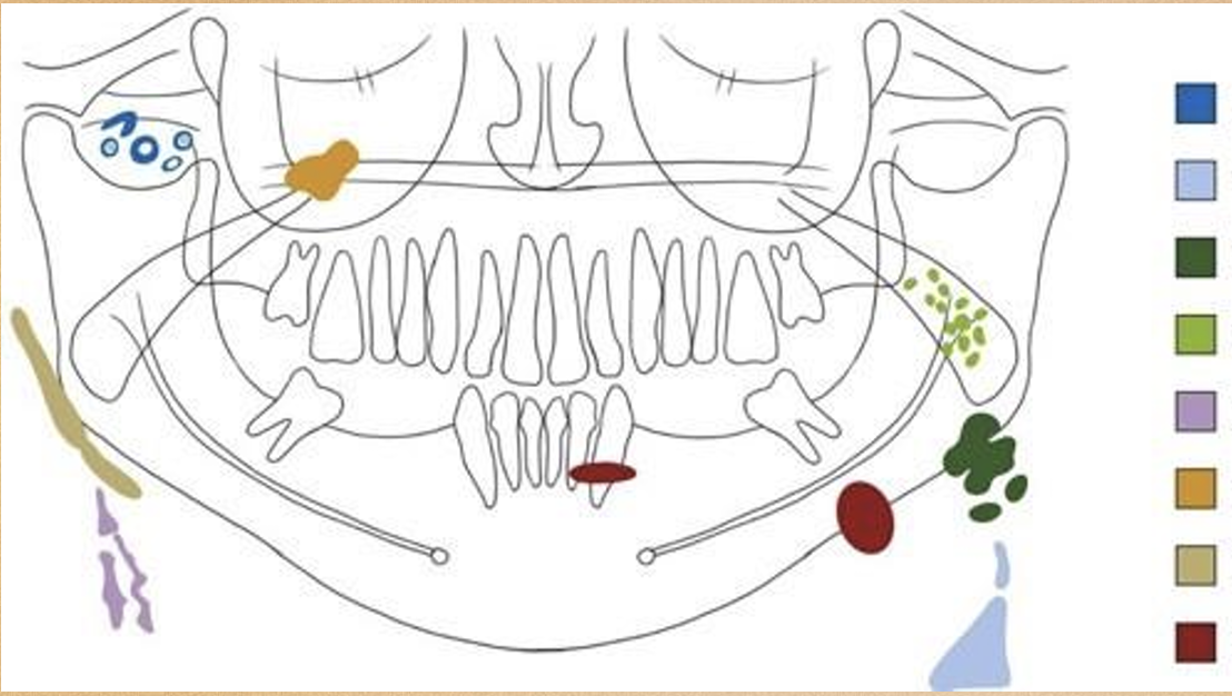
light green
tonsilloliths

purple
calcified atheromatous plaque

orange
antrolith

beige
ossified stylohyoid ligament

red
sialoliths

what’s goin on here?
long standing cyst that has residual calcifications
what is this?
dystrophic calcification of the tonsils
what is this?
dystrophic calcification of lymph nodes
what is this?
calcification of the carotid artery
what is this?
calcifications of the carotid artery on MDCT
in the 5 pixels this image is comprised of, what do you see?
calcification of the facial artery
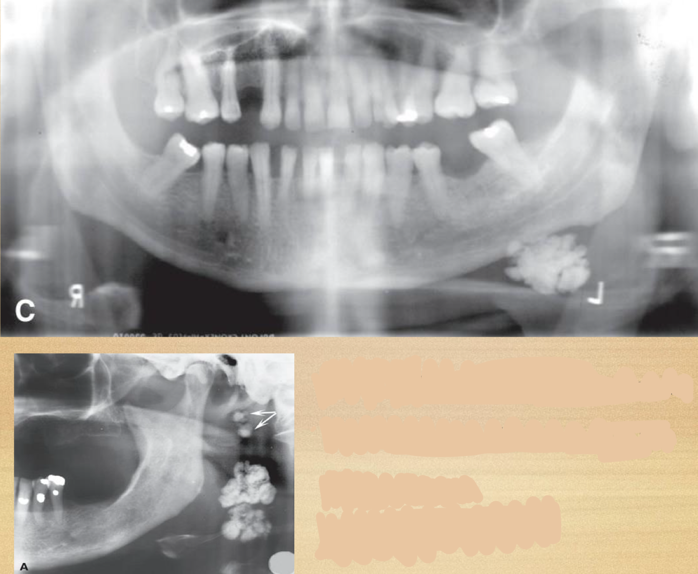
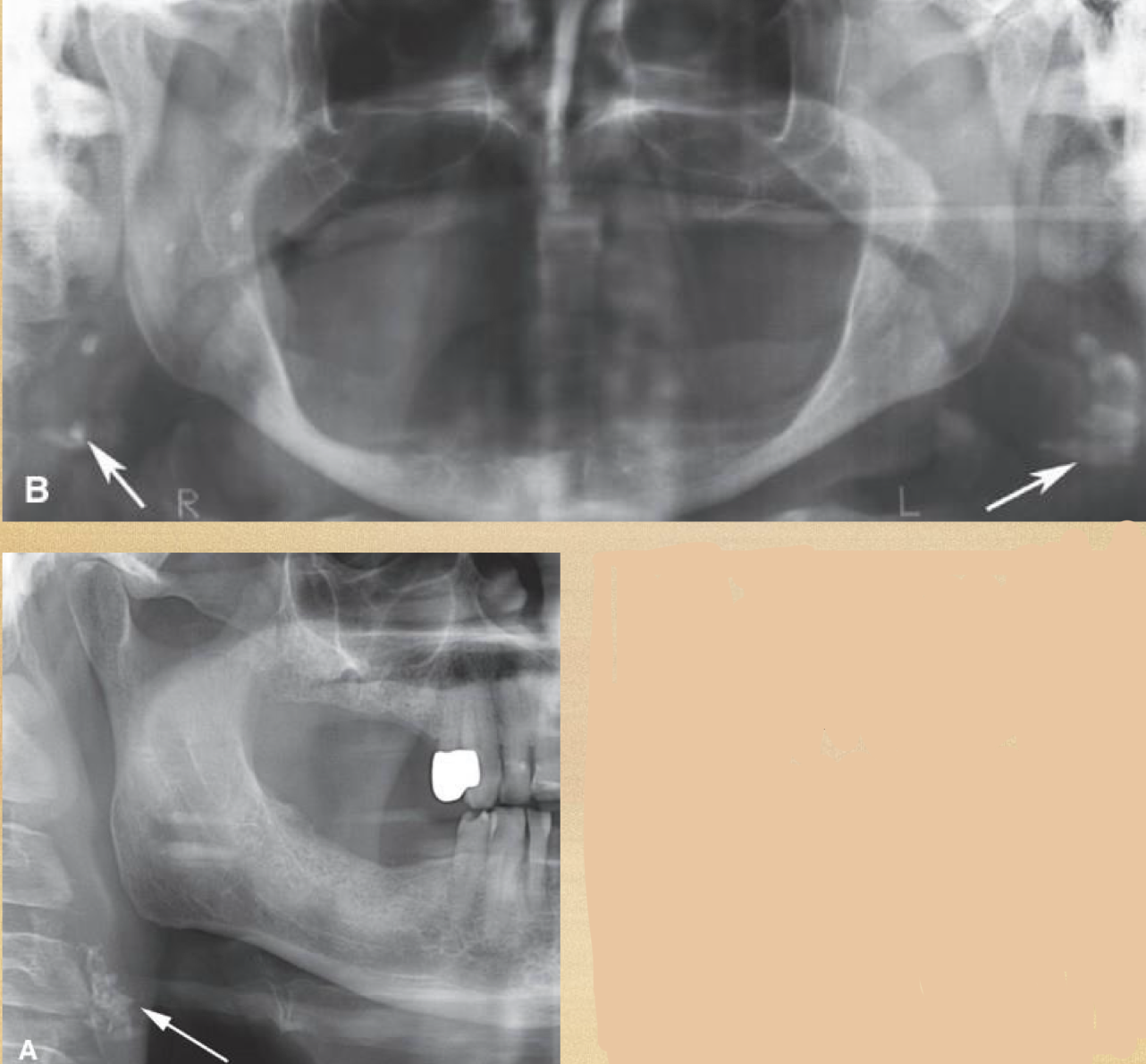
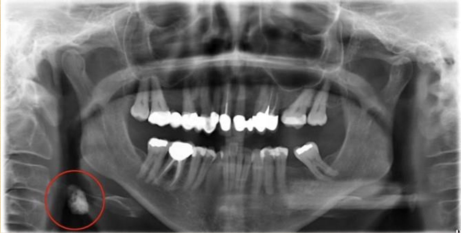
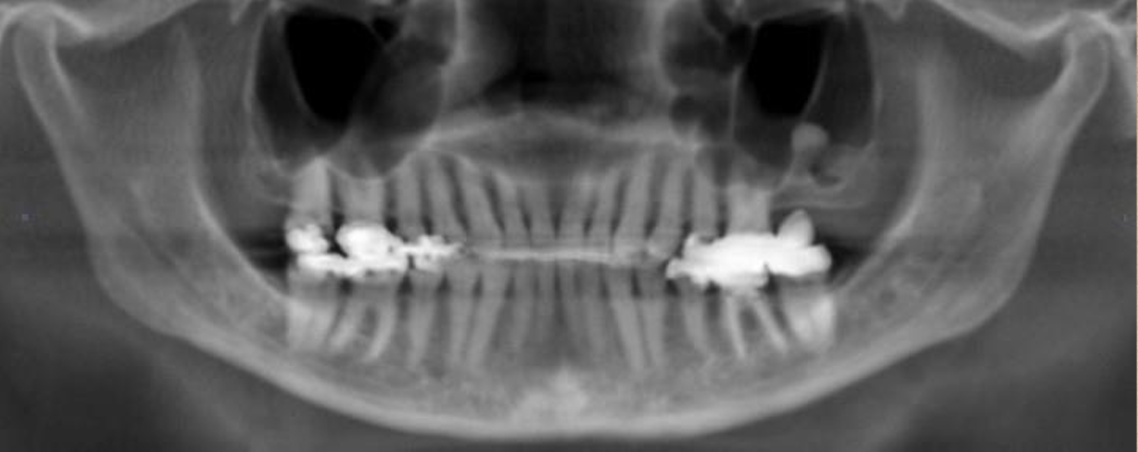
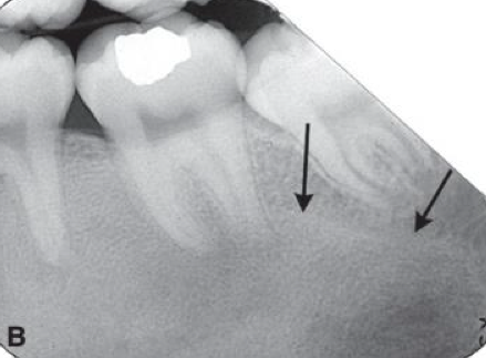
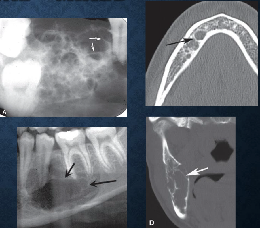
what is this?
phleboliths
what is this?
phleboliths
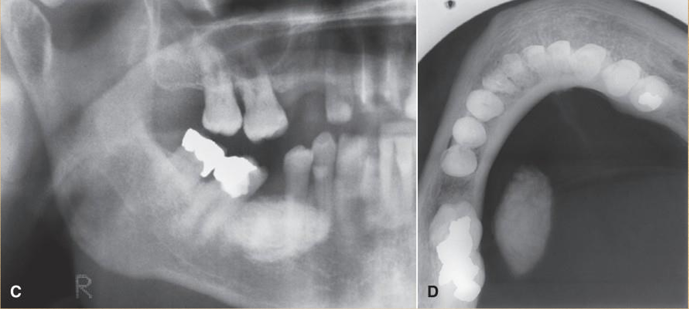
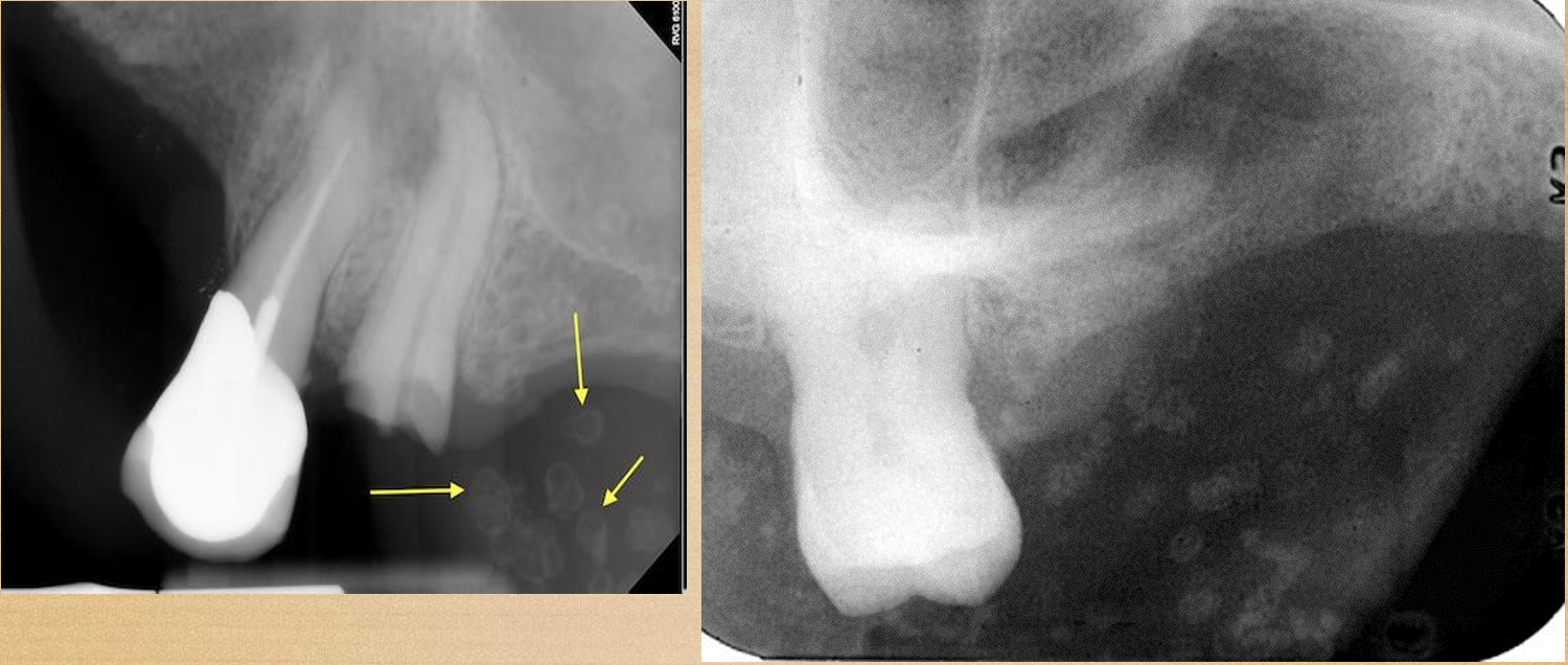
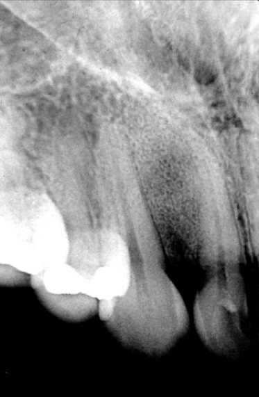
what is this?
sialolith
what is this?
sialolith
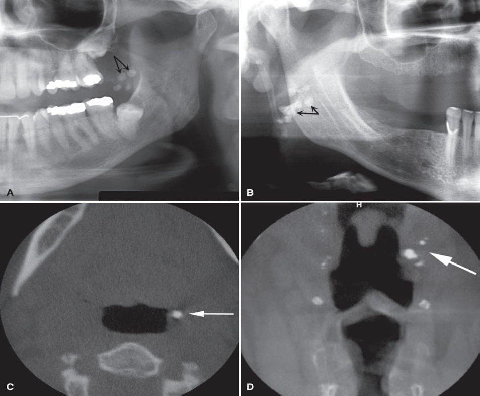
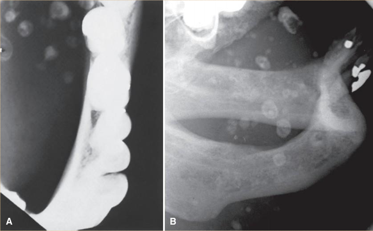
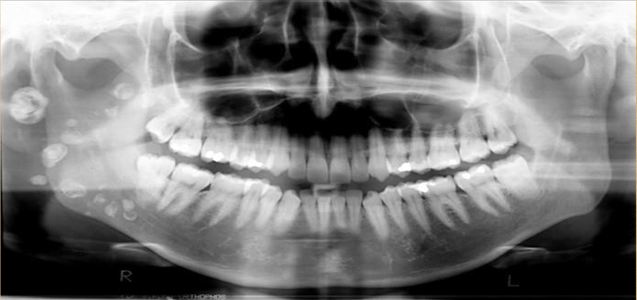
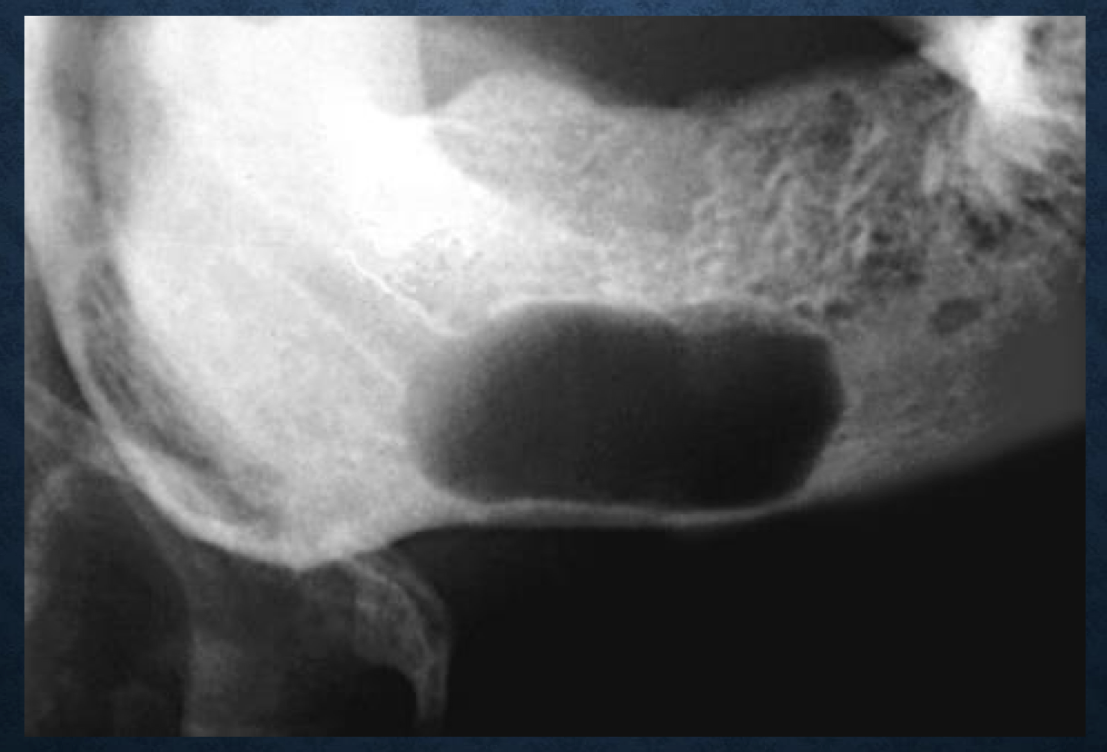
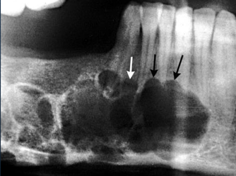
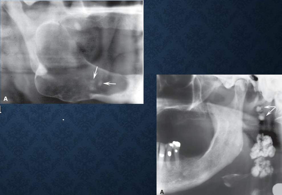
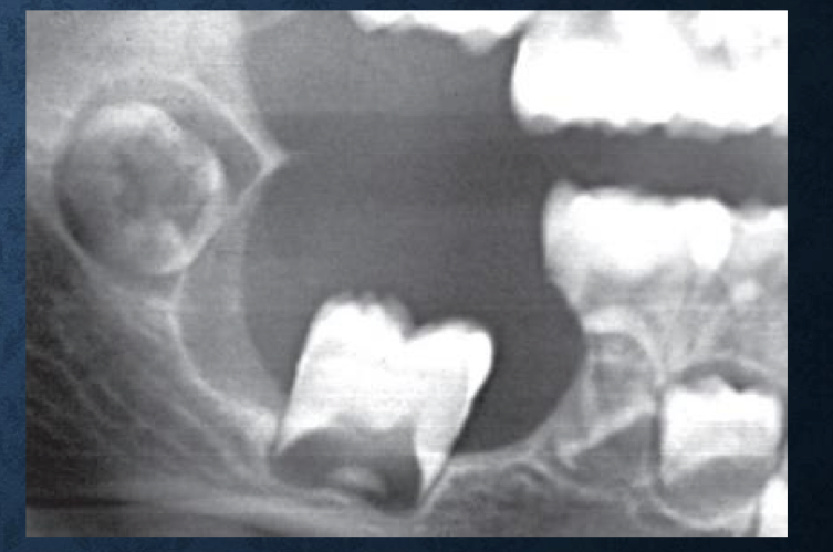
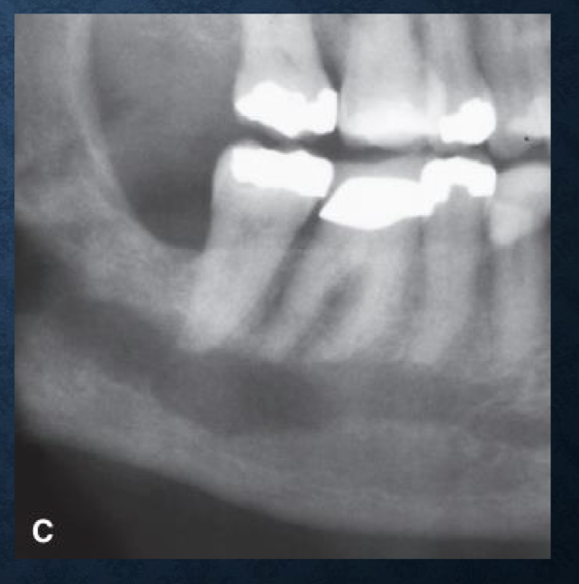
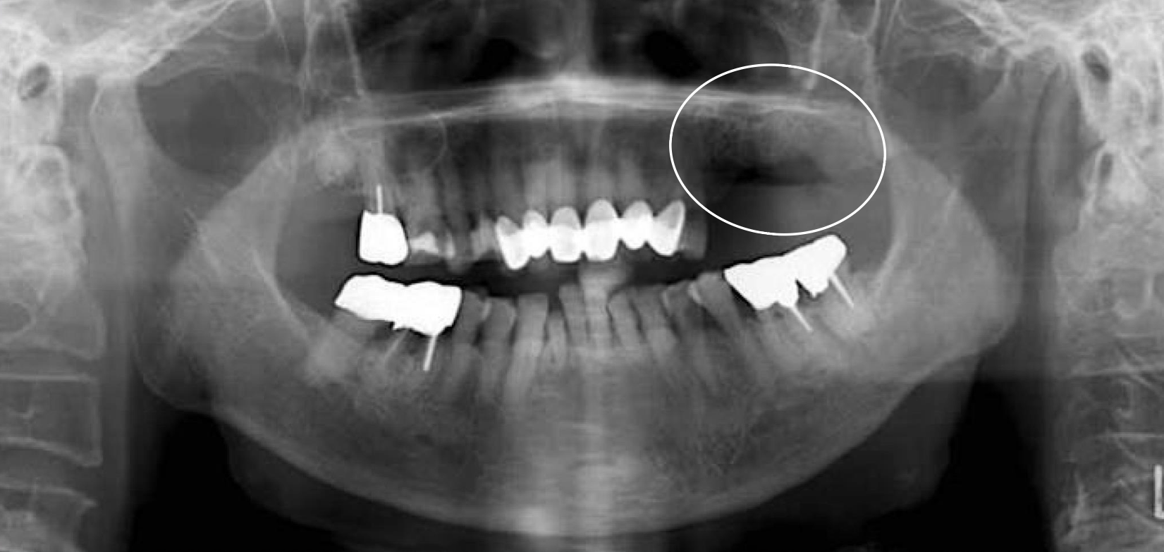
what do you see in this ~reconstructed pano~
antrolith
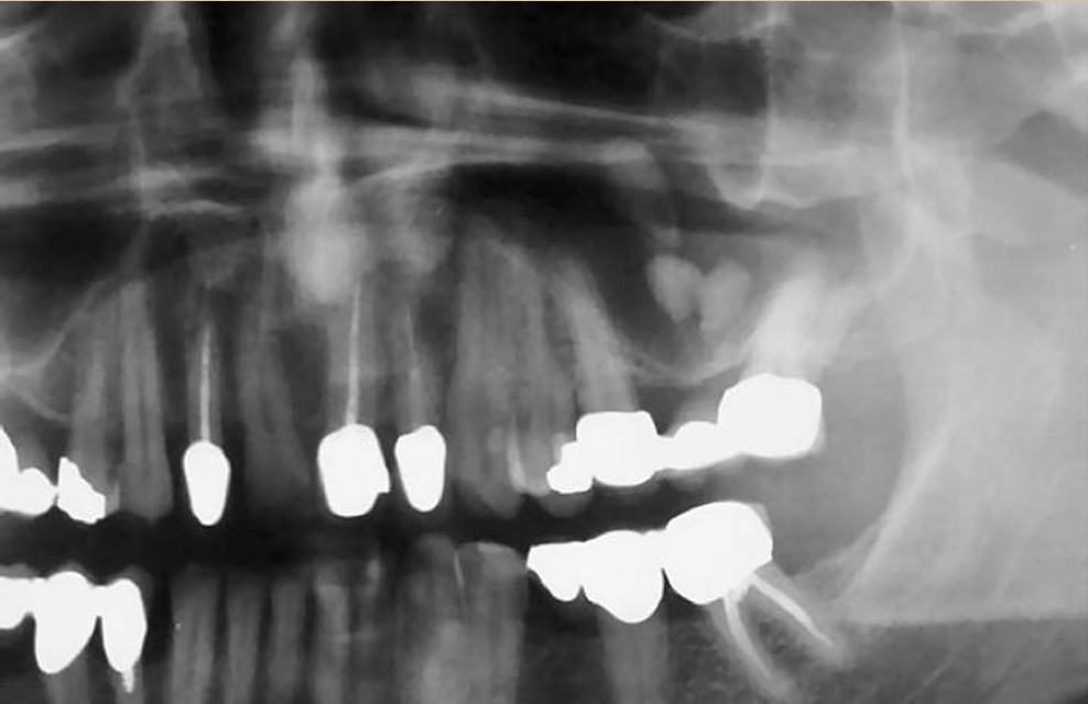
what is this?
antrolith
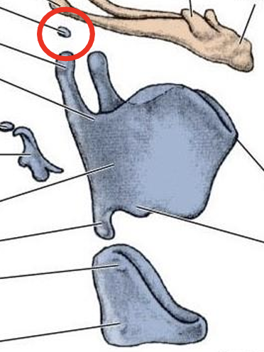
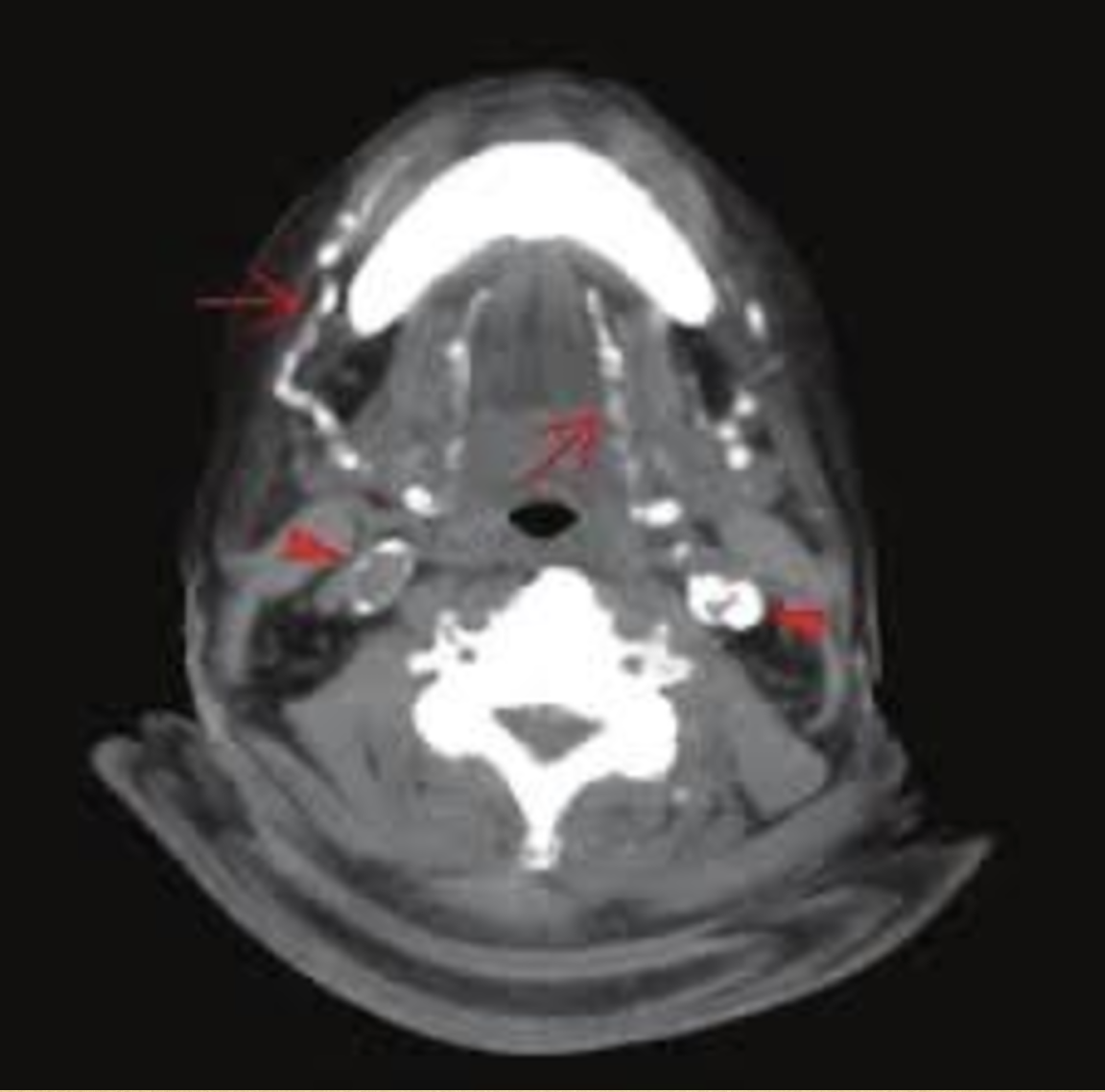
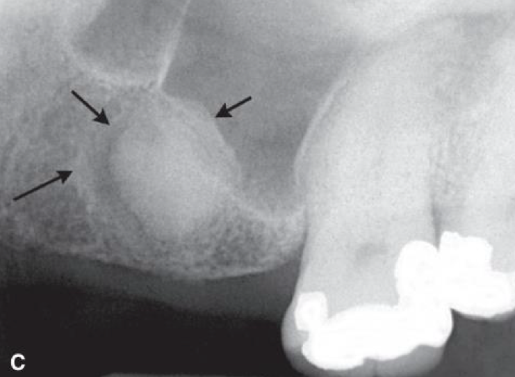
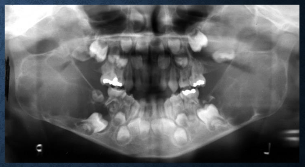
what is circled in red?
triticeal cartilage
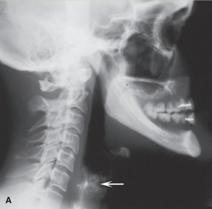
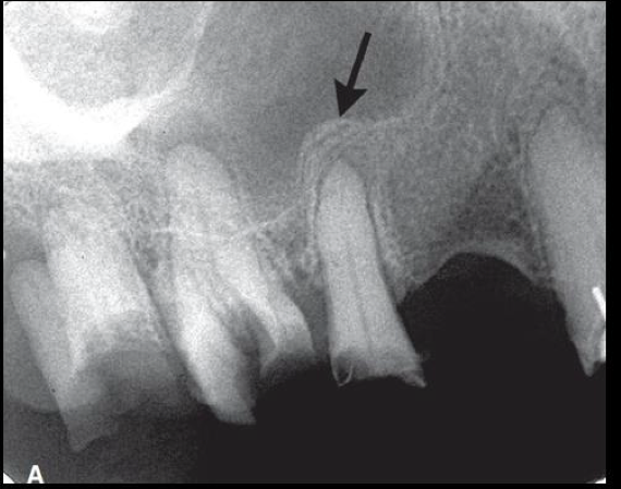
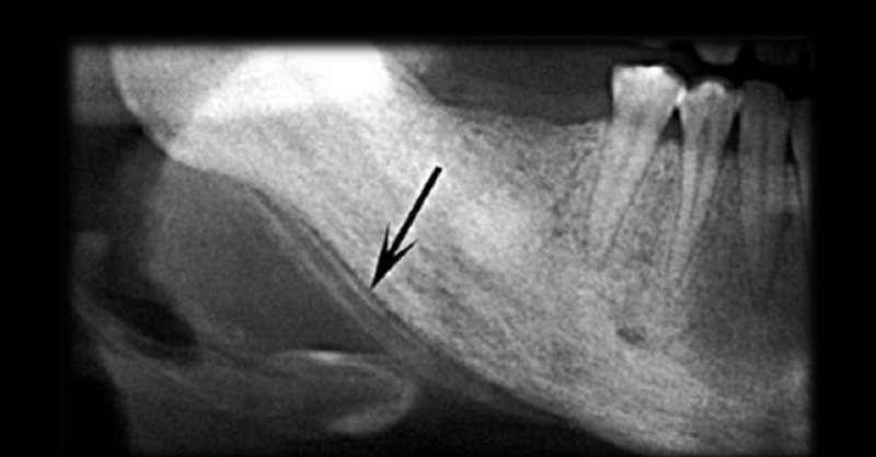
what is the white arrow pointing to?
calcified thyroid cartilage

what is A?
calcification of triticeous cartilage

what is B?
calcification of thyroid cartilage
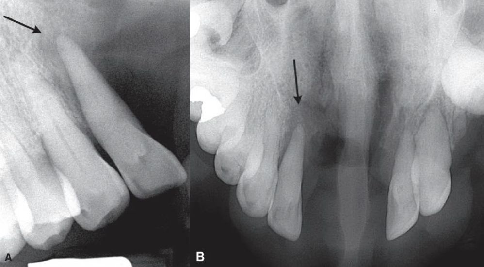
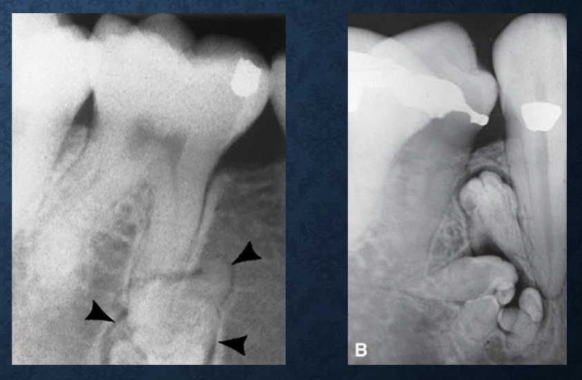
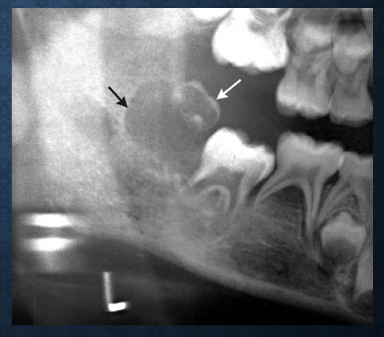
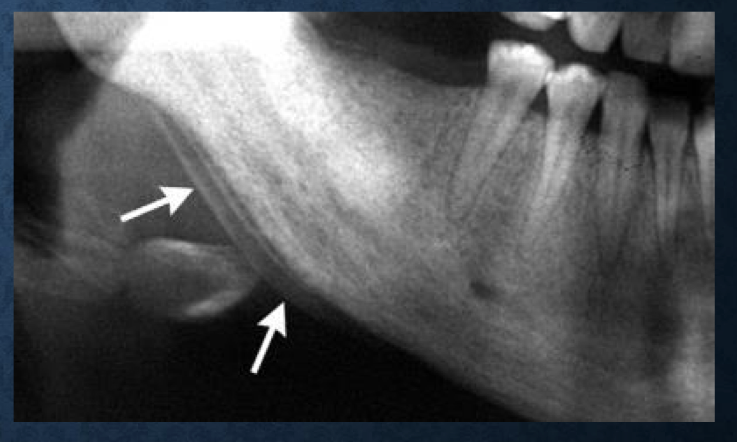
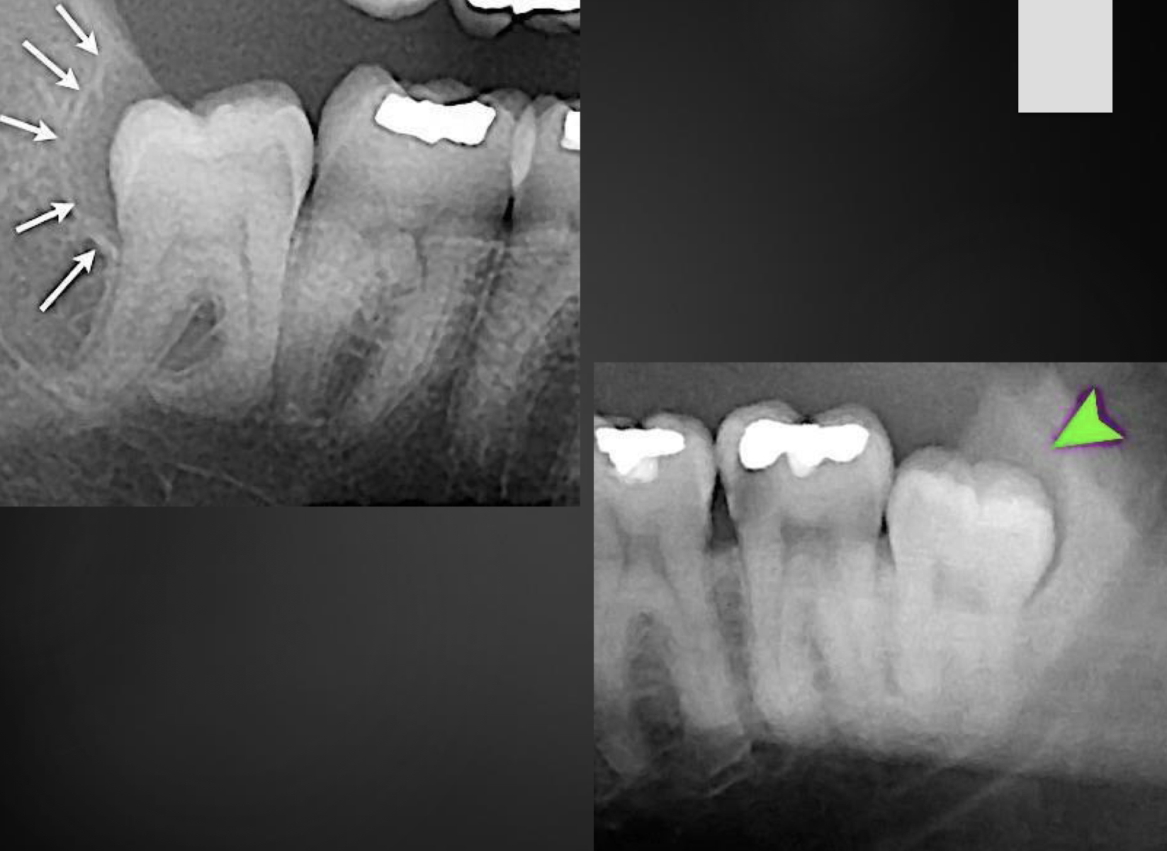
what are the white arrows pointing at?
calcified thyroid cartilage
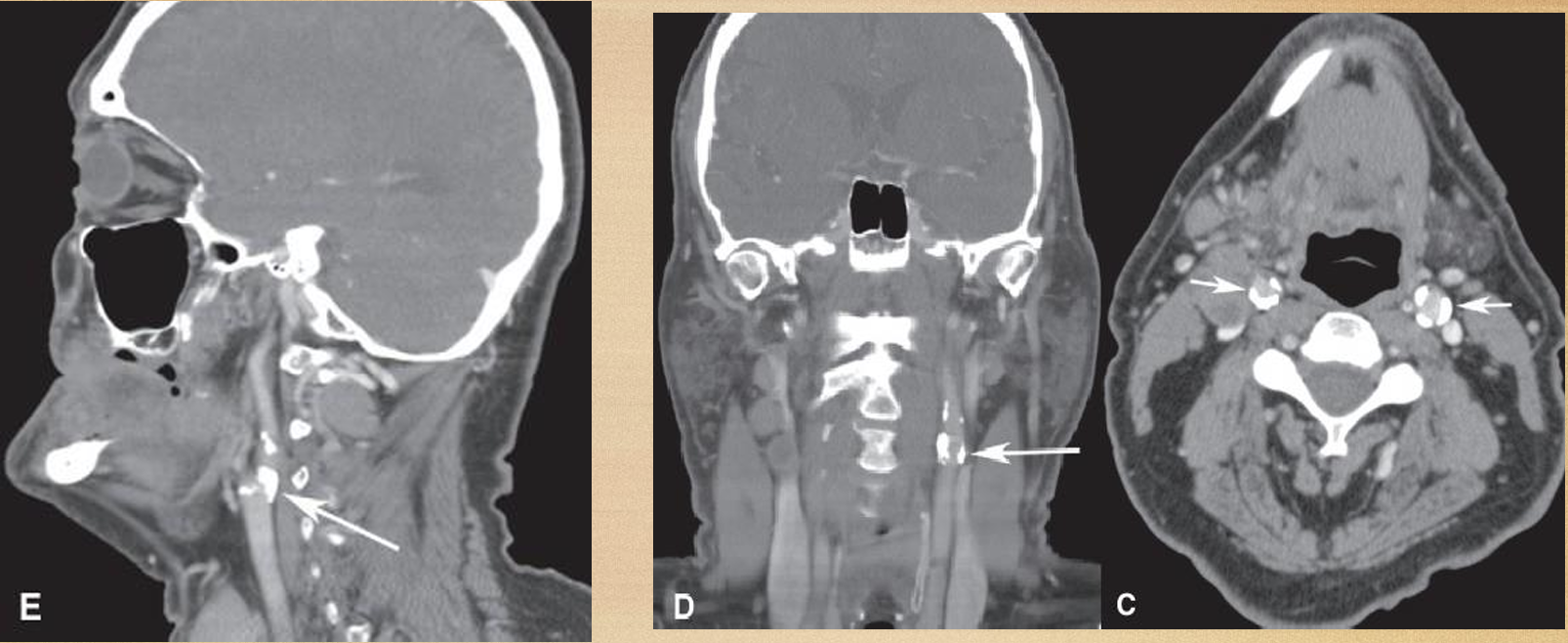
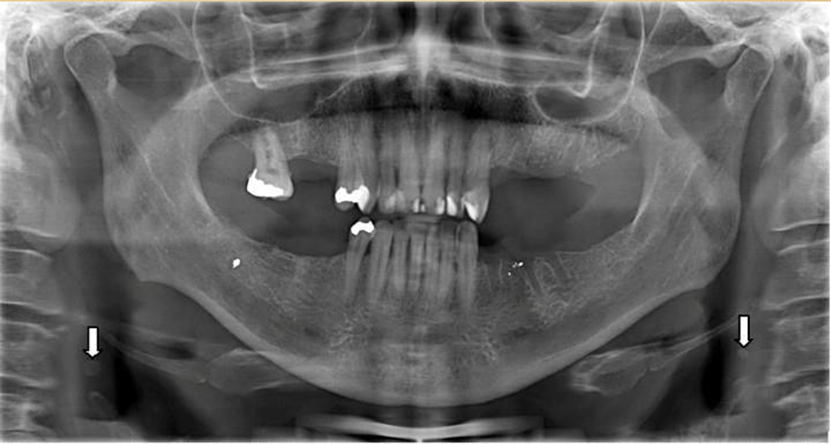
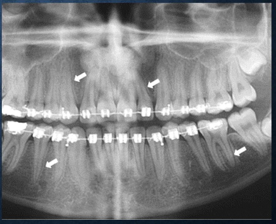
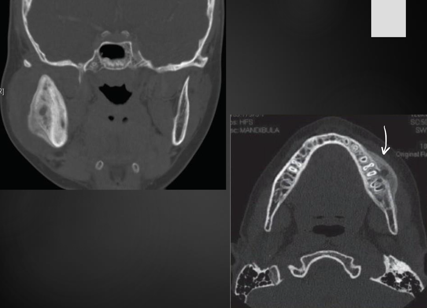
what are the white arrows pointing at?
ossification of the stylohyoid ligament
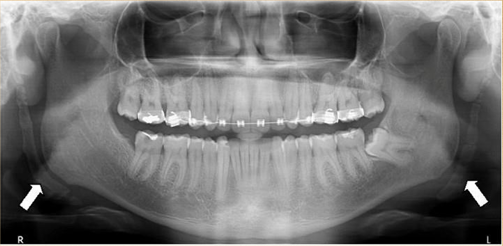
what are the white arrows pointing at?
ossification of the stylohyoid ligament
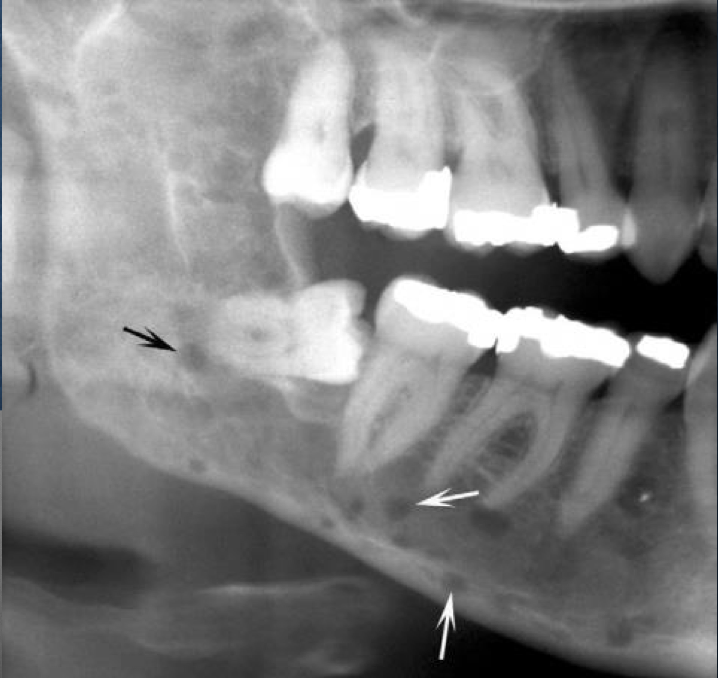
what is this?
osteoma cutis
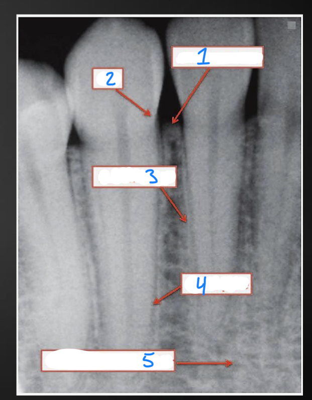
1?
alveolar crest

2?
CEJ

3?
lamina dura

4?
PDL space

5?
alveolar bone proper
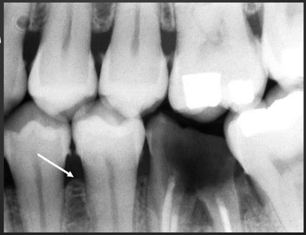
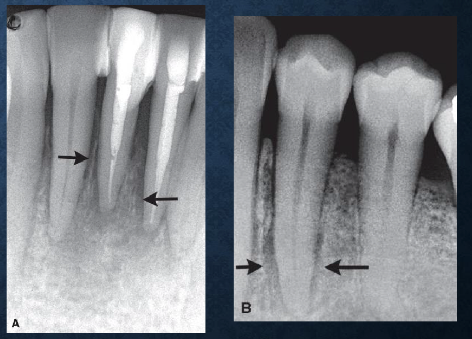
what is the arrow pointing to?
early bone changes
what kind of bone loss is this?
horizontal
what kind of bone loss is this?
vertical
what are the arrows pointing at?
interdental craters
what can be seen in these images?
buccal/lingual cortical plate loss
what are these arrows pointing at?
osseous deformities in furcations
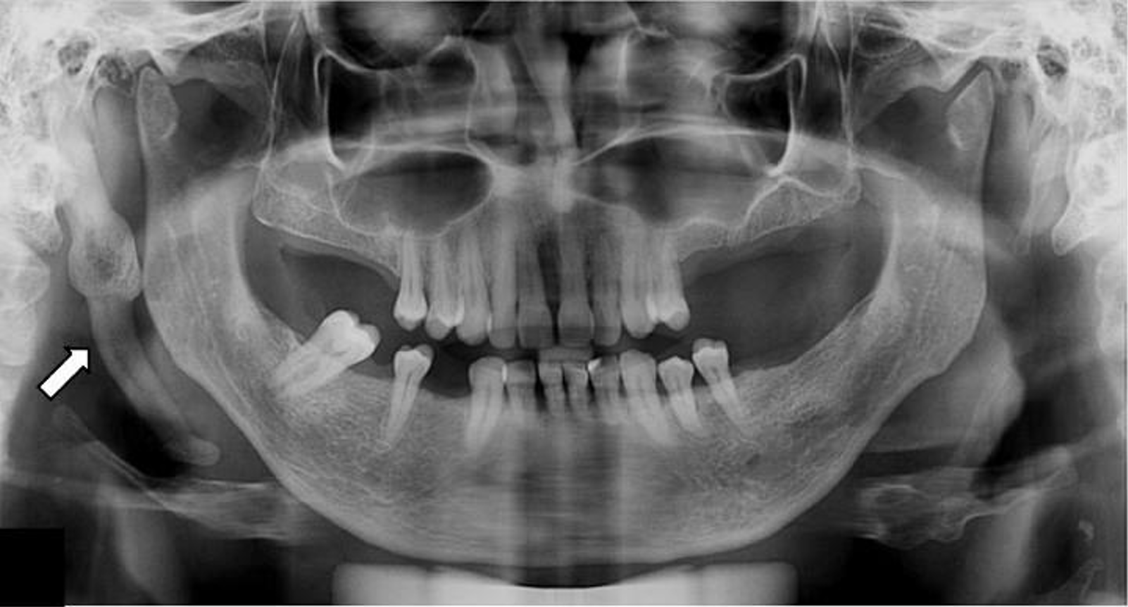
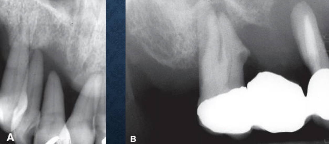
what is the arrow pointing at?
periodontal abscess
what’s going on here?
aggressive periodontitis

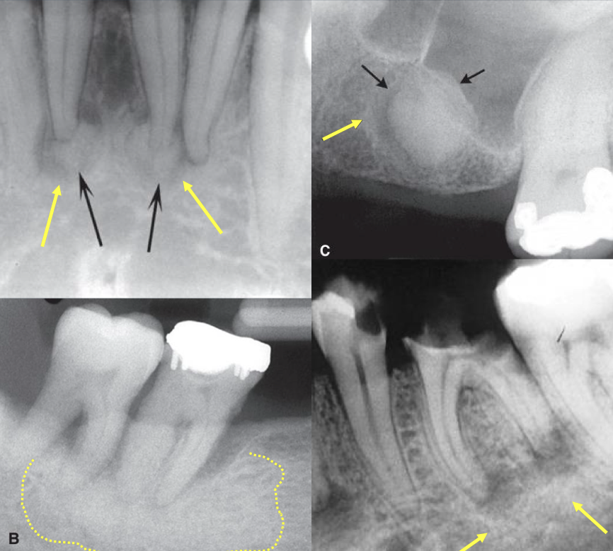
what is a, and how can you tell?
squamous cell carcinoma- irregular bone destruction and this is the only area affected

what is b, and how can you tell?
malignant tumor- loss of the floor of the maxillary sinus and irregular PDL widening (metastasis)
what is c, and how can you tell?
langerhans’ cell histiocytosis- bone destruction with epicenter at the midroot region, scooped out, usually no other pathology
what is the epicenter of this lesion and what does that tell you?
within the IAN
neural or vascular origin
punched-out
sharp boundary but no radiopaque line
surrounding bone appears normal
sclerotic
thick radiopaque border of reactive bone
indicates slow growth, potential of the lesion to stimulate production of bone
soft tissue capsule
radiolucent at the periphery
usually indicates a benign tumor
blending
gradual transition between adjacent normal and abnormal bone
focus on trabecular, not marrow
invasive
area of few or no trabeculae
focus on marrow spaces creating finger like projections
rapid growth, malignancy
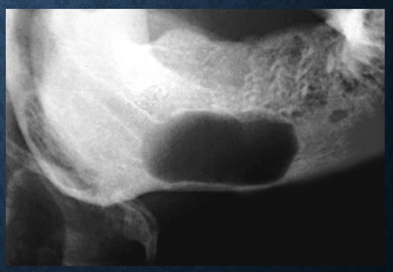
what shape is this lesion?
scalloped
how would you describe these lesions?
well defined, irregular

a?
air/gas

b?
fat

c?
fluid

e?
bone marrow
blue line?
soft tissue

purple line?
enamel

green line?
metal

pink line?
cortical bone

red line?
trabeculae
what trabeculation pattern is this?
ground glass
what trabecular pattern is this?
orange peel
how would you describe this trabecular pattern?
increased number or size of trabeculation
internal septation
strands of bone, can divide lesion into compartments- multilocular
can be curved or straight, coarse or fine
dystrophic calcification
occurs in damaged soft tissue like lymph nodes or chronically inflammed cysts
amorphous bone
homogenous, dense amorphous structure
tooth structure
enamel, dentin, pulp
a lesion that apically displaces teeth is typically…
follicular cyst or odontomas
a lesion that anteriorly displaces teeth is typically…
cherubism
a lesion that superiorly displaces teeth is typically…
lymphoma, leukemia, langerhan’s
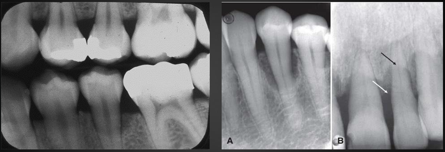
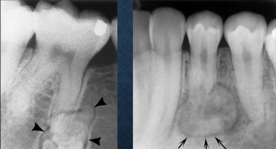
uniform widened PDL with intact lamina dura is typically…
orthodontic movement
irregular widened PDL with destruction of lamina dura is typically…
malignancy, quickly grows throughout PDL space
if you see tooth floating in the air on a radiograph, it is likely…
malignancy
superior displacement of the IAN is typically associated with…
fibrous dysplasia
widening of the IAN is typically associated with…
benign neural or vascular lesion
irregular widening of the IAN with cortical destruction is typically associated with…
malignancy
ossifying fibroma grows…
concentrically
fibrous dysplasia…
enlarges bone but maintains bone shape
periosteal reaction
exudate from inflammatory lesion lifts periosteum off of the surface
when it occurs multiple times, can result in onion-skin appearance
most commonly seen with osteomyelitis
less common but can be seen in leukemia and langerhan’s
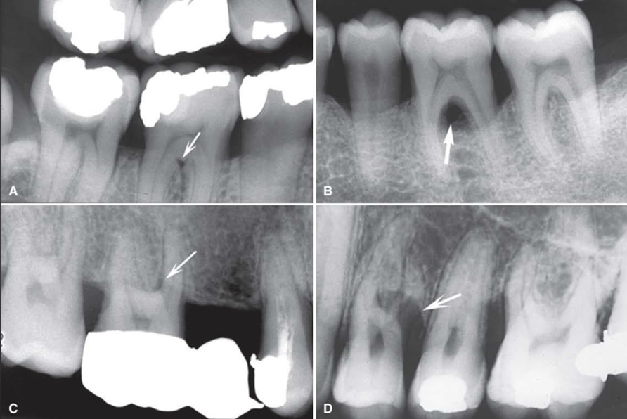
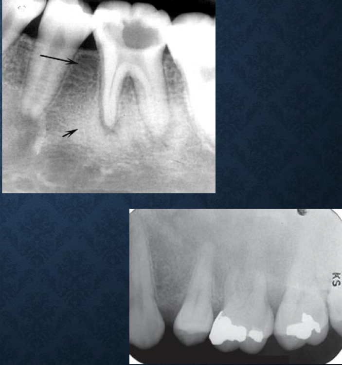
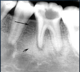
what is this?
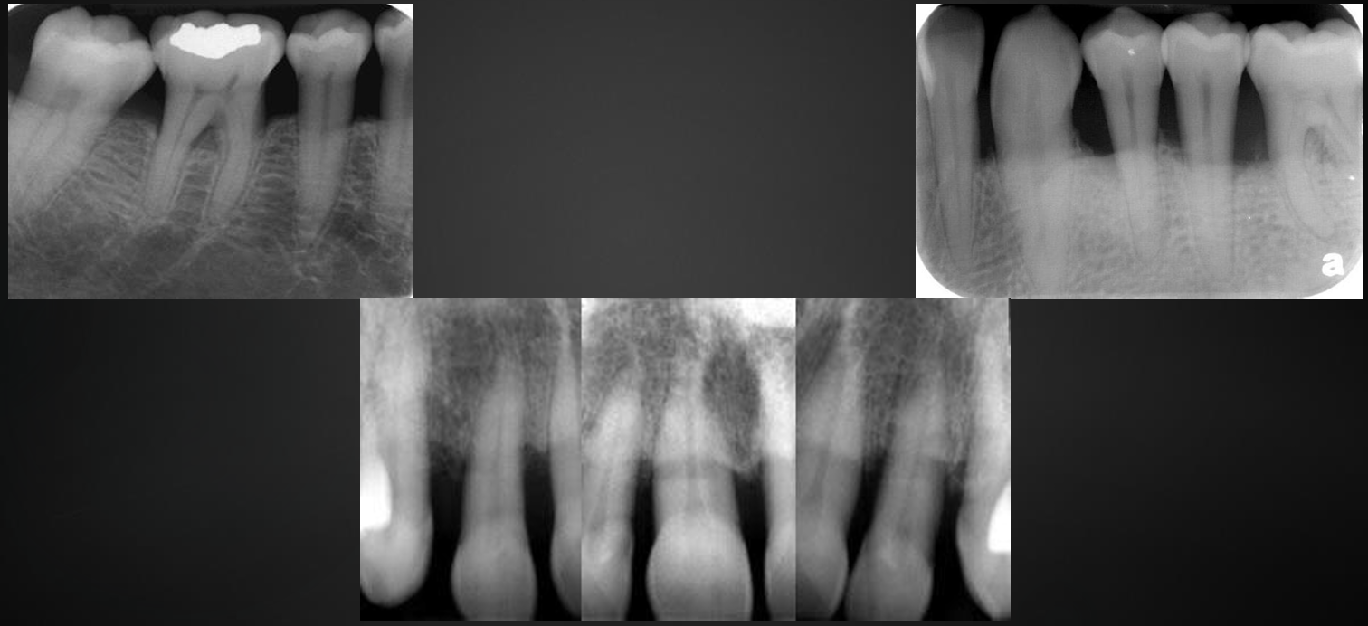
periapical inflammatory lesion
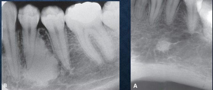
what is this?
periostitis
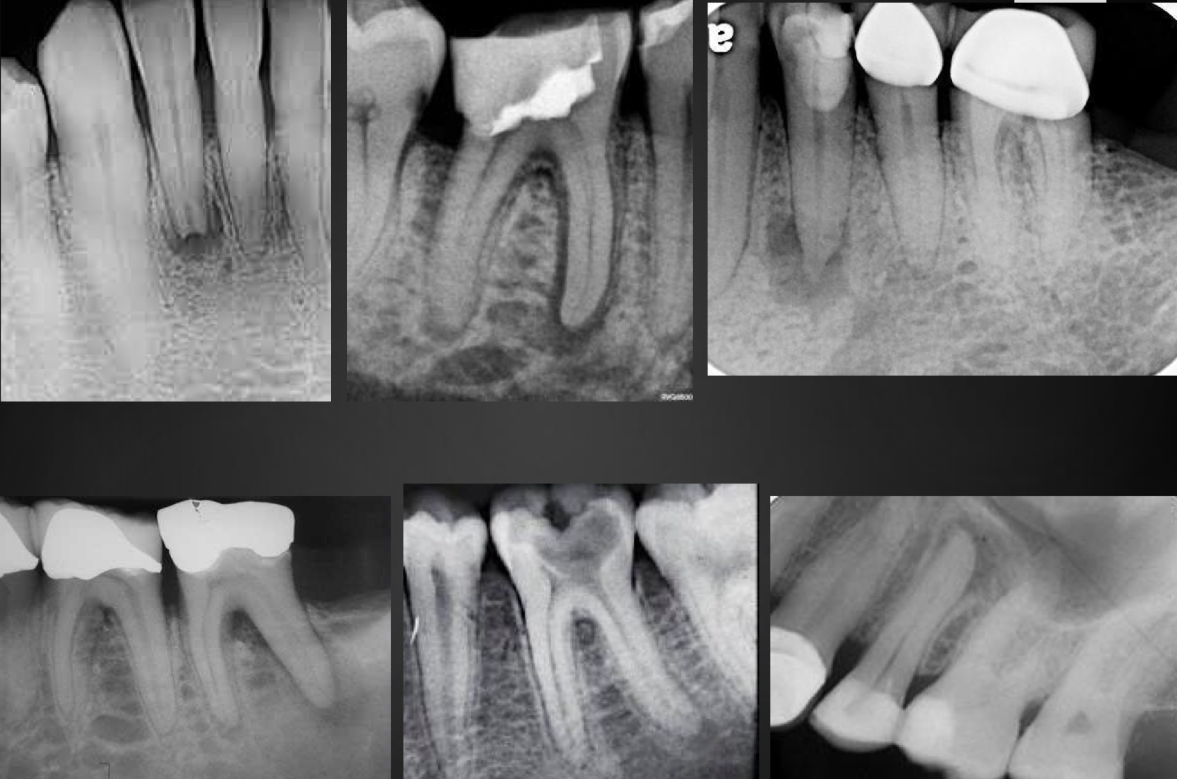
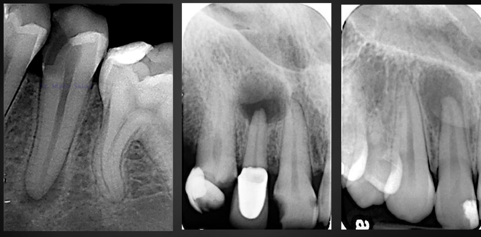
what do all of these images have?
periapical inflammatory lesion
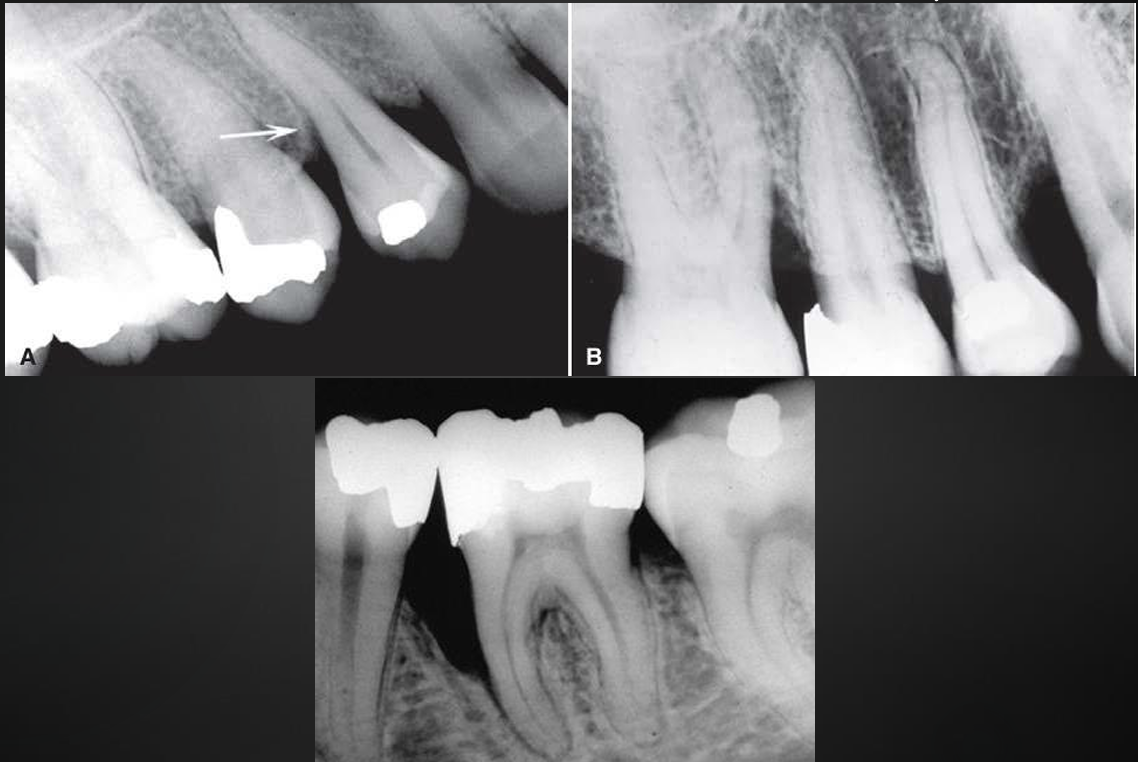
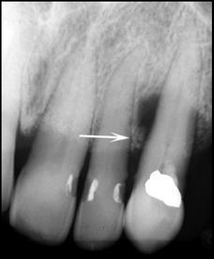
what is this?
acute phase osteomyelitis
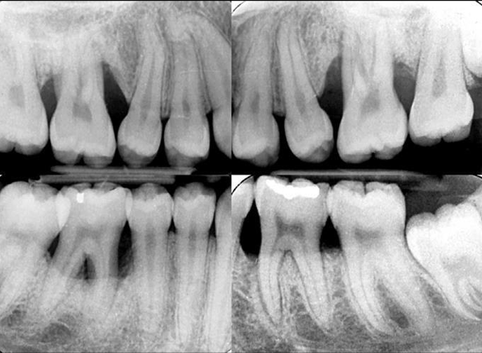
what is this?
periosteal bone reaction
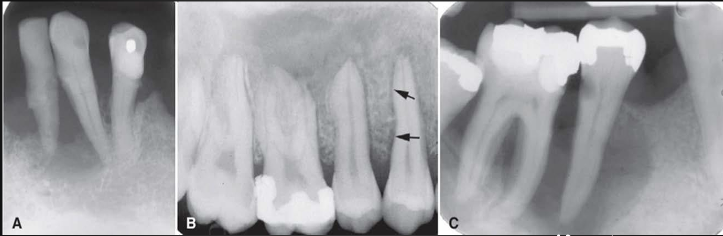
what is this? (there are more pics so look at the ppxt)
osteomyelitis
the two images on the left show…. while the two images on the right show…
radiographic manifestations of malignancies
radiographic manifestations post radiation treatment
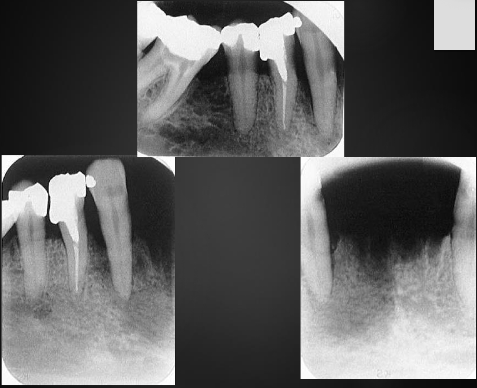
what is this? (there are more pics so look in the ppxt)
osteoradionecrosis
what is this? (there are more pics so look in the ppxt)
antiresorptive agent- induced ONJ
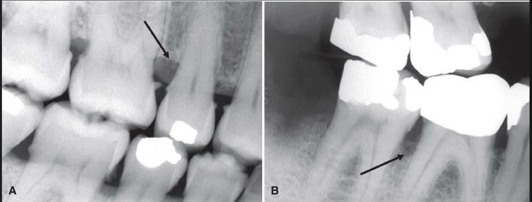
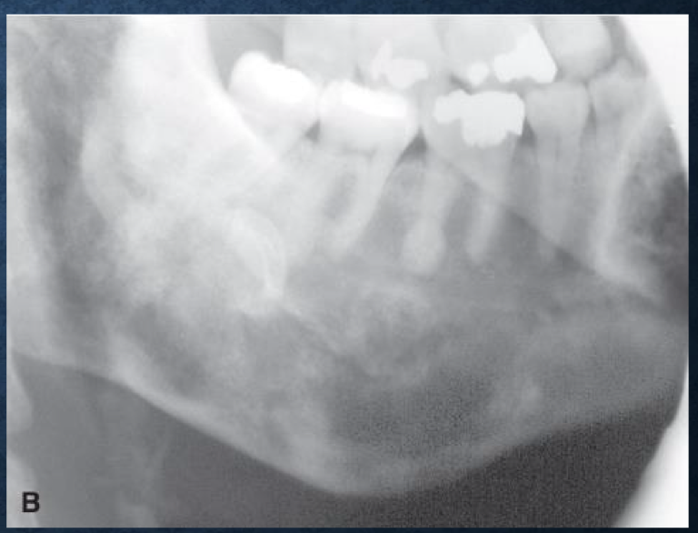
what is this?
pericoronitis
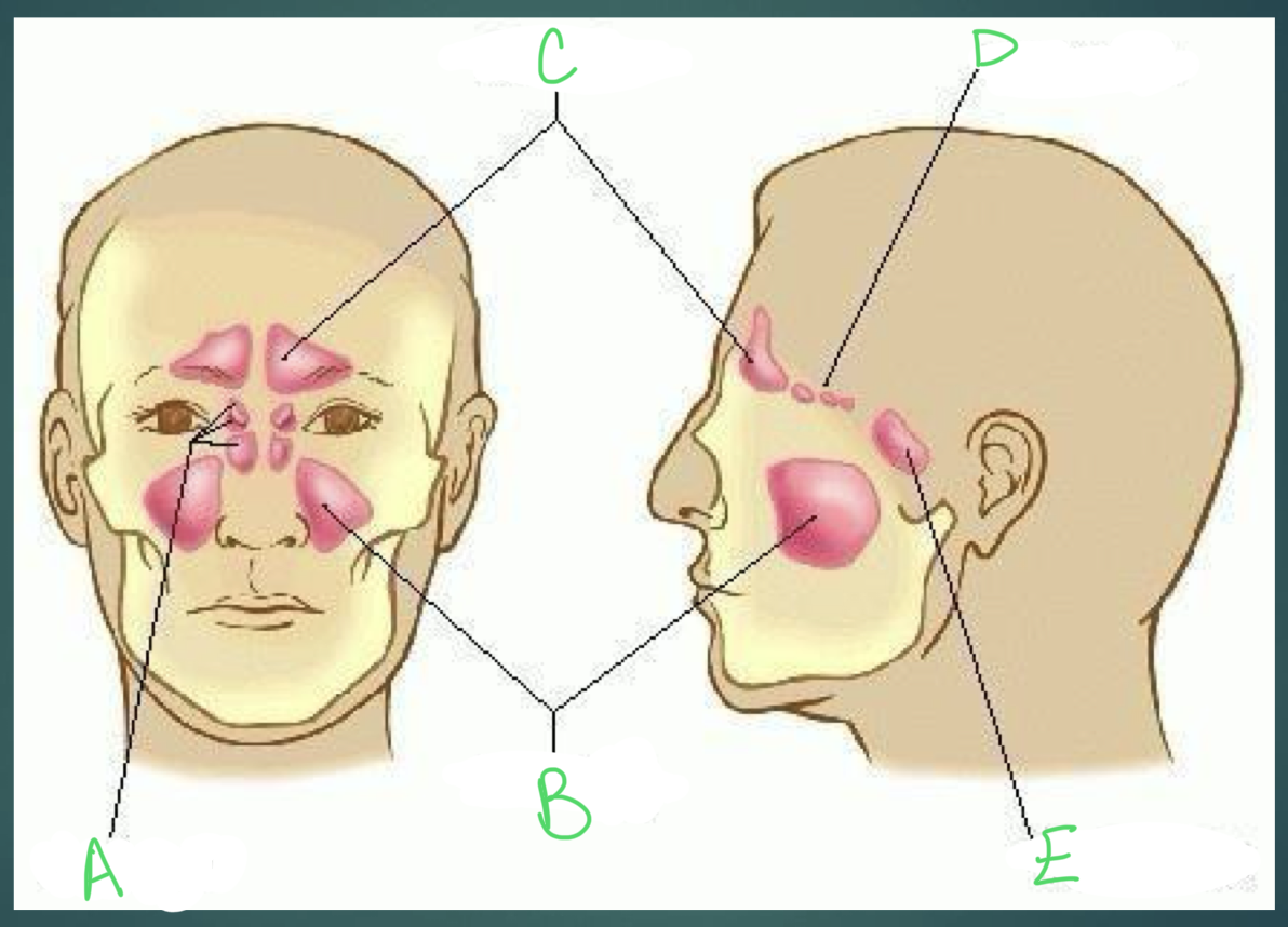
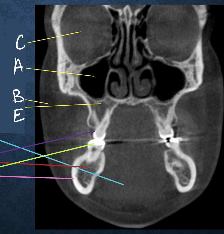
a and d?
ethmoidal sinuses

b?
maxillary sinuses

c?
frontal sinuses

e?
sphenoidal sinuses

a?
hiatus semilunaris

b?
middle meatus

c?
ethmoid bulla
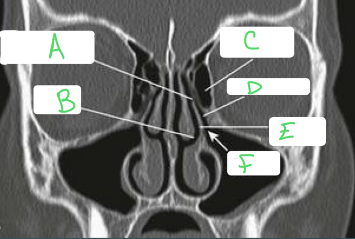
d?
infundibulum

e?
uncinate process

f?
maxillary ostium