Lab 8 PowerPoint Questions
1/119
Earn XP
Description and Tags
Anatomy and Phisiology 1
Name | Mastery | Learn | Test | Matching | Spaced | Call with Kai |
|---|
No analytics yet
Send a link to your students to track their progress
120 Terms
Which type of muscle tissue is highly branched, possess intercalated discs, and is comprised of cells having a single nucleus?
cardiac
Which cellular component/feature do skeletal, cardiac, and smooth muscle share?
large number of mitochondria
Which type of muscle tissue is comprised of multinucleated, striated cells that are under voluntary control?
skeletal
Why do skeletal muscle cells contain high numbers of mitochondria?
to supply the ATP needed for skeletal muscle function
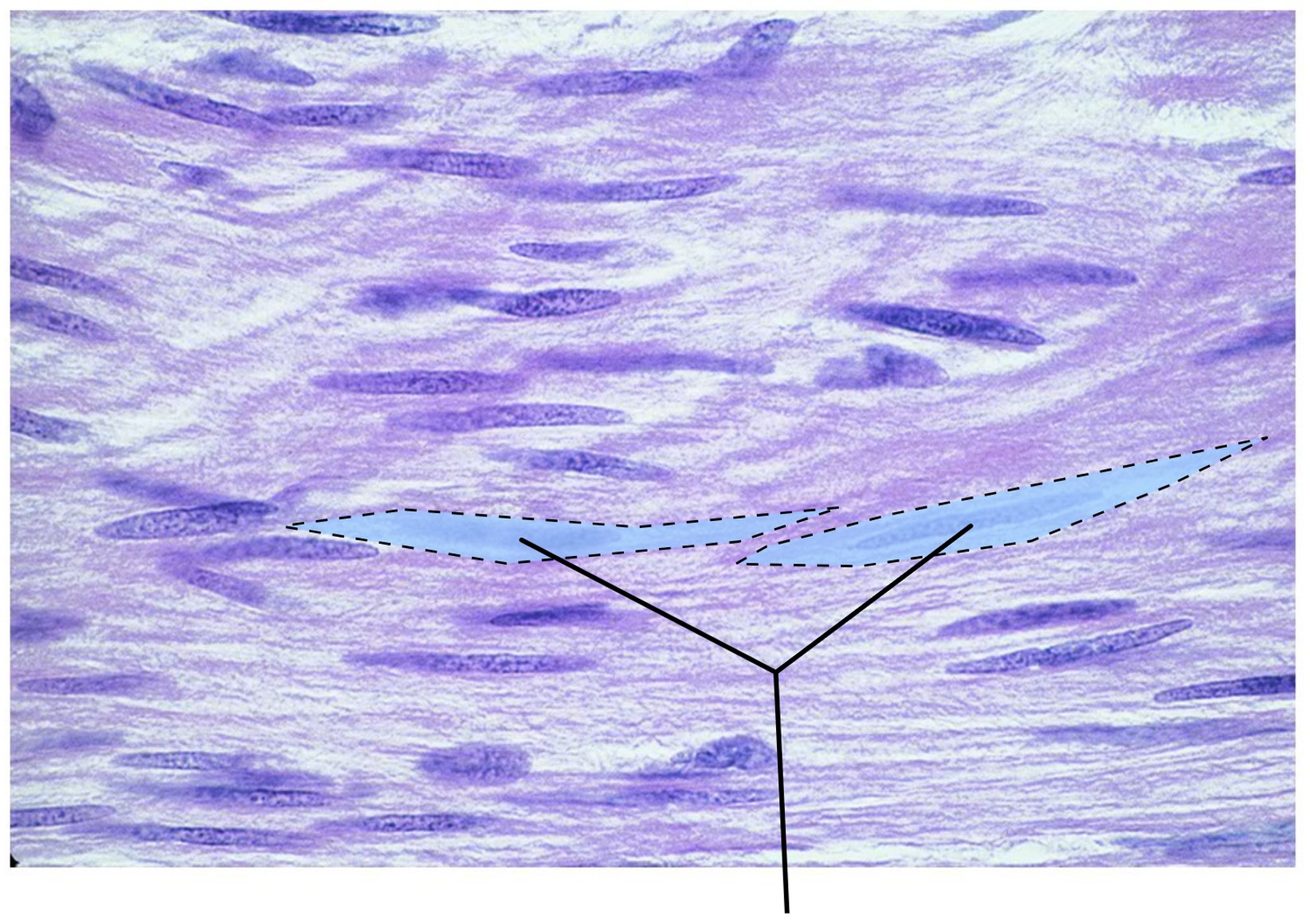
Which type of muscle tissue is comprised of cells that lack striations?
smooth
What tissue is highlighted in this image?
smooth muscle

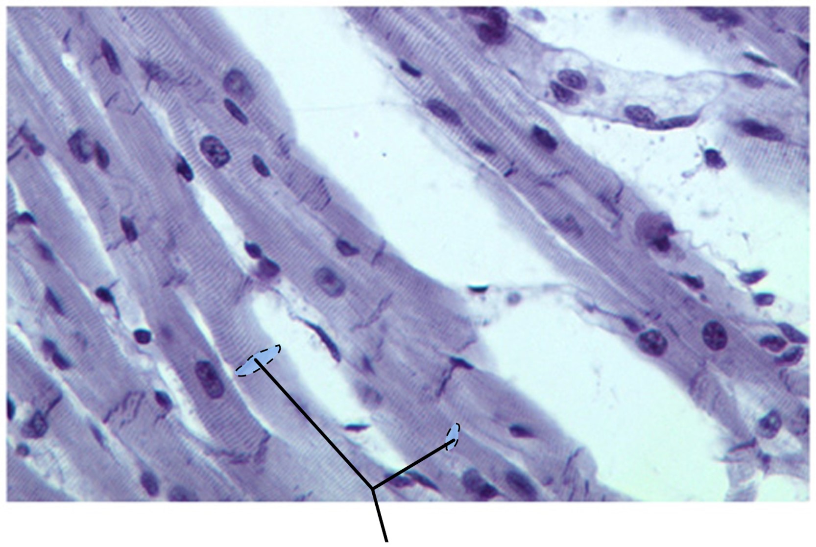
What structures are highlighted in this image?
intercalated discs
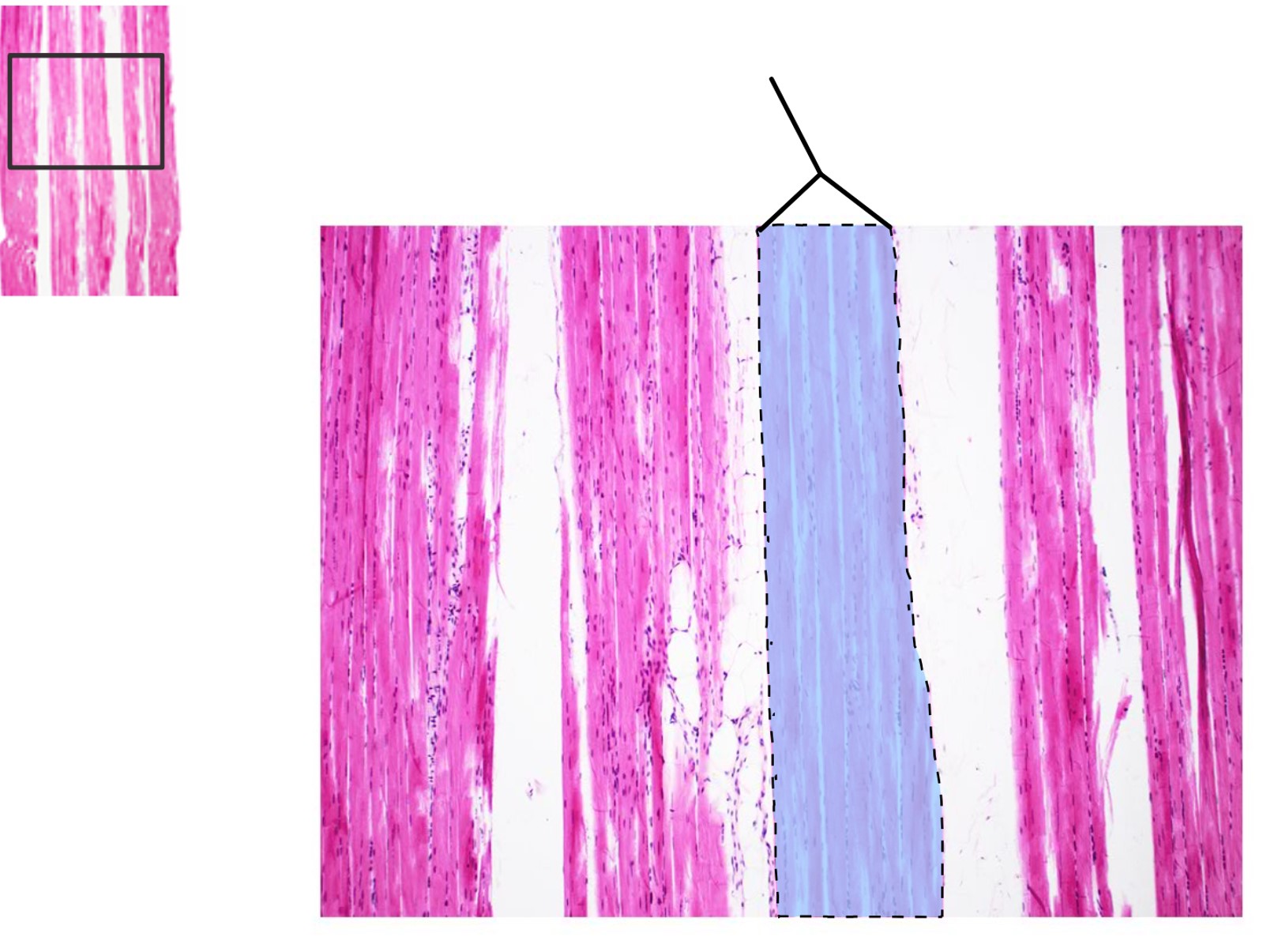
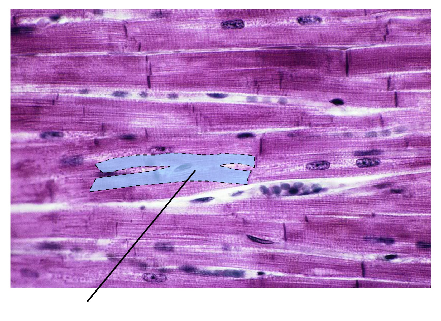
What muscle structure is highlighted?
fasicle
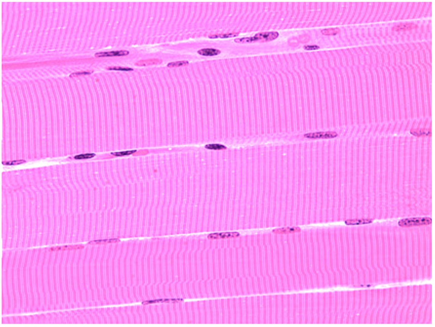
What tissue is shown in this image?
skeletal muscle

What tissue is highlighted in this image?
smooth muscle
What structure is highlighted in this image?
striations
What type of cell is highlighted in this image?
cardiac muscle cell
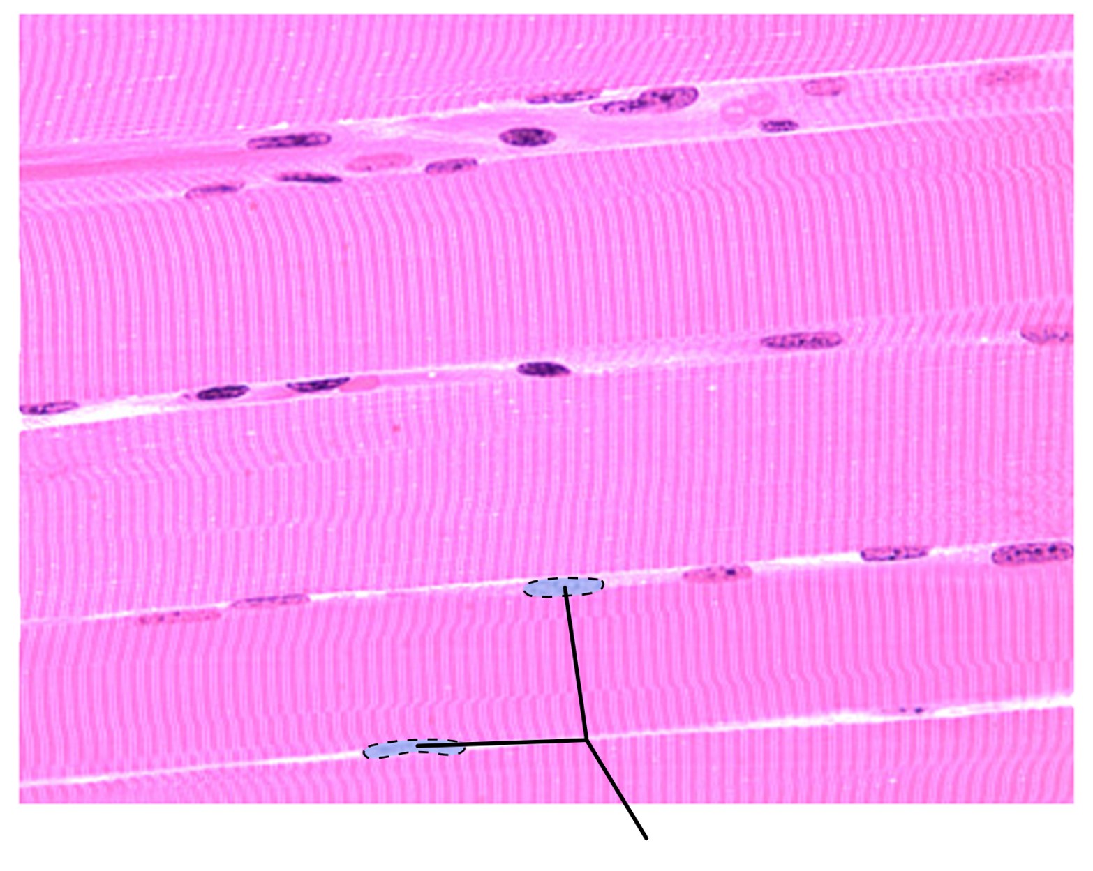
What structures are highlighted in this image?
nuclei
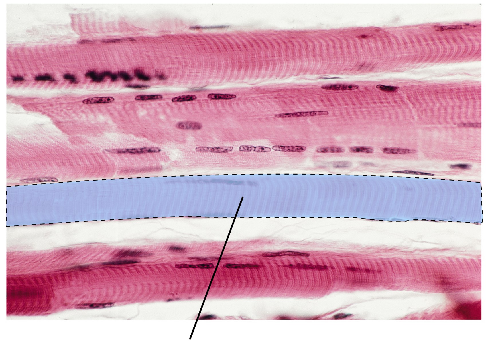
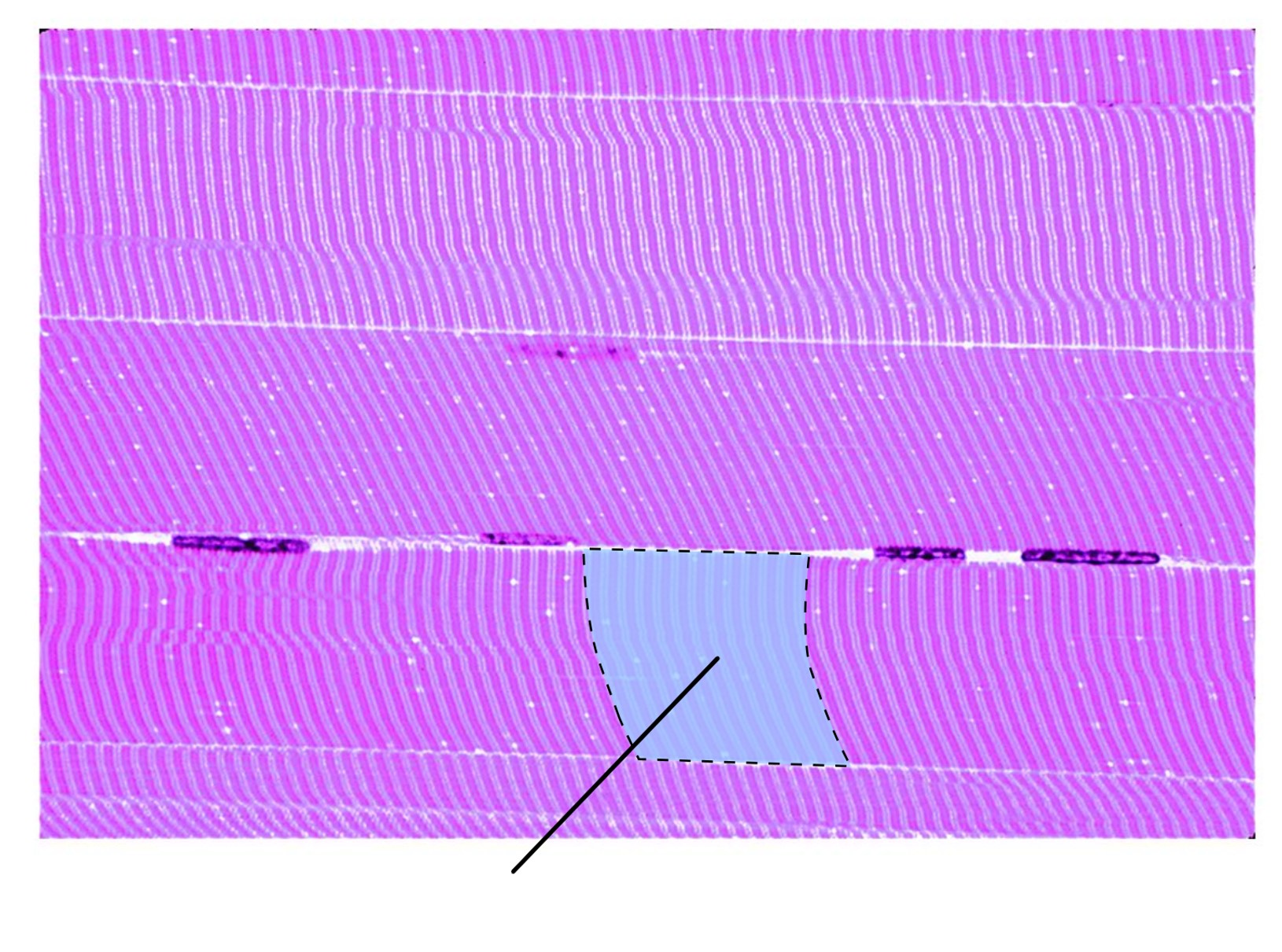
What structure is highlighted in this image?
skeletal muscle fiber
What kind of muscle has the highlighted structures?
cardiac muscle
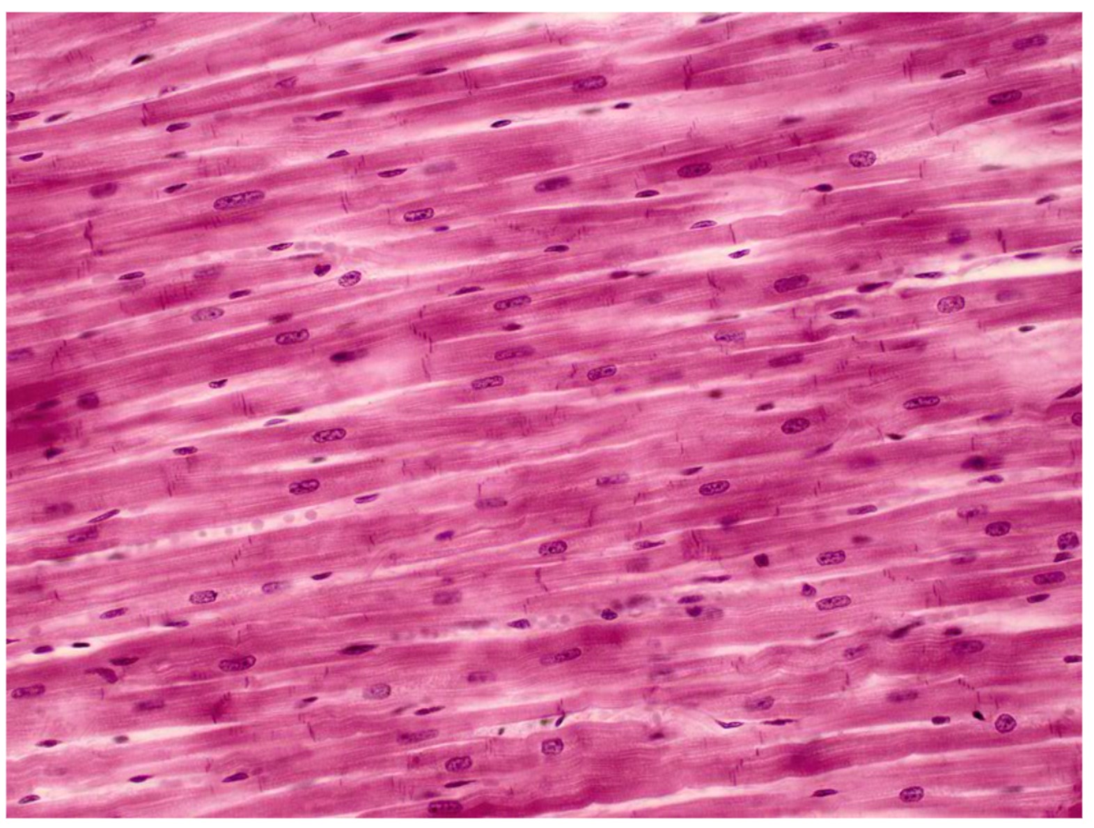
What tissue is shown in the image?
cardiac muscle
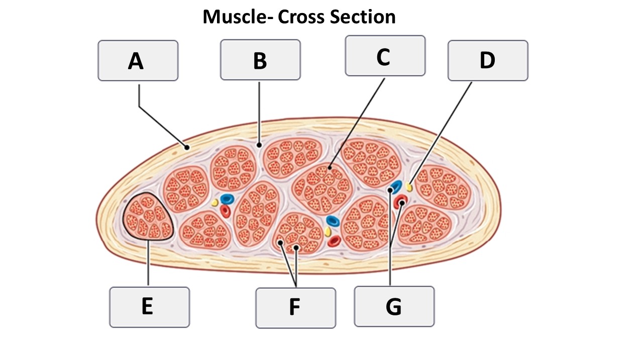
epimysium
A

perimysium
B

endomysium
C

muscle fasicle
E

muscle fibers
F

blood vessels and nerves
F

endomysium
C
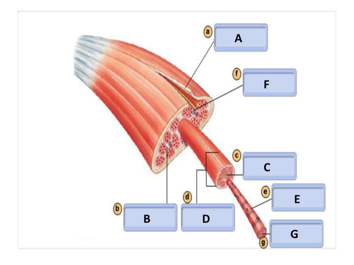
epimysium
A

muscle fiber
E

myofibril
G

muscle fascicle
D

perimysium
B
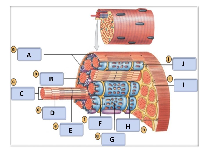
terminal cisterna
J

sarcoplasma
I

triad
F

T tubules
H

sarcoplasmic reticulum
G

thin filament
D

thick filament
E

myofibril
C

mitochondria
A

sarcolemma
B

zone of overlap
I

M line
H

thick filament
C

saracomere
A

thin filament
B

I band
F

A band
G

Z line
D
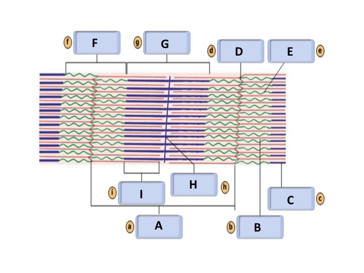
titin
E

skeletal muscle
A

muscle fiber
C
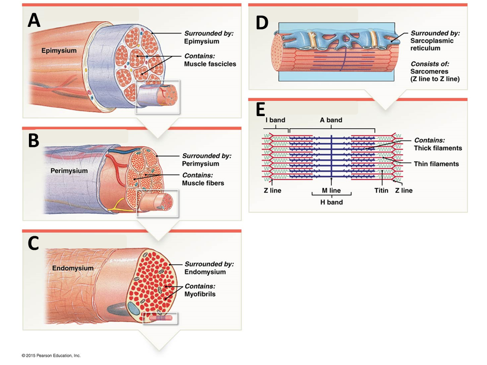
muscle fasicle
B

myofibril
D

sarcomere
E
What transport process is used when acetylcholine (ACh) is released into the synaptic cleft?
exocytosis
Acetylcholine is a(n) __________, which is a type of chemical released by neurons that changes the plasma membrane permeability or other properties of another cell.
neurotransmitter
What are two ways that ACh is removed from the synaptic cleft?
diffusion and break down by acetylcholinesterase
During contraction, the entire skeletal muscle shortens and produces a pull on the tendons on either end of the muscle. This is called ___________.
tension
When an action potential travels along the sarcolemma, it triggers the release of calcium ions by the sarcoplasmic reticulum (SR). What part of the SR releases calcium?
terminal cisternae
Interactions between thin and thick myofilaments of the sarcomere are responsible for ________.
muscle contraction
_______ covers the active sites of actin prior to calcium binding to troponin.
tropomyosin
“Cocking” the myosin head requires energy. What molecule acts as an ATPase to break down ATP for the energy needed to cock the myosin head?
myosin
Binding of neurotransmitter to the receptors on the motor end plate opens what type of ion channels?
chemically-gated channels
Once an action potential is propagated down the t-tubules, the terminal cisternae release ___________.
calcium ions
During events in the cross bridge cycle, which molecule has a binding site for calcium ions?
troponin
During events in the cross bridge cycle, which molecule covers the actin binding site?
tropomyosin
During events in the cross bridge cycle, which molecule has a binding site for myosin heads?
actin
What molecule must bind to the myosin head in order for it to disconnect with actin?
ATP
Hydrolysis of ATP returns the myosin molecule to the _________________________ conformation
high-energy

axon terminal
1
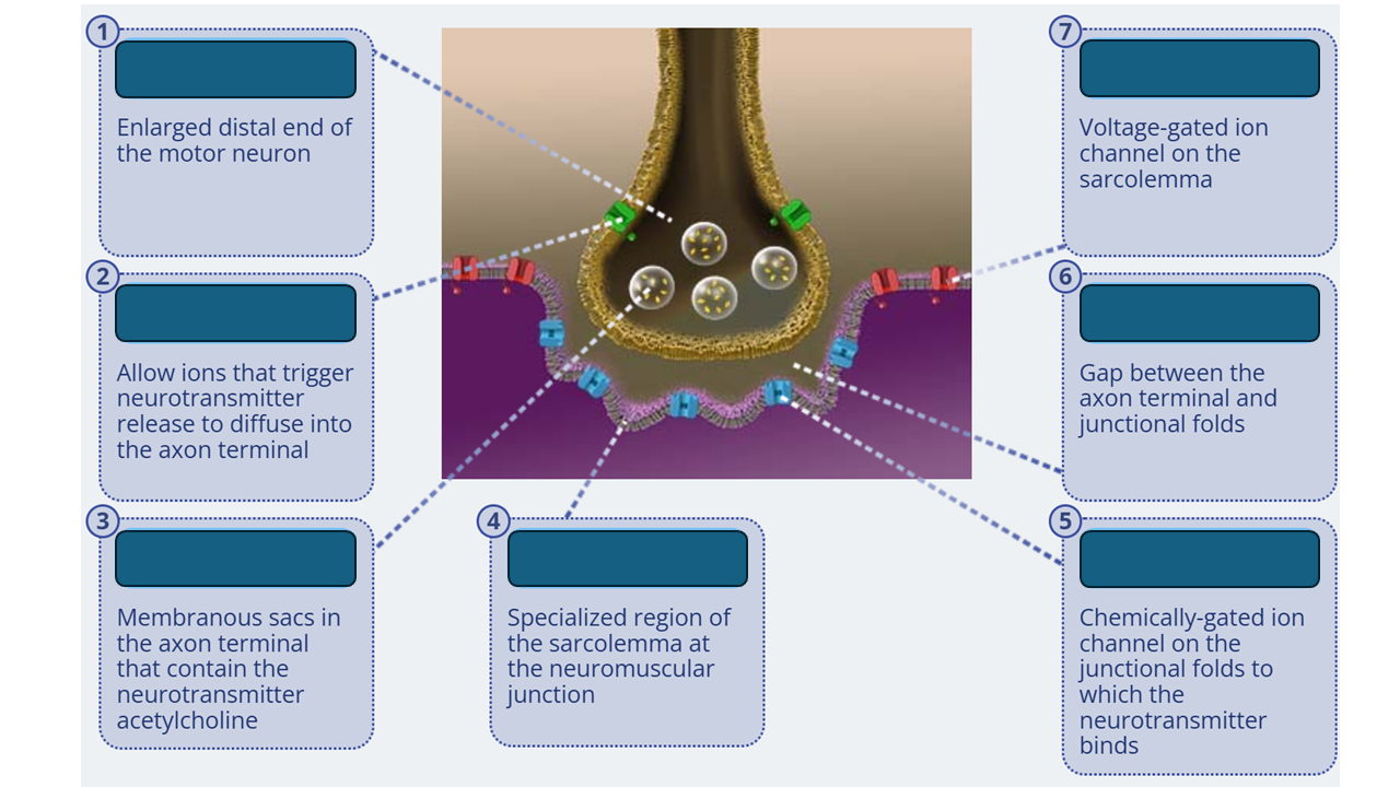
calcium channels
2

synaptic vesicle
3

junctional fold
4

acetylcholine receptor
5

synaptic cleft
6

sodium channel
7
What will change as an action potential travels along the axon before it reaches the axon terminal?
The axon depolarizes, and the charge on the inside becomes positive
Binding of acetylcholine to the acetylcholine receptors on a muscle fiber causes chemically-gated ion channels to
open
Which ion diffuses into a muscle fiber through an open acetylcholine channel at the junctional fold?
sodium
What effect does the diffusion of sodium ions through chemically-gated ion channels have on the membrane potential across the junctional folds?
The voltage becomes less negative on the inner surface
Does the depolarization of the junctional folds cause the voltage-gated sodium channels to open or close?
open
Botulinum toxin prevents the release of acetylcholine from the axon terminal. What happens when botulinum toxin prevents the release of acetylcholine from the axon terminal?
The membrane potential of the junctional folds will remain unchanged and the muscle fiber will not contract
Curare is a plant extract that can bind to acetylcholine receptors, but in a way that does not cause the chemically-gated channels to open. What happens when an action potential reaches the axon terminal if curare is present?
The membrane potential of the junctional folds will remain unchanged, and the muscle fiber will not contract
Neostigmine and related drugs prevent the breakdown of acetylcholine by acetylcholinesterase. What happens when neostigmine and related drugs prevent the breakdown of acetylcholine by acetylcholinesterase?
The junctional folds will remain depolarized and the muscle fiber will remain contracted
An action potential at the axon terminal of a motor neuron opens what type of ion channels?
Voltage-gated calcium channels
Which one of the following components stimulates contraction of a skeletal muscle?
motor neuron
Which structure of the neuromuscular junction is a chemically-gated ion channel that the neurotransmitter binds to?
acetylcholine receptor
How does neurotransmitter binding to it's receptor activate a muscle fiber?
It opens chemically-gated ion channels that allow sodium ions to diffuse into the junctional folds
Where in the neuromuscular junction do you find voltage-gated calcium ion channels?
axon terminal
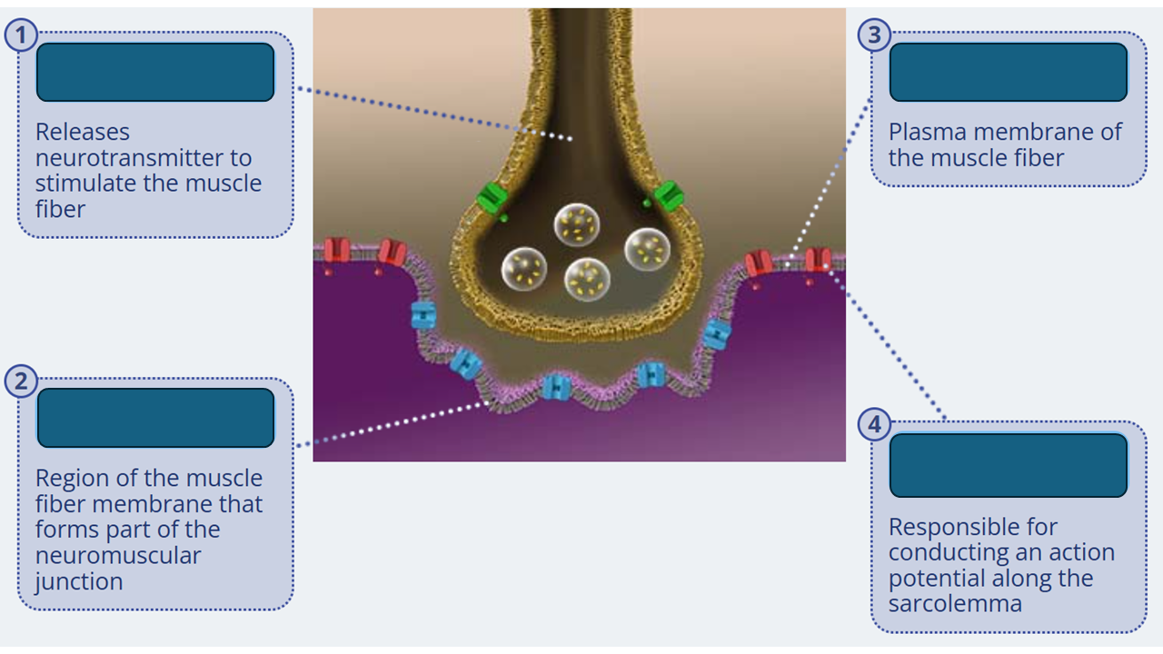
motor neuron
1

junctional folds
2

sarcolemma
3

voltage-gated sodium channels
4
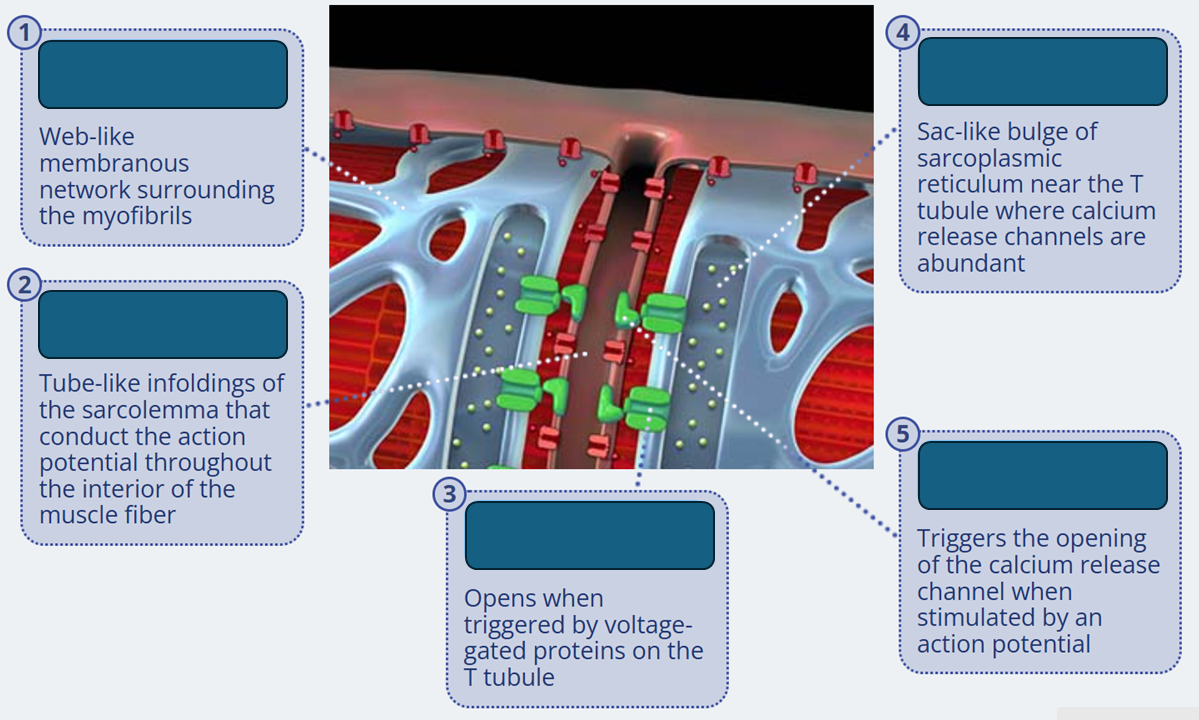
sarcoplasmic reticulum
1

T tubule
2

calcium release channels
3

terminal cistern
4

voltage-gated protein
5

thin filament
1

myosin bindin site
2

thick filament
3

tropomyosin
4