[SPECON] Wound Assessment and Management
1/41
There's no tags or description
Looks like no tags are added yet.
Name | Mastery | Learn | Test | Matching | Spaced | Call with Kai |
|---|
No analytics yet
Send a link to your students to track their progress
42 Terms
This is the largest organ of the body making up 15% of the total body weight. It serves as an external coverage of the body.
Follow Up Question: What is the importance of this organ?
Skin or integument
The skin serves as the barrier protecting us from the outside.
What are the three layers of the skin?
Epidermis
Dermis
Subcutaneous tissue or hypodermis
What are the main functions of the skin?
Protection (acid mantle, defensins, moisture barrier/water retention)
Immunity (Langerhans cells, macrophages, interleukin I)
Thermoregulation (excretion of sweat, vasodilation/vasoconstriction)
Sensation
Sweat production
Storage and synthesis (excretion/absorption/synthesis of vitamin D, storage of lipids and water, blood reservoir)
The skin has an acid mantle. What is its pH level?
Follow Up Question: What is the purpose of this acidity and why is it there?
4-6.5 pH
Suppress bacterial growth; it is there because of sweat and oil glands
These are anti-microbial peptides or proteins that exude an immunomodulatory and chemotactic functions. It is likely to affect skin infection, inflammation, and wound healing.
Follow Up Question: How does it inhibit bacterial growth?
Defensins
It inhibits bacterial growth through the use of direct membrane membrane dysfunction.
How does the skin help with water retention and keeping homeostasis in the body?
The skin prevents the evaporation of our fluids in the body.
A unique population of tissue resident macrophages (WBCs) that form a network of cells across the epidermis of the skin. They can migrate from the epidermal surface to the lymph nodes.
Follow Up Question: How do these cells help with the immune system?
Cells of Langerhans
They are a dense network of immune system cells that help determine the appropriate immune response and interpret the microenvironmental context where they encounter foreign substances. They act as the first line of defense when there is a wound.
One of the parts of the cytokines that have a role in the regulation immune and inflammatory responses due to sterile insults.
Follow Up Question: What forms these cells?
Interleukin I (IL1)
Keratin cells
How does Interleukin I aid in the inflammatory processes?
Introduces vasodilation to increase blood flow
Helps with the production of platelets of wound clotting
Helps with the production if nitrous oxide (vasodilator)
How does the skin help in thermoregulation of the body?
Excretion of sweat
Vasodilation (dissipate heat) or vasoconstriction (conserve heat) of blood vessels
Aside from its role in thermoregulation, what else does sweat production do?
It is another way we release toxins and excess fluids from our body.
Our skin is able to convert sunlight into vitamin D when it comes into contact with the skin. What is the importance of vitamin D?
It is for the production of calcium.
What are the layers of the skin?

This is the outermost and initial protective barrier of our skin.
Follow Up Question: What are the layers of the epidermis?
Epidermis
Stratum corneum
Stratum lucidum
Stratum granulosum
Stratum spinosum
Stratum basale

This layer consists of dead keratinocytes and is for waterproofing of the skin.
Stratum corneum

This is a layer of translucent cells only seen in the palms and soles of the feet.
Stratum lucidum

This layer is where the Langerhans cells are located and is for water retention.
Stratum granulosum

This layer contains keratinocytes and Langerhans cells and provides an additonal protective layer to the skin.
Stratum spinosum

This is a single layer of epidermal cells with melanocytes that can regenerate.
Stratum basale

This layer is mostly composed of collagen, elastin fibers, and contains a network of nerve endings, sensory receptors, capillaries, and sweat and sebaceous glands.
Follow Up Question: What are the functions of this layer?
Dermis
Supports the structure of the skin
Provides mechanical strength because of the papillary layer

What are the two layers of the dermis?
Papillary and reticular layers

This layer is an extensive series of ridges that increases the surface area of the skin.
Papillary layer
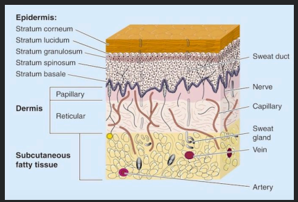
This layer attaches to the subcutaneous tissue via connective tissues.
Reticular layer

This layer is composed of adipose and connective tissues and is where the major blood vessels are located.
Follow Up Question: What are its functions?
Subcutaneous fatty tissue
Thermal insulation
Calory storage
Mechanical shock absorber

This is a breakdown of the protective function of the skin and the loss of continuity of the epithelium, with or without loss of underlying connective tissue (i.e. muscle, bones, nerves). This can occur following injury to the skin or underlying tissue or organs caused by surgery, a blow, a cut, chemicals, heat or cold, friction or shear force, pressure, or as a result of disease, such as leg ulcers or carcinomas.
Follow Up Question: How are wounds classified?
Wound
Acute or chronic
This is the natural physiologic reaction to tissue injury, consisting of interplay between numerous cell types, cytokines, mediators, and the vascular system.
Wound healing
These cells help with inflammatory process by increasing the amount of cells needed to be in the area and increasing the blood flow to the area.
Cytokines and mediators
What are the phases of wound healing?
Inflammatory phase
Proliferative phase
Maturation and remodeling
What occurs immediately after the injury?
There is outpouring of lymphatic fluid and blood, arterial vasoconstriction to the damaged endothelial lining, and then aggregation of platelets.
What is the main goal immediately after injury?
Goal is to achieve hemostasis.
This test is used to determine how long it takes for the blood clot.
Follow Up Question: How long does it take for the blood to clot here?
Prothrombin time test
10-13 seconds
[FILL IN THE BLANKS] In wound healing, ______ is a short-lived process that is soon followed by ______, which allows the influx of ______ and ______.
In wound healing, vasoconstriction is a short-lived process that is soon followed by vasodilation, which allows the influx of white cells and more thrombocytes.
This phase of wound healing:
Consists of hemostasis (regulation of blood loss) and chemotaxis (cell mediators creating hormones that help with the inflammatory phase)
WBCs and thrombocytes speed up the inflammatory process
Mediators and cytokines promote collagen degradation
Includes interleukins (IL1, IL6), tumor necrosis factor-α (TNFα), platelet-derived growth factor (PDGF), and fibroblast growth factor-2 (FGF2)
Serotonin and histamine
Inflammatory phase
When does the inflammatory phase occur?
0-25 days
This is the formation of new blood cells.
Follow Up Question: Which phase of wound healing does it occur?
Neovascularization
Inflammatory phase
This is the reparation of the actual skin that occurs during the inflammatory phase.
Re-epithelialization
This is responsible for the contribution of the defense of the wound through the stimulation of acute phase responses, hematopoiesis, and immune reactions.
Follow Up Question: What is hematopoiesis?
Interleukin 6 (IL6)
This is a monocyte-derived cytokine that is a highly-conserved known to play a major role in the pathogenesis in gram negative shocks.
Tumor necrosis factor-α (TNFα)
This helps to heal wounds, repair damage to blood vessel walls, helps blood vessels grow, and help with angiogenesis and neovascularization.
Follow Up Question: What releases this?
Platelet-derived growth factor (PDGF)
Platelets
This aids in the repair and regeneration of tissues and promotes fibroblast proliferation (or the scaffolding of the wound).
Fibroblast growth factor-2 (FGF2)
This promotes cellular viability, the proliferation and migration of fibroblasts and keratinocytes, and other functions important in wound healing.
Serotonin
This exerts a vasodilatory effect and enhances the amount of blood going to the wound. It also participates in wound healing as well and promotes inflammation as well.
Histamine