HUMB C8
 Call Kai
 Learn
Learn Practice Test
 Spaced Repetition
 Match
Match Flashcards
 Knowt Play
1/81

There's no tags or description
Looks like no tags are added yet.
Name | Mastery | Learn | Test | Matching | Spaced | Call with Kai |
|---|
No analytics yet
Send a link to your students to track their progress
82 Terms
Neuron (nerve cell)
basic structural and functional unit of the nervous system
Neuroglia
major supporting cells of neurons
Axon
nerve fibre
Nerve
bundle of axons and their sheaths
Sensory receptors
separate specialised cells which detect temperature, pain, touch, light, sound, odour and other stimuli
Action potential
electrical signal that transmits information along neurons and allows communication between neurons and effectors
Effector
the organ, tissue, or cell in which an effect or action takes place
Interneurons
Connect sensory neurons to motor neurons inside the CNS
Synapse
junction of a neuron with another cell
Ganglia
collection of neuron cell bodies/somas outside CNS
Plexuses
extensive network of usually axons outside CNS
Nervous System Functions
Receive internal and external sensory input
Integrate information
Motor output
Maintaining homeostasis
Establish and maintain mental activity
Nervous system – Structural Classification
Central nervous system (CNS): decision maker, protected by skull and vertebrae
Brain
Spinal Cord
Peripheral nervous system (PNS): messengers that collect info and take it to CNS, allow for motor output
Sensory receptors
12 pairs of Cranial nerves arising from brain
31 pairs of spinal nerves arising from spinal cord
Ganglia
Plexuses
Nervous system – System Classification
PNS and CNS in constant communuication with each other
PNS divided into Somatic NS, Autonomic NS, Enteric NS - all with a sensory (afferent) and motor (efferent) component
Autonomic NS motor component produces sympathetic and parasympathetic responses
Nervous system – Functional Classification
PNS split inito sensory division and motor division
Only the motor division can be classified as somatic, autonomic, or enteric
Autonomic can be further divided into parasympathetic and sympathetic
Somatic NS
Enables voluntary contraction of skeletal muscles
Single neuron system, one synapse
Cell bodies in ventral horn
Myelinated axons
Autonomic NS
Involuntary control
Action potentials in motor neurons travel from the CNS to smooth or cardiac muscle, or glands
Two-neuron system, two synapses (preganglionic and postganglionic motor neurons)
Preganglionic are myelinated, postganglionic are unmyelinated
Preganglionic somas in lateral horn in CNS, postganglionic somas in autonomic ganglion in PNS
Target tissues stimulated or inhibited (organ activity can be increased or decreased)
Enteric nervous system - Functions
Controls digestive system:
Stimulate/inhibit smooth muscle contraction and gland secretions
Detect changes in content of lumen
Enteric nervous system - Anatomy
Sensory neurons connect digestive tract to CNS
Autonomic motor neurons connect CNS to digestive tract
Enteric neurons confined to enteric plexuses within digestive tract wall
Sensory/afferent division
Specialised (sensory) receptors detects external and internal environmental stimuli
Collects and transmits input as electrical signals from sensory receptors to CNS (nerves)
sensory neuron’s cell body is outside CNS (cluster forms dorsal root ganglion)
Motor/efferent division
Transmits electric signals from CNS to effector (e.g., muscle, gland)
Motor neuron’s cell body located inside CNS
Autonomic NS Motor Division - Sympathetic Response
Fight or flight
arises from thoraco-lumbar region
Primes the body to act in threatening situations
Shorter neuron pathway, quicker response
Complementary to parasympathetic
E.g: increased heart rate and respiratory rate, sweating, pupil dilation
Autonomic NS Motor Division - Parasympathetic Response
Rest and digest/feed and breed
Arises from cranio-sacral region
Relaxes the body inhibiting high energy functions
Longer neuron pathway, slower response
Complementary to sympathetic
Neuron
Structural and functional unit of NS
≈100 billion in body
Receive info, integrate it, and send appropriate response to target
Classified by structure and/or function
Highly modified cell
Electrically excitable
Unidirectional impulse flow
Neuron parts
cell body (soma) - contains all major organelles except centrioles, protein synthesis, energy production, signal integration
Dendrites - branched extensions of soma, Generate electric current when stimulated that flows from dendrites’ tips to soma
Axon - vary from mm to m, can be collateral (branched), often arise from axon hillock
Axon parts
trigger zone = axon hillock + inital segment,
presynaptic terminal/axon terminals (axon endings)
terminal boutons/synaptic knobs (swollen ends of axon terminals)
myelin sheath (in PNS sheath made of Schwann cells)
node of Ranvier (gaps between Schwann cells)
Neurons – structural classification
Multipolar
bipolar
unipolar/psuedo-unipolar
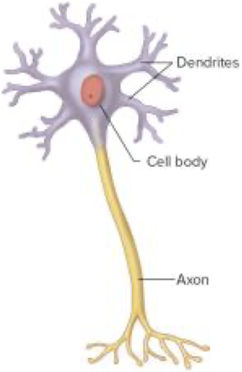
Multipolar neuron
One cell body
Many dendritic processes and an axon
E.g motor neurons, interneurons
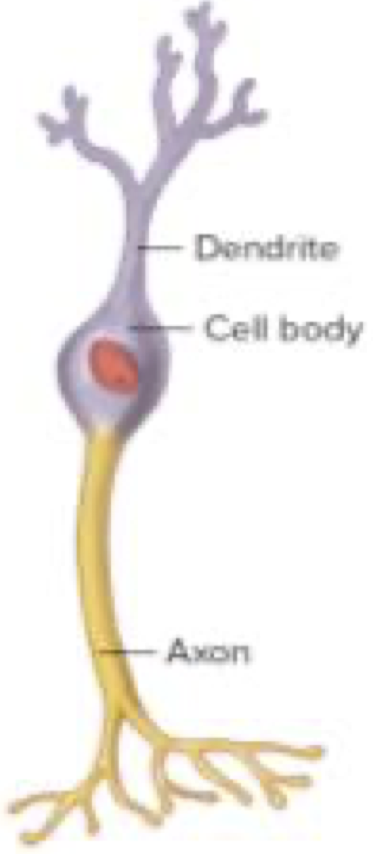
Bipolar neuron
One cell body and two processes, a dendrite and an axon
Specialised sensory neurons in retina of eye and nasal cavity
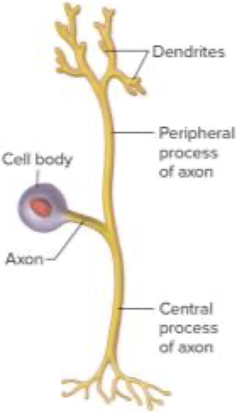
Psuedo-unipolar neuron
One cell body and only one process, axon - this bifurcates into peripheral process (dendtrites) and central process (axon terminals)
Dendrites connected to sensory receptors, axon terminals to CNS
Sensory neurons
Neurons – functional classification
Sensory neuron
Motor neuron
Inter-neuron
Sensory neuron
Conduct action potential (AP) from sensory receptors to CNS
Motor neuron
Conduct AP away from CNS towards muscles/glands
Inter-neuron
Conduct AP within CNS from one neuron to another
Neuroglia
10-50 times more abundant than neurons
Supporting cells
6 different types - Astrocytes, Ependymal, Microglial, and Oligodendrocytes in CNS, Schwann and Satellite in PNS
CNS Neuroglia - Astrocytes
Star shaped
Branched cytoplasmic processes with end feet
Functions - Scaffold CNS cells and capillaries, control blood-brain barrier permeability, synaptic support, and CNS homeostasis
CNS Neuroglia - Ependymal cells
Line brain ventricles and central canal of spinal cord
Ciliated to circulate cerebrospinal fluid (CSF), simple cuboidal and columnar
Function - Produce and regulate CSF, Protection, and hormone transport
CNS Neuroglia - Microglial cells
Oval body with many projections
Become active, mobile, & phagocytic in response to inflammation, otherwise resting
Function - Phagocytose foreign substances, necrotic tissue, and pathogens
CNS Neuroglia - Oligodendrocytes
Have cytoplasmic extensions that wrap around axon, forming myelin sheath
Each cell can enclose multiple axons
Function - Insulate CNS axons, saltatory conduction
PNS Neuroglia - Schwann cells
Have cytoplasmic extensions that wrap around axon, forming myelin sheath
Each cell forms part of sheath around one axon
Function - Insulate PNS axons, saltatory conduction of action potential (AP propagates by jumping from one Node of Ranvier to next)
PNS Neuroglia - Satellite cells
Surround soma in ganglia
Function - Provide support and nutrition to soma, Protects neurons from heavy metal poisons (Pb, Hg)
Myelinated axons
Schwann cells (PNS) and oligodendrocytes (CNS) wrap repeatedly around axon
Nodes of Ranvier
Saltatory conduction of electrical impulses
Faster nerve impulse conduction
Unmyelinated axon
Axons rest in invaginations of Schwann cells (PNS) and oligodendrocytes (CNS)
electrical impulses travel as continuous wave
Thinner
Slower nerve impulse conduction
Grey matter
Consists of somas, dendrites, axon terminals, unmyelinated axons, glial cells, and synapses
On periphery (outer cortex) in brain but in middle of spinal cord as H-shape
Forms ganglioins in PNS
White matter
White due to myelin sheath
Very few somas
Forms nerves in PNS and nerve tracts in CNS
On inner side in brain but periphery in spinal cord
Electrical impulses
allows responses to environment (external & internal), emotions, conscious thoughts, memory, and action of glands & muscles
Membrane potential
measure of electrical properties of cell membrane
Determined by:
Ionic concentration difference across plasma membrane
Permeability of plasma membrane
Cell membrane – ionic concentration
Higher concentration of Na+ and Cl- extracellularly
Higher concentration of K+, proteins, and PO43- intracellularly
if total number of cations and anions in extracellular and intracellular fluid is equal, cell is electrically neutral
Cell excitability depends on:
ion movement across cell membrane
differences in ionic concentration across cell membrane
Na+/K+ pump
Cell membrane – ion channels
Non-gated/Leak ion channels:
Ion specific
Always open
More K+ and Cl- leak ioc channels that Na+ ones
Gated ion channels:
require signals to open
Ion specific: ligand-gated open when chemicals attach, voltage-gated open with charge difference, and other-gated open with temp, pressure, etc
Resting membrane potential (RMP)
difference in charge across cell membrane in a resting cell caused by leak ion channels and Na+/K+ pump
Measured in mV
RMP of neurons is -70mV
-ve if intracellular side is more negative
Establishing resting membrane potential
Intracellular fluid becomes more -ve because:
More K+ leak ion channels moving K+ out than Na+ ones moving Na+ in
Anionic proteins remain inside cell as they’re too large and charged to pass cell membrane
Na+/K+ pump moves 3Na+ out but only 2K+ in
Action Potential (AP) - Def and Steps
how neuron transmits info, as electrical signals, away from soma, down axon, to effector
Steps:
Resting membrane potential
Depolarisation
Repolarisation
Threshold
membrane potential a neuron needs to reach to trigger an AP
-50 to -55mV for neurons
AP - Resting membrane potential
All gated Na+ and K+ channels are closed
K+ leak channels open, moving K+ out
Na+/K+ moving 3Na+ out and 2K+ in
-ve RMP
AP - Depolarisation
Cell is stimulated
Na+ voltage gated channels open, moving Na+ inside
K+ voltage gated channels remain closed so less K+ leaves cell
Membrane potential becomes more positive
AP - Repolarisation
Na+ voltage gated channels close, preventing Na+ from entering
K+ voltage gated channels open, moving K+ out
Membrane potential becomes more negative
AP - Hyperpolarisation (afterpotential)
Na+ voltage gated channels close, preventing Na+ from entering
K+ voltage gated channels close slowly so K+ continues to leave the cell, producing afterpotential
Membrane potential becomes very negative
AP - Resting Membrane Potential
Na+ and K+ gated channels are closed
RMP reestablished by Na+/K+ pump actively (against concentration gradient) moving 3 Na+ out and 2 K+ in
Graded potential
short-lived, localised (occur in small region) changes in membrane potential that occur in dendrites or soma
if the stimulus, and thus graded potential, is large enough, AP fired
Can summate (add together)
Decremental (signal weakens as it spreads so cant transfer info over long distance)
All-or-none principle
If the stimulus, and thus graded potential, is large enough to cross threshold, the magnitude of AP is same every time
If threshold is not crossed, no AP fired
Stronger stimuli increase frequency not magnitude
Refractory period
Absolute refractory period - during depolarisation and most of repolarisation, no more AP can be generated
Relative refractory period - during late repolarisation and afterpotential, only very strong stimuli can generate another AP
Propagation of axon potentials
Only in unmyelinated axons
one axon membrane segment is depolarised, causes subsequent segment to be depolarised, then the initial segment is repolarised
Unidirectional (soma to axon terminal)
Saltatory conduction
only in myelinated axons
AP jumps from one Node of Ranvier to next
Faster impulse transmission
Synapse - Def and Types
Junction where a pre-synaptic neuron communicates with a post-synaptic cell (neuron, muscle fibre, gland celletc)
A neuron can have thousands of synapses
Electrical synapse – less common, electric signal transmits AP, in cardiac muscles & some smooth muscles
Chemical synapses – most common, uses neurotransmitter (chemical messenger) to transmit AP
Synaptic Cleft:
space between pre-synaptic membrane and post-synaptic membrane
Chemical Synapse
Pre-synaptic terminal has Ca2+ voltage gated channels in wall that open when AP recahes them
Ca2+ moves into cell and signal synaptic vesicles to move towards and merge with pre-synaptic membrane and via exocytosis, releases neurotransmitters into synaptic cleft
Neurotransmitters attach to receptor sites to ligand-gated Na+ channels, causing them to open and allow Na+ to diffuse into post-synaptic cell
If Na+ movement is enough to reach threshold, AP generated in next cell
Overstimulation avoided by enzymnes in synaptic cleft breaking down neurotransmitters, closing channels, to be reabsorbed into presynaptic terminal
Spinal cord
Extends from brain at foramen magnum to second lumbar vetebra
Shorter than vetebral column as it grows slower
Protected by meninges and bony veterbral column
Anterior median fissure (deeper, wider) and posterior median sulcus
Spinal Cord - Rootlets to Nerves
Rootlets arise posteriorly and anteriorly on sides
6-8 rootlets combine to form posterior and anterior root
Dorsal root ganglion
Posterior and anterior root join to form spinal nerve
Grey matter
On inside of spinal cord as butterfly/H shape
Posterior, lateral, and anterior horn
Gray commissure connects left and right sides, contains hole in middle called central canal where CSF circulates
White matter
Posterior, lateral, and anterior column
On outside of spinal cord surrounding grey matter
White commissure connects left and right sides
meninges
connective tissue membrane surrounding brain and spinal cord
Organisation of neurons in spinal cord
Sensory neurons (bipolar and pseduo-unipolar) enter through posterior root
Motor neurons (mulipolar) enter through anterior root
Spinal nerve is ‘mixed’ because have both sensory and motor neurons
Organisation of sensory neurons in spinal nerves
dendrites are located where sensory receptors are
axon travels along spinal nerve
soma is in dorsal root ganglion
central process of axon travels through dorsal root and into grey matter
collateral branches ascend and descend into white matter
synpases with motor neuron or interneuron in grey matter
Organisation of motor neurons in spinal nerves
Dendrites and soma in grey matter
Leave through anterior rootlet
Travel through anterior root and spinal nerve to reach effector
Reflexes
Automatic response to a stimulus, without higher brain centre involvement or mental processing
homeostatic (help maintain homeostasis)
Produces same stereotyped, immediate response from spinal cord
Rapid, predictable, and unlearnt
Somatic vs Autonomic Reflexes
somatic - skeletal muscle, protect from harm, e.g. pulling back when touching hot surface
autonomic - smooth muscle, cardiac muscle, and glands, maintain bodily functions, e.g. blood pressure
Monosynaptic Vs Polysynaptic
Monosynaptic - simplest, one synapse (sensory neuron to motor neuron)
Polysynaptic - slower, multiple synapses (involve interneurons)
Reflex Arc def
neuron pathway that facilitates a reflex
Reflex Arc
Sensory receptors detects stimulus
Sensory neurons conduct AP to spinal cord via posterior root
sensory neuron synapses interneuron or motor neuron
Motor neuron axon conducts AP to effector via anterior root
Reflex Arc - Brain
Sensory information sent to brain via ascending tract and returned via descending tract
Reflex does not need brain to occur, but can be supressed or exaggerated by brain
Reaction
Voluntary response to sensory stimulus
Slower than reflex
Involves brain and spinal cord
Reaction time improves through repetition
e.g catching/dodging a ball