Canine PE
1/23
There's no tags or description
Looks like no tags are added yet.
Name | Mastery | Learn | Test | Matching | Spaced | Call with Kai |
|---|
No analytics yet
Send a link to your students to track their progress
24 Terms
Example of Signalment
Name (optional), age, sex (if altered), Color (optional), breed, species
“Sammy is a 6-year-old neutered male black labrador mix canine”
Oral Mucous Membranes
Healthy = Normal pink & moist
Capillary Refill Time
Normal- Returns to original color in 1-2 seconds
Skin Tent
Less than 5 % dehydrated: No detectable clinical signs
Dog and cat normal temp
Dog: 100-102.5 F
Cat: 100-102.5 F
Dog normal HR
Large: 60-120 bpm
Small: 120-160 bpm
Dog and Cat normal resp rate
Both 10-30 rpm
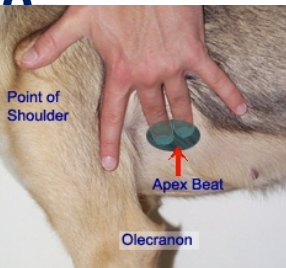
Common Heart Rate collection sites:
Auscultate OR palpate Apex beat (mitral valve) on Left hemithorax
Palpate- femoral artery pulse
Common Respiratory Rate collection sites:
Visualize thorax and abdomen move with each breath from a distance
Palpate- Place hand on caudal thorax/cranial abdomen and palpate each inhalation
Auscultate- place stethoscope on thorax or trachea
If patient is panting the majority of the time, state as “Panting,” but also provide rate. (will be fast, tachypneic)
Peripheral Lymph Nodes
Describing Healthy Peripheral Lymph Nodes:
1. Symmetric (to contralateral LN)
2. Soft
3. Mobile
4. Non-painful
5. No heat
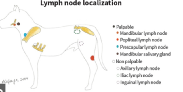
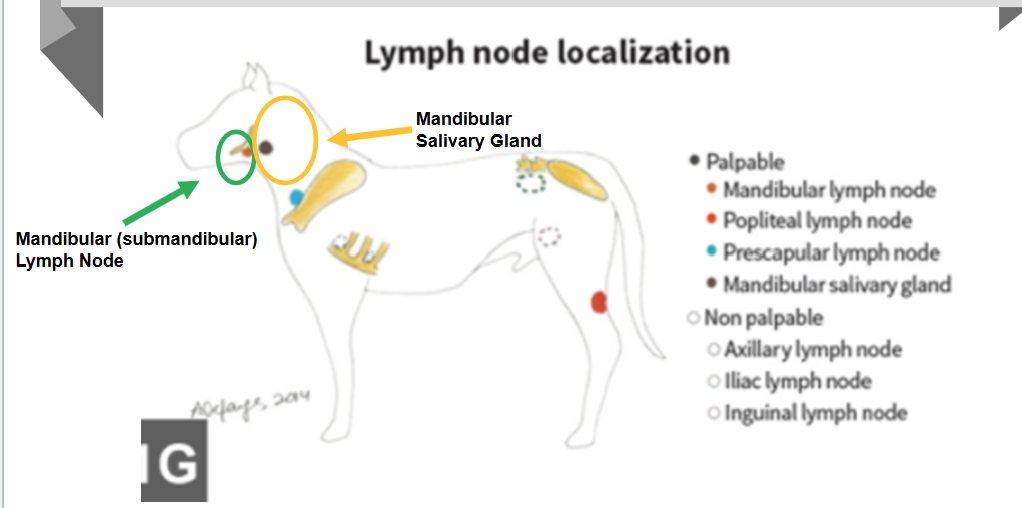
Mandibular Lymph Node & Salivary Gland Locations
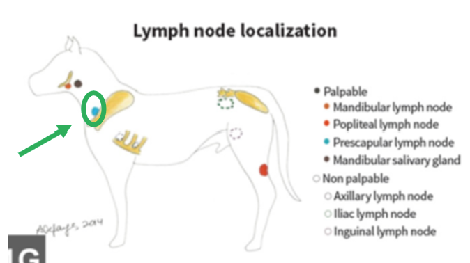
Prescapular Lymph Node Location
(aka Superficial Cervical LN)
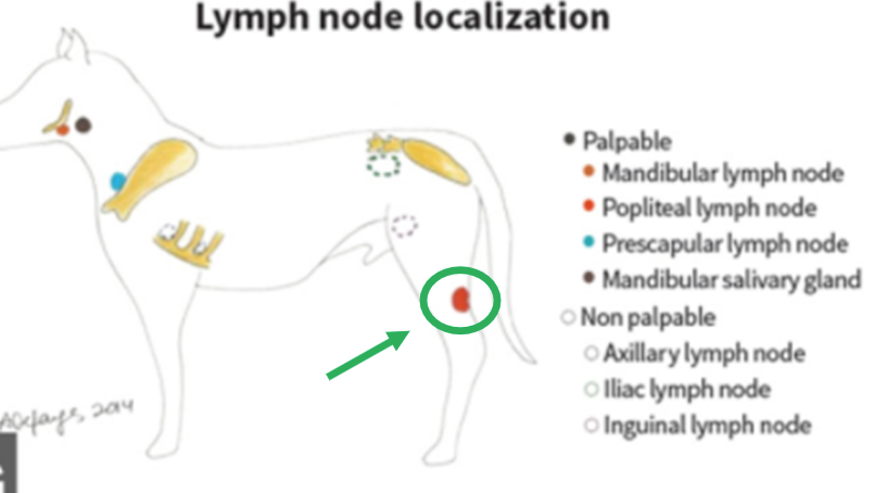
Popliteal Lymph Node Location
Integument (Skin)
Run hands over entire surface of the skin
Describe coat quality
Conscious Proprioception
Evaluates patient’s awareness of their limbs in space
AND their ability to correct any abnormal positioning.
Spine
Palpate along each dorsal spinous process/transverse process
• Cervical (neck) caudally to Coccygeal (tail)
• Pain, crepitus, asymmetry?
Cardiovascular Evaluation
1. Collect heart rate if not already performed
2. Auscultate Left AND Right sides of the heart
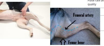
3. Assess Femoral Pulse
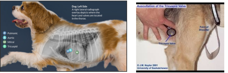
Cardiac Auscultation
Left side of heart
• Mitral valve (5th Intercostal Space (ICS)), Aortic valve (4th ICS), Pulmonic (3rd ICS)
Right side of heart
• Tricuspid valve (4-5th ICS)
Describing a healthy cardiac auscultation: Strong, no murmurs, no arrhythmias auscultated
Femoral Pulse
Is the pulse strong and immediately follows heartbeat (aka synchronous)?
Yes= normal
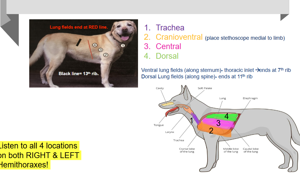
Respiratory System Evaluation
1. Assess respiratory effort
2. Auscultate trachea to appreciate referred upper airway sounds
3. Auscultate 3 locations on Left and Right Hemithoraxes
Cranioventral, Central, Dorsal
Listen for 1-2 breaths per location
ormal lung sounds = normal bronchovesicular sounds
Abdominal Palpation
Direction: Slide Cranial → Caudal & Slide Dorsal → Ventral.
Hand position
• Fingertips/palms pointing dorsally (sensitive nerves in fingertips)
• Small cat or dog: 1 hand
• Med-large dog: 2 hands in “prayer” position, recommend 1 hand in between
pelvic limbs, and other hand placed laterally
• Palpate urinary bladder with 1 hand
Describing Abdominal Palpation Findings:
• Soft/compliant vs these/hard (non-diagnostic)
• Non-painful vs painful
• Abdominal masses
• Organomegaly
• Urinary bladder: Describe how full
ie)Turgid/distended, mildly, moderately, non-
palpable (empty)
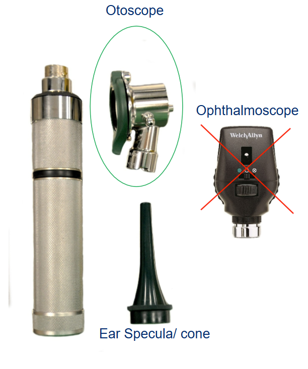
Otoscope
Cone sizes:
• Small - this size works well for cats or dogs under 20lbs
• Medium - this size works well for dogs 20-80lbs.
• Large - this size works well for dogs > 80 lbs or for dogs under anesthesia.
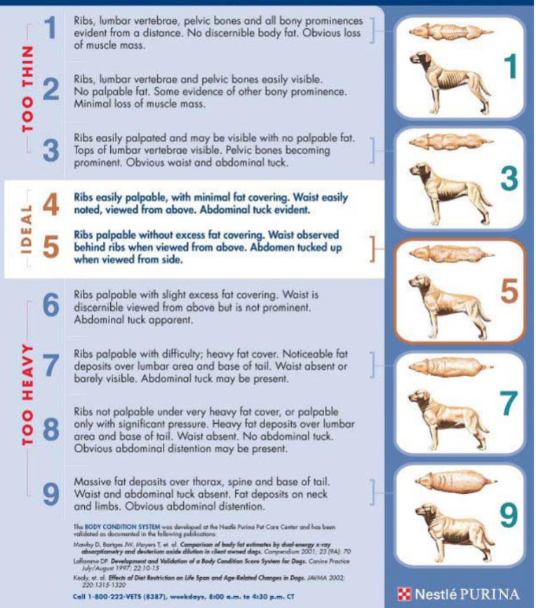
Canine BCS Score
1-9 scale
4-5 is ideal - ribs easily palpable, minimal fat covering, waste easily noted/ viewed from above. Abdominal tuck