Anterior Thoracic wall (EXAM 1- Anatomy)
1/60
There's no tags or description
Looks like no tags are added yet.
Name | Mastery | Learn | Test | Matching | Spaced | Call with Kai |
|---|
No analytics yet
Send a link to your students to track their progress
61 Terms
Describe the Jugular notch
The visible dip at the superior border of the manubrium
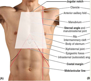
Describe the sternal angle and its location
Palpable horizontal ridge where the manubrium meets the body of the sternum
Marks the level of various significance
2nd costal cartilage (at level 2 rib)
corresponds to T4-T5
Bifurcation of the trachea
Beginning and end of the aortic arch
The superior border of the heart
Separation of superior and inferior mediastinum

Describe the costal margin and its location
The curved lower boundary of the thoracic cage, formed by the cartilages of ribs 7 to 10.

Identify the location infrasternal angle
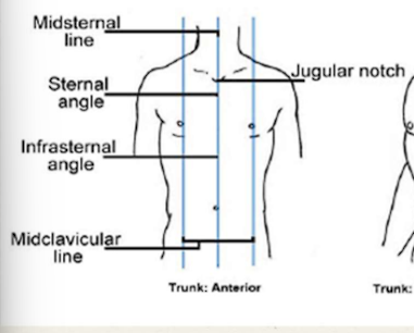
Identify the location midclavicular line

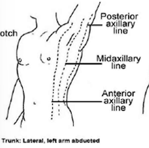
Identify the location Axillary lines
Anterior axillary line
Midaxillary line
Posterior axillary line
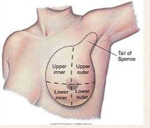
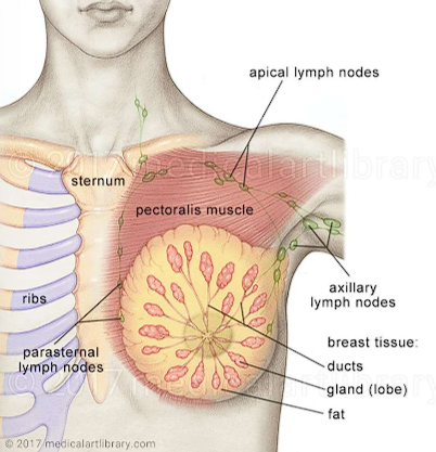
what are the borders of the breast?
Sternum to mid axillary line
axillary tail/ tail of spence
Clavicle to 5th-6th intercostal space
what is the retromammary space?
Fascia that separates the breast from the pectoralis major and serratus anterior muscle
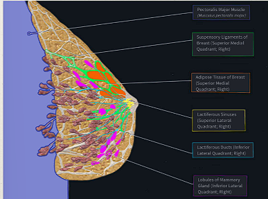
Describe the mammary glands and what it contains?
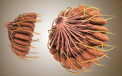
Functional unit of the breast responsible for milk production
Each breast contains 15-20 lobes
Each lobe is made up of mammary lobules
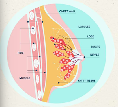
Describe the lactiferous ducts and what it contains?
Milk from each lobule drains into the lactiferous ducts
Ducts aim towards the nipple
Near nipple, each duct enlarges → lactiferous sinus → small reservoir for milk
Describe the areola and what it contains?
circular pigmented area of the skin surrounding the nipple
light pink→ dark brown
Sebaceous glands → lubricates/protect during breastfeeding
Smooth muscle fibers → pucker areola and erect nipple
Describe the nipple and what it contains?
Prominent, conical projection at center of areola
Exit point for milk
15-20 openings from the lactiferous ducts
No hair or sweat glands
T4 dermatomal layer
Describe the suspensory ligaments (Ligaments of cooper) and what it contains?
Fibrous connective tissue bands that extend from dermis →pectoral fascia
Maintain breast shape and structural integrity
age or disease→ ligaments stretched or pulled causing skin dimpling
Describe the primary lymphatic drainage of the breast
Both breast drain into axillary lymph or parasternal lymph
75-90% Axillary lymph nodes
10-15% Parasternal lymph
Where does Metastasis most commonly occur?
Axillary nodes, with palpation they become hard and fixed
Describe the location of axillary lymph nodes and how Right/Left axillary nodes travel?
Lateral breast tissue
Right axillar nodes → right lymphatic duct →right internal jugular vein
Left axillary nodes → thoracic duct → subclavian vein
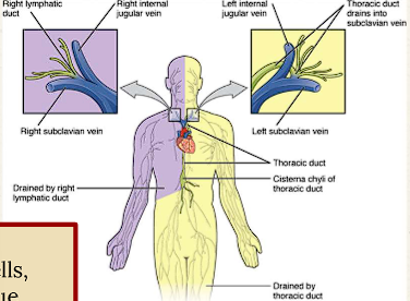
Describe the location of the parasternal lymph
Medial breast tissue
Deep behind sternum
harder to palpate which is why medial tumors=silent
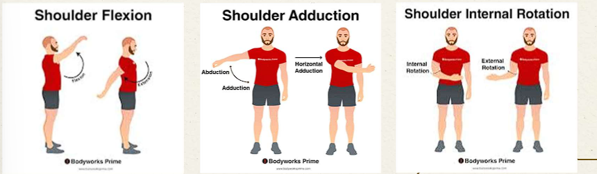
What are the corresponding actions of the Pectoralis Major?
Flexion
Adduction and medial rotation of the arm
Horizontal adduction of the arm
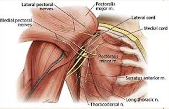
What is the innervation of the Pectoralis Major?
Medial pectoral (C8-T1) and Lateral pectoral nerves (C5-C7)
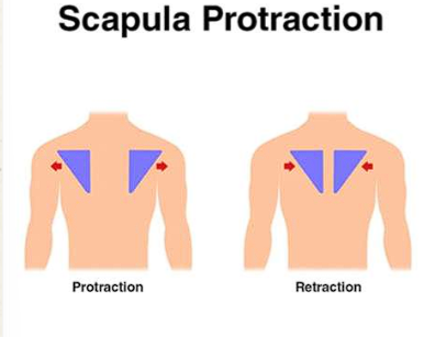
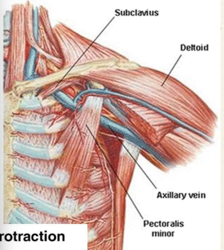
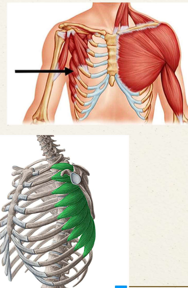
What are the corresponding actions of the Pectoralis Minor?
Protraction of scapula
Medial rotation of scapula
Downward movement of scapula
What is the innervation of the Pectoralis Minor?
Medial pectoral nerve (branches will pierce pectoralis minor to innervate pectoralis major)
What are the corresponding actions of the serratus anterior muscles?
Protraction of the scapula
Upward rotation of the scapula
Stabilization of scapula against the thoracic wall
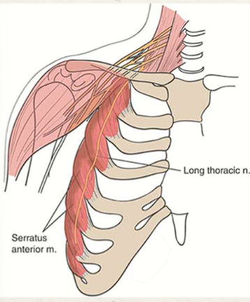
What is the innervation of the serratus anterior muscles?
Long thoracic nerve
What are the corresponding actions of the subclavius?
Stabilizes and depresses the clavicle
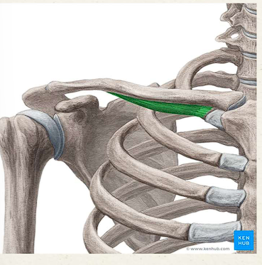
What is the innervation of the subclavius?
subclavian nerve
What are the corresponding actions of the intercostals?
Elevates ribs
Prevents intercostal space blowing out or retracting in during respiration
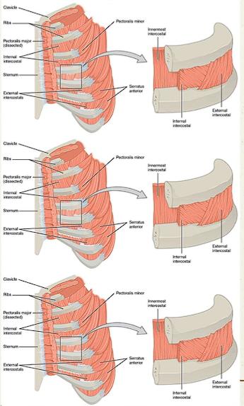
What is the innervation of the intercostals?
intercostal nerves T1-T11
Describe the functions of the thoracic cage
Protection of viscera
Thoracic: Heart, lung, great vessels
Abdominal organs due to dome shape of diaphragm: liver, spleen, stomach, kidneys
Mechanics of respiration
changing the internal volume to create pressure changes
Attachment point for musculature
Upper limb movement, postural support, abdominal wall, accessory muscles of respiration
what do the true ribs (1-7)/ Vertebrosternal ribs articulate with?
Both the vertebrae and the sternum
(connect directly via their own individual costal cartilages= sternocostal joint)

What do the false ribs (8-10)/ Vertebrochondral ribs articulate with?
vertebrae and the costal cartilage above it
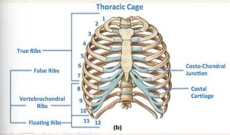
What do the floating ribs (11-12) articulate with?
NO interchondral joints or costochondral joints

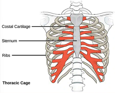
Describe the costal cartilage and what articulates with it
Bones of hyaline cartilage that prolong the ribs forward and provide chest elasticity (acts as flexible bridge that attaches anterior end of rib to sternum)
false ribs (8-10)/ Vertebrochondral ribs articulate
true ribs (1-7)/ Vertebrosternal ribs articulate
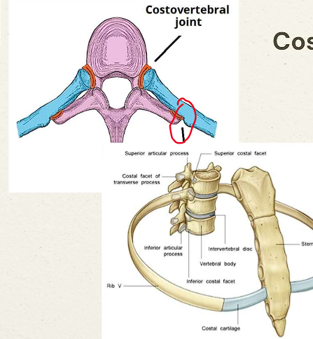
Describe the costovertebral joint and what articulates with it
Hinge joint attaching the head of the rib to the thoracic vertebral bodies
Typical ribs 3-9 articulate
Superior- attaches to vertebrae above
Inferior- attaches to its own vertebrae
Rib 1, 10 (sometimes), 11 and 12 only articulate w/ inferior vertebrae
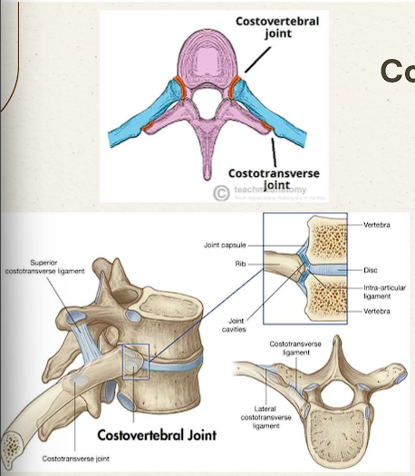
Describe the costotransverse joint and what articulates with it
Articulation of the rib’s tubercle with the transverse process of the same numbered vertebrae
Describe the Costochondral joint and what articulates with it
NO FUNCTIONAL MOVEMENT
Cartilaginous joint allows thoracic cage to expand and recoil during breathing
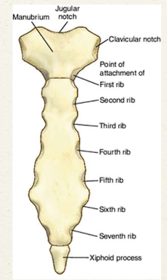
The sternum contains what structures?
Manubrium
Sternal angle
Sternal body
XIphoid process

Describe the Manubrium
Superior portion containing jugular notch
JN: Internal jugular vein runs deep)

Describe the sternum body
Middle portion of the sternum

Describe the xiphoid process
Lowest portion of sternum, used as a surgical landmark but can break during CPR
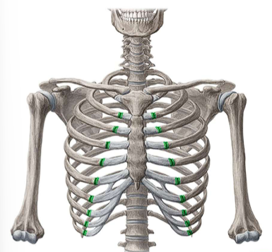
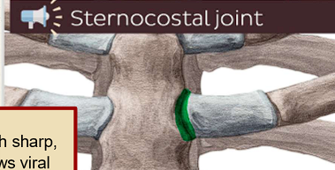
Describe the sternocostal joint and articulations
Articulations between costal cartilages and sternum
Both synovial (2-7) and synchondrosis (1)
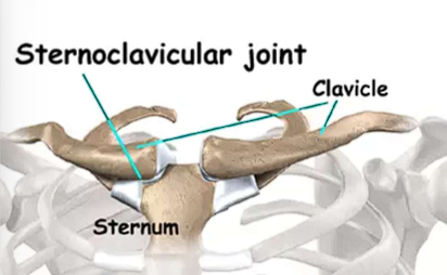
Describe the sternoclavicular joint
Synovial joint
provides stability
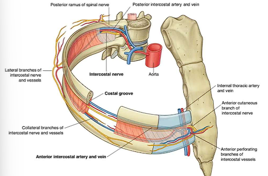
Describe intercostal nerves and what do they innervate?
Exits intervertebral foramina of the spine
Goes through intercostal space between internal and innermost intercostals
Motor innervation= intercostal muscles in thorax and abdominal muscles
Sensory innervation= overlying skin, pleura and parietal peritoneum
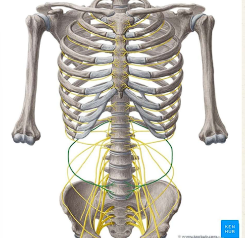
Describe subcostal nerves and what do they innervate?
inferior to 12th rib
along posterior wall→ abdominal regions
Motor innervation= abdominal muscles
Sensory innervation= hip, lateral gluteal region, lower anterior abdominal wall
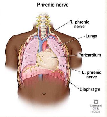
Describe the phrenic nerve and what do they innervate?
Ventral Rami/ C3-C5 (C5 keeps diaphragm alive)
Anterior root of lung → along pericardium
Motor innervation: diaphragm
Sensory innervation: diaphragm, pleura, pericardium, inferior vena cava, and peritoneum
Describe the long thoracic nerve and what do they innervate?
Posterior to brachial plexus→ superficial to serratus anterior
Motor innervation: serratus anterior muscle
What are the arteries and veins associated with the vasculature of the anterior thoracic wall?
Internal thoracic artery
Internal thoracic vein
Intercostal artery
intercostal vein
Subcostal artery
subcostal vein
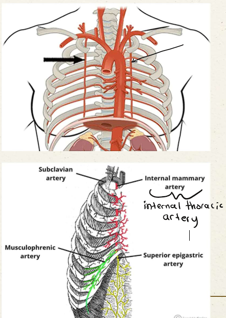
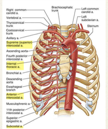
Describe the internal thoracic artery
Branch off subclavian artery
Vertically, bilaterally, lateral to sternum along internal surface of rib cage
Branches to: Anterior intercostals, musculophrenic arteries, superior epigastric artery
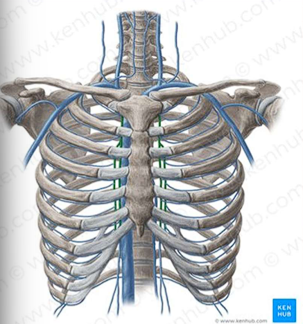
Describe the internal thoracic vein
Formed by the confluence of superior epigastric and musculophrenic veins
Runs next to the internal thoracic artery
Empties into the brachiocephalic vein
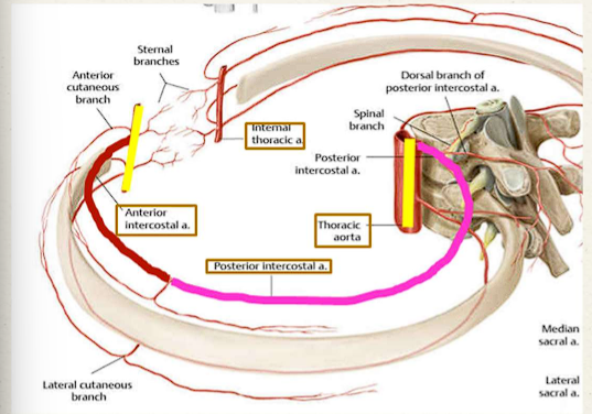
Describe the Intercostal arteries and veins
Anterior and posterior intercostal artery/vein meets in the middle and anastomosis (connection/bridge)
Supplies: intercostal muscles, parietal pleura, skin, breast tissue (in 2nd, 3rd and 4th)
Describe the subcostal artery/vein
last paired branch of the thoracic aorta
Supplies some abdominal wall muscles
Veins drain into azygos system
Describe the structure of the diaphragm
Separates thoracic cavity from abdominal cavity
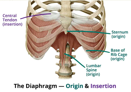
Describe the apertures of the diaphragm
Three separate hiatus for vessels:
Aorta
Inferior vena cava
Esophagus
what is the innervation of the diaphragm?
Phrenic nerve
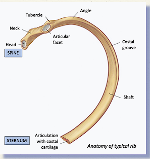
Bony landmarks of typical ribs (3-9)
Head - Connects with the thoracic vertebrae costal facets
Neck
Tubercle
Articular facet - connects with the thoracic vertebrae transverse costal facet
Costal Angle
Costal Groove
Shaft / Body
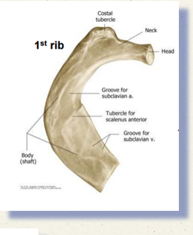
Bony landmarks of Rib1 (Atypical rib)
Only 1 articular facet
Groove for subclavian artery
Groove for subclavian vein
Attachment of subclavius muscle
Scalene tubercle
Bony landmarks for Rib 2 (Atypical rib)
Similar landmarks to typical ribs
Serratus anterior tuberosity
bony landmarks for Rib 11-12 (Atypical ribs)
Only one large articular facet
Tubercle is absent
Costal Groove is absent
Distal end is pointed and capped in cartilage
Where do Posterior intercostal artery/vein in space 1-2 arise from?
subclavian artery
Where do Posterior intercostal artery/vein in space 3-11 arise from?
thoracic aorta
Where do Anterior intercostal artery/vein in space 1-6 arise from?
Internal thoracic artery
Where do anterior intercostal artery/vein in space 7-9 arise from?
Musculophrenic artery