Spinal cord physiology
1/34
There's no tags or description
Looks like no tags are added yet.
Name | Mastery | Learn | Test | Matching | Spaced | Call with Kai |
|---|
No analytics yet
Send a link to your students to track their progress
35 Terms
Function of white matter tracts
Highway for motor and sensory nerve impulse propagation
Function of grey matter
Receiving and integrating incoming/outgoing information
Reflex
Fast, involuntary, unplanned sequence of actions that occur in response to particular stimulus
Where do sensory neurones enter the spinal cord?
Dorsal horn - cell bodies in dorsal root ganglion
Where do motor neurones enter the spinal cord?
Ventral horn - cell bodies in ventral horn
Location of sensory neurone cell bodies vs motor neuorne cell bodies?
sensory = dorsal root ganglion
motor = ventral horn of spinal cord
Location of UMN
Cerebral cortex and brainstem
axons remain in the CNS & synapse with LMN directly/indirectly
Location of LMN
In the brainstem and spinal cord
axons leave the CNS to synapse with muscle fibres, making up final common pathway
What kind of neurotransmitter do alpha motor neurones relase?
ACh - acetylcholine
Where are alpha motor neurones found?
Ventral horn of spinal cord
Where do alpha motor neurones receive and integrate signals from?
Muscle spindles (Ia afferents)
Golgi tendon organs (Ib afferents)
Cutaneous receptors
spinal interneurons
UMNs
Cause of MND?
Degeneration of a-motor neurones in the upper brainstem & lower spinal cord
Describe the basic reflex pathway
stimulus
sensory receptor
afferent sensory neurone
integration centre (CNS/spinal cord)
Efferent motor neurone
effector organ
response
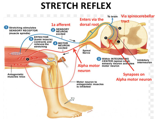
Stretch reflex
Reflex causing contraction of skeletal muscle in response to stretching of the muscle (detected by muscle spindles)
Monosynaptic reflex
Single synapse between muscle sensory fibre and a-motor neurone
eg; stretch refWhat is teh lex
What is the orientation of muscle spindle fibres to the muscle they are in?
They run parallel with muscle
Describe structure of muscle spindle
intrafusal muscle fibres
Ia sensory fibres
y-motor neurone
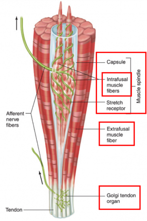
Result of muscle spindle stretching
Increases Ia afferent activity
Where do Ia afferent sensory neurones synapse with the a-motor neurone?
Ventral horn of spinal cord
Roe of y-motor neurone in spinal reflex?
To regulate the sensitivity of sensory afferents to stretch
Example of stretch reflex?
Patella tendon reflex
Reciprocal inhibition
Inhibition of antagonist muscle to allow contraction of agonist muscle during stretch reflex
What is the inverse stretch reflex pathway protective against?
Muscle overload - preventing damage to muscles and tendons
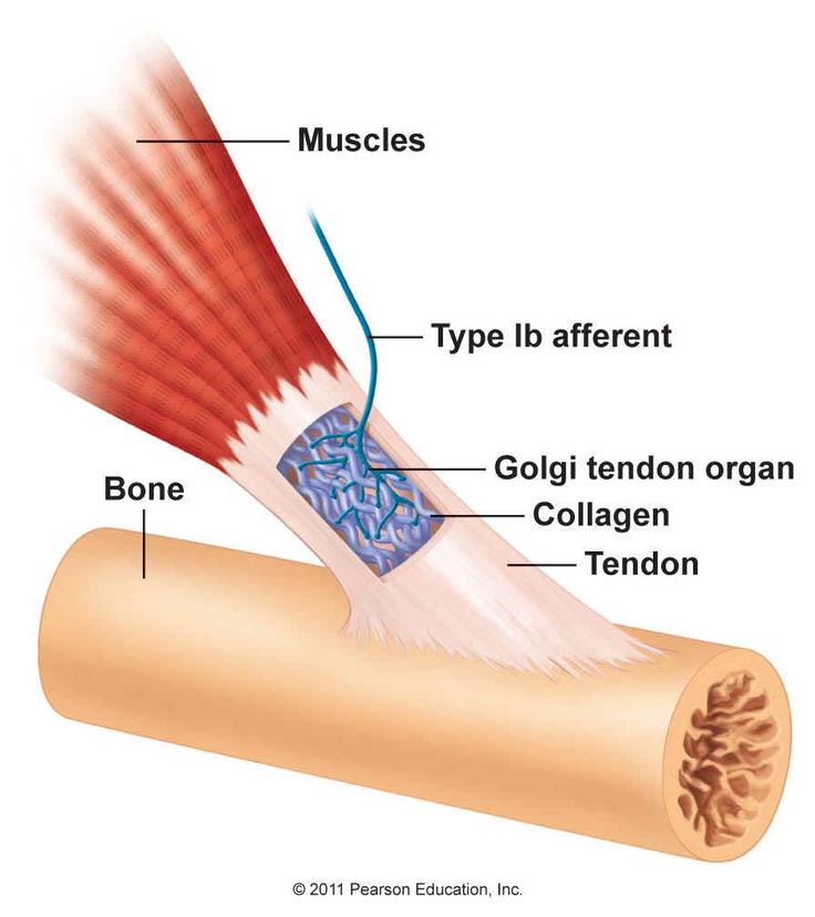
Function of Golgi tendon organ?
Encodes and regulates muscle tension - protecting them form damage
Golgi tendon organ location?
Junction of muscles and tendons - running in series with extrafusal fibres
Innervation of Golgi tendon organs?
Ib afferent neurones
What activates Golgi tendon organs?
Force generated by muscle contraction acts directly on the tendon - increasing the tension of collagen fibrils in golgi organ.
this causes compression of intertwined sensory receptors and increased activity of Ib afferents
In the inverse stretch reflex, what does the Ib afferent synapse with in the spinal cord?
Inhibitory interneurons
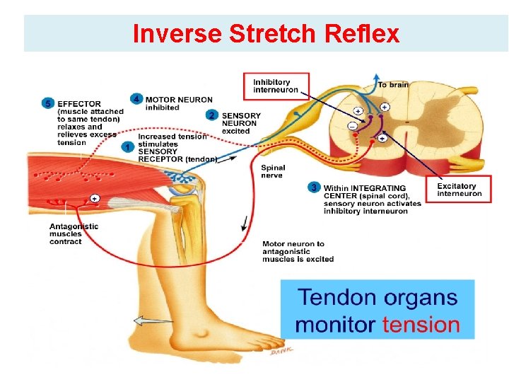
In the inverse stretch reflex, what impact does Ib activation have on a-motor neurones?
Decreases the activity of a-motor neurones
What is the basic principal or the inverse stretch reflex?
Up to a point, the harder a muscle stretched the stronger the contraction
if tension is too much - contraction will stop suddenly and muscle will relax
Inverse stretch reflex
Golgi tendon organ stimulated by high muscle tension
Signals sent along Ib afferent fibres to spinal cord
Ib synapse with inhibitory Ib interneurons = inhibiting a-motor neurones = muscle relaxes
Simultaneously, excitory signals are sent to antagonist muscle promoting its contraction

What kind of reflex is the inverse flexor reflex?
Polysynaptic
what kind of neurone supplies afferent sensory information from nociceptor?
alpha-delta neurone
What are CPGs?
Produce complex rhythmic movements (eg: walking & running) without input from higher centres.
neural networks that produce rhythmic patterned outputs without sensory feedback
Where are CPGs found?
Spinal cord and brainstem - consisting of sensory neurones, interneurons and motor neurones