Head review
1/103
There's no tags or description
Looks like no tags are added yet.
Name | Mastery | Learn | Test | Matching | Spaced | Call with Kai |
|---|
No analytics yet
Send a link to your students to track their progress
104 Terms
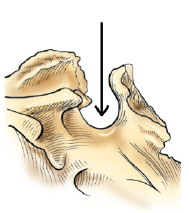
sella turcica
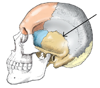
squamosal
the posterior half of the base of the skull is formed by which bone
occipital
the tickest and densest portion of bone in the cranium is the
petrous portion of the temporal bone
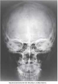
the CR angle for the PA axial Caldwell projection of the skull is
15 degrees caudad
the petromastoid portion is a part of which bone
temporal

projection
PA
the suture located between the occipital bone and the parietal bones is the
lambdoidal
CR angle and line
perpendicular to IOML
how was the CR directed
perpendicular
for an SMV projection of the cranial base the CR should always be perpendicular to the
infraorbitomeatal
petrous portion
which bone has condyles that articulates with the atlas of the cervical spine
occipital
the superior aspect of the sphenoid bone contains a deep depression that contains the
pituitary gland
which of the following lines is placed perpendicular to the image receptor plane for the AP axial Towne projection
orbitomeatal line
all of the following are cranial bones except the
maxillae
the cr and center of the image receptor position for a lateral projection of the skull is ? Inches ? the EAM
2 above
which bone in the skull contains the auditory organs and the organs of hearing
temporal
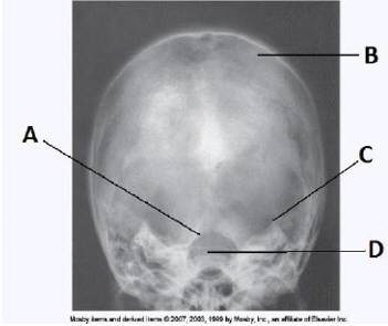
AP axial towne
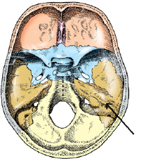
the large aperture in the occipital bone through which the medulla oblongata and spinal cord exits the
foramen magnum
which of the following skull types is considered average in size and shape
mesocephalic
all of these structures are demonstrated on an AP axial towne projection of the skull except
frontal bone
the base of the anterior portion of the occipital bone contains two large opening that allow blood vessels and nerves to pass through. these two openings are called the
jugular foramina
parietal
which of the following is perpendicular to the image receptor plane for a Caldwell projection of the skull
orbitomeatal line
the six areas of incomplete ossification in a newborn infants skull are called
frontanels
if the patient cannot flex the neck to place the orbitomeatal line perpendicular to the image receptor for an AP axial towne projection which line should be placed perpendicular
infraorbitomeatal line
the cranial bones are rigidly joined together by articulations called
sutures
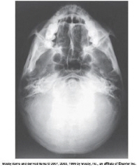
which line should be placed parallel to the plane of the image receptor for the SMV projection of the cranial base
infraorbitomeatal
PA axial Caldwell
which plane of the head is placed parallel to the plane of the image receptor for a lateral projection of the skull
midsagittal
Towne
Which projection of the skull requires MSP be positioned parallel and interpupillary line perpendicular to IR plane
Lateral
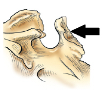
dorsum sellae
which skull type is narrow from side to side
for the SMV (skull, mandible, and sinuses) the CR is directed perpendicular to what line
infraorbitomeatal
zygoma
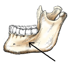
body
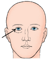
outer canthus
axiolateral oblique of the mandibular body
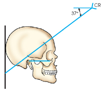
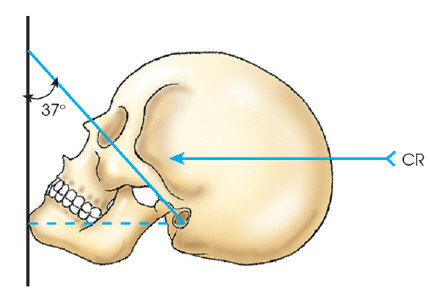
what is the CR angle for the AP axial modified towne method of the TMJ’s if the OML is aligned perpendicular to the IR
35 caudad
how is the head positioned to demonstrate the body of the mandible in the axiolateral oblique projection
30 degrees toward the IR
which of the following is placed perpendicular to the front edge of the IR for a lateral projection of the facial bones
infraorbitomeatal line
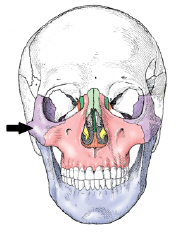
the maxillary sinus is located in which bone
maxilla
which of the following is centered to the image receptor for a parietoacanthial projection of the facial bones
acanthion
the largest sinus is the
maxillary
which sinus is located immediately below the sella turcica
sphenoidal
all of the following are facial bones except the (ethmoid, maxillae, mandible, zygomatic bones)
ethmoid
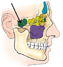
sphenoidalw
which projection will best demonstrate the frontal and anterior ethmoidal sinuses
PA caldwell method

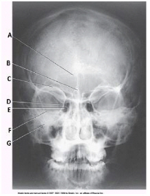
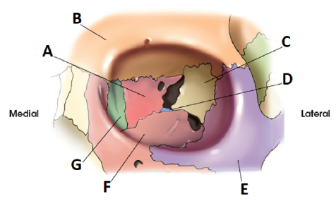
letter G
lacrimal
the orbit is made up of __ cranial bones and __ facial bones
3, 4
the parietoacanthial projection of the facial bones is commonly called the __ method
Waters
all of the following are clearly demonstrated on the parietoacanthial projection (Waters method) except (foramen magnum, orbits, zygomatic arches, maxillae)
formen magnum

letter C
maxillary sinuses
all of the following bones contain air sinuses except ( frontal, parietal, ethmoid, sphenoid)
parietal
which of the following positioning lines is placed perpendicular to the image receptor for a parietoacanthial waters method
MML
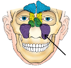
maxillary
if the patient cannot flex the neck to place the OML perp to the image receptor for an AP Axial Towne projection which line should be placed perp to the IR and include an adjustment for the CR angle
IOML
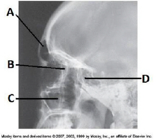
letter D
sphenoid
how many bones make up the face
14
which facial bone contains a foramen through which tear duct passes
lacrimal
ethmoid
which positioning line must be aligned perp to the IR for the PA mandible
OML
projection (method)
parietoacanthial (waters)
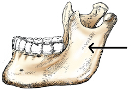
ramus
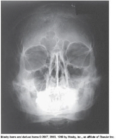
which of the following must be projected below the maxillary sinuses for the parietoacanthial projection (waters method) of the sinuses
petrous pyramids
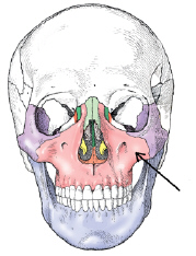
maxilla
how many bones compose the bony orbit
7
how must the head be positioned to best demonstrate the ramus of the mandible in an axiolateral projection
lateral
for a lateral projection of the facial bones the CR is directed to the
zygoma
which projection best demonstrates the maxillary sinuses
parietoacanthial (waters)
for a lateral projection of the facial bones, the CR will enter
halfway between the outer canthus and the EAM
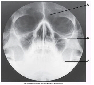
which paranasal sinuses are labeled with the letter A in this image
frontal
waters
parietoacanthial (modified waters)
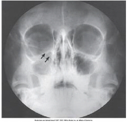
where are the petrous ridges seen on an accurately positioned image of the parietoacanthial waters method projection of the paranasal sinuses
inferior to the floor of the maxillary sinuses

letter B
frontal
the zygomatic arches are a part of which bone
temporal
the radiographic appearance of the erosion of bony rib margins is a possible indication of
osteomyelitis
which of the following features makes the cervical vertebra unique as compared with other vertebrae of the spine (transverse, overlapping vertebral bodies, bifid on spinous processes)
all
what is the joint space between the manubrium and body of sternum called
sternal angle
which aspect of the rib articulates with the thoracic vertebral body
head
flexing the neck
which 2 projections should be performed for an injury to the right anterior upper ribs
PA and RAO
which ribs are considered to be true ribs
1-7
the condition of the lumbar spine in which there is anterior displacement of one vertebra over another is termed
spondylolisthesis
the intervertebral foramina of the cervical spine open
45 anteriorly and 15 inferiorly
a radiograph of an AP open mouth projection reveals that the base of the skull is superimposed over the dens
excessive extension of the skull
which articulation involves the tubercle of a rib
costotransverse
the zygapophyseal joints of the cervical spine are clearly demonstrated on which projection
lateral
where is the articular pillar located on a cervical vertebra
between the superior and inferior articular processes
the z joints for the typical cervical vertebra lie at an angle of __ in relation to the midsagittal plane
90
avulsion fracture of the spinous process of any vertebra C6-T1
clay shovelers
this fracture extends through the pedicles of C2
hangmans
fracture of the anterior and posterior arches of C1
jefferson