Nucleur Medicine imagery
1/17
There's no tags or description
Looks like no tags are added yet.
Name | Mastery | Learn | Test | Matching | Spaced | Call with Kai |
|---|
No analytics yet
Send a link to your students to track their progress
18 Terms
what are the two types of radiation
non ionising radiation
energy to make molecule/atoms vibrate to produce head
not energetic enough to detach electrons from atoms/molecules
mri uses non ionising radiation
ionising radiation
energy to detach electrons from atoms/molecules = make ions
we use in nuclear medicine
what are the two key ways ionising radiation is used
ion directly damage DNA which we can use for treatment
ionising radiation has enough energy to pass through body so we can use for imaging
e.g. x rays such as x ray and CT imaging
what is half life?
types of radioactive decay:
half life ( t1/2) = amount of time for radioactive decay(decays per second) to drop by half.
Alpha decay
Higher damaging but stopped easily
Good for treatment, bad for imaging
Beta decay
Fairly damaging fairly easily stopped
Gamma decay
Less damaging and very penetrating, great for imaging, bad for treatment
what is difference between radiopharmaceutical ( aka tracer) vs contrast agents
tracers administered in lower conc and dont affect the process they are targeting
radio-tracers radioactive, contrast agents aren’t
( if we can detect radiation we can build a picture of where tracer has gone in body and tracks the function it targets)
draw spider diagram for when we use each ‘thing’ in nuclear medicine in context of treatment and diagnosis
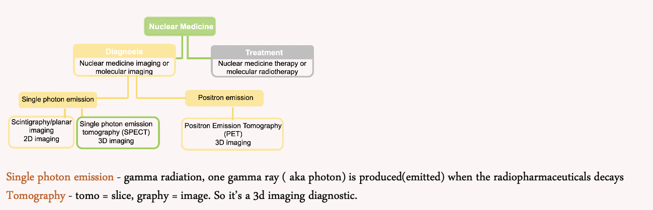
SPECT ( single photo emission computed tomography)
include radiopharmaceutical used
spect acquisition
gamma ray produced when radiopharmaceutical decays creating 3D image
Iodine (I)123 and Technetium (Tc)99m.
spect aquisition
Patient is injected with radiotracer
Gamma camera takes a 2d image projection
SPECT takes projections from different directions around patient
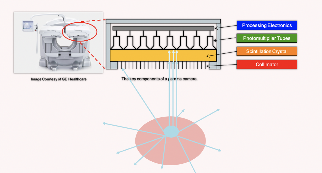
SPECT: the gamma camera
include 4 key components and what they do
draw rough diagrams
what is a sinogram
gamma camera components r - how to make 3d images
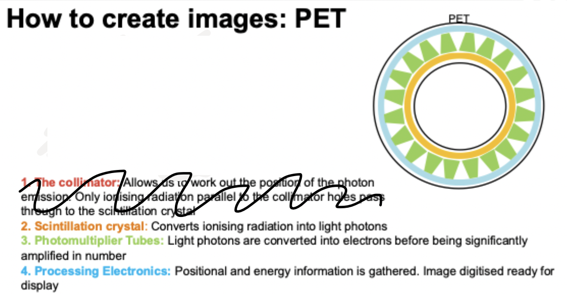
(1) the Collimator - allows us to work out position of photon emission. Only ionising radiation parallel to collimator holes pass through scintillation crystal
(2) Scintillation crystal - converts ionising radiation into light photons
(3) Photomultiplier Tubes - light photons are converted into electrons before being significantly amplified in number
(4) Processing Electronics - positional and energy info is gathered. Image digitalized ready for display
sinogram = stack of projections taken at different angels
PET ( Positron Emission Tomography)
PET acquisition
radiopharmaceuticasls used
PET acquisition
Patient lies inside the PET ring of detectors
Radiotracer within the body emits a positron which annihilates (destroys) producing back-to-back photons
Photons are measured by detectors in the PET ring
If 2 photons are detected within a certain time frame then this is called a coincident event and a line of response (LOR) is drawn between them
PET radiopharmaceutical =Fluorine (F)18
PET:
include key components
NOTE : PET USES SAME COMPONENTS AS GAMMA CAMERA BUT ARRANGED DIFFERENTLY
differences between PET and SPECT
PET | SPECT |
Positron emission | Gamma emission |
2 gammas detected | Single gamma detected |
PET scanner is a ring of detectors | Gamma camera is 1 or 2 heads which rotate around patient |
Position determined by line between 2 gamma rays | Position determined using collimators |
Gamma rays 511keV | Gamma rays normally around 140keV |
state and explain 2 examples of PET
glucose metabolism imaging ( how fast brain uses sugar)
Radiopharmaceutical: F18 FDG = glucose analogue ( meaning body treats same as real glucose)
used especially in epilepsy and dementia
amyloid imaging
Amyloid beta: neurotoxic protein deposited in the grey matter in Alzheimer’s disease
Radiopharmaceutical: F18 florbetapir
f18 emits positrons we can measure and florbetapir sticks to amyloid beta plaques in brain
state and explain 2 examples of SPECT
perfusion imaging
perfusion= blood flow to brain
radiopharmaceutical : Tc99m ECD
Tc99m emits gamma rays which we can measure
ECD delivered to the brain in proportion to the amount of blood flow to the brain region. it crosses blood brain barrier and gets trapped in brain cells
DATscan
Radiopharmaceutical - I123 Ioflupane
I123 emits gamma rays which we can measure (with gamma camera)
Ioflupane targets presynaptic dopamine transporter
often used to diagnose parkinson’s
name 4 measures of image quality
noise
contrast
spacial resolution
artefact
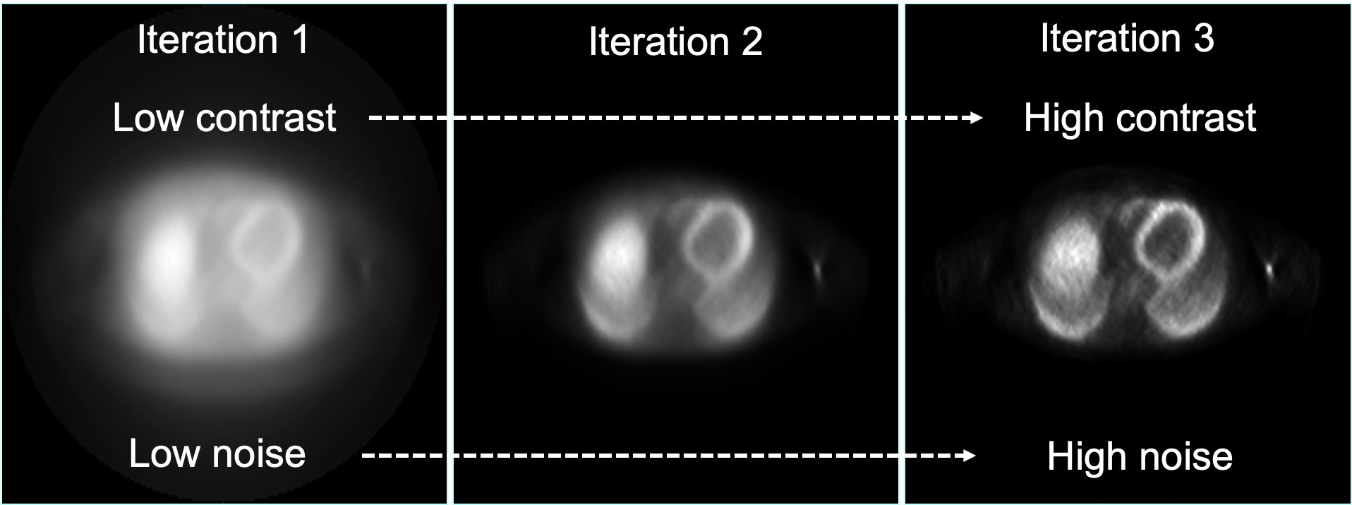
noise - what is it and how to reduce it ?
what is it?
random statistical fluctuations make the image blurry
how to reduce it?
detect more photons - i.e. longer scan or bigger dose of radioactivity
contrast - what is it and how to increase it?
what is it?
difference between high and low areas of radiopharmaceutical uptake
how to increase it?
more iterations - although have to optimise for noise also
spatial resolution
what is it
how it impacts image quality in SPECT
name 4 factors which affect spacial resolution in PET
what is it?
Amount of blurring in the image
how it impacts image quality in SPECT
determined by collimator geometry and distance to patient - angle of acceptance
name 4 factors which affect spacial resolution in PET
- crystal width
- anger logic
- photon noncollinearity
- positrons range
name two artefacts that are most prevalent in nuclear medicine imaging
motion
patients moving during scans
to correct you have to align the SPECT projections before reconstructing
attenuation
The gamma photons emitted from the tracer need to reach the detector
Attenuation = photons being absorbed or scattered in the body
hybrid imaging - CT
include advantages and disadvantages of PET MRI over PET CT
pet CT and spect CT because most are sold with a CT scanner attached. imaging is sequential for both.
advantages and disadvantages PET MRI over PET CT
✓Great soft tissue contrast
✓scan simultaneously
✗worse for attenuation correction