Caren test 2 part 1
1/100
There's no tags or description
Looks like no tags are added yet.
Name | Mastery | Learn | Test | Matching | Spaced | Call with Kai |
|---|
No analytics yet
Send a link to your students to track their progress
101 Terms
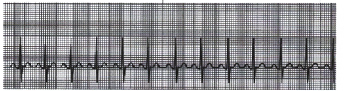
Atrial Flutter
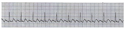
Sinus Tachycardia
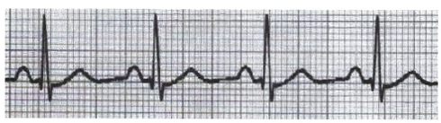
Sinus Bradycardia
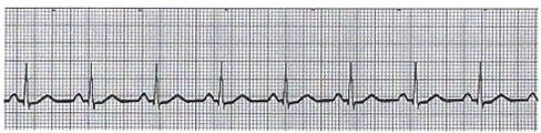
Normal Sinus Rhythm
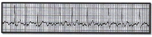
Atrial Fibrillation (A-Fib)
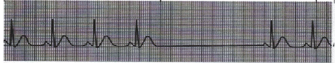
Sinus Pause / Arrest
What is the blood flow pathway starting from the SVC, IVC, and Coronary Sinus?
Blood enters the right atrium → passes through the tricuspid valve → enters the right ventricle → goes through the pulmonic valve → into the main pulmonary artery → then travels to the lungs.
What is the blood flow pathway from the lungs back to the body?
Blood returns from the lungs through the 4 pulmonary veins → enters the left atrium → passes through the mitral valve → enters the left ventricle → goes out through the aortic valve → into the ascending aorta → aortic arch → descending aorta → abdominal aorta → to the body.
Where is the heart located in the thoracic cavity?
In the middle of the thoracic cavity (mediastinum) behind the sternum, between the lungs, and above the diaphragm.
What structure surrounds and protects the heart?
The pericardium, a protective sac that protects Friction, trauma, and infection, attached to the thorax through the great vessels.
What forms the apex of the heart?
It is slightly larger than a man’s fist.
How many chambers does the heart have, and what are they?
The heart has 4 chambers — 2 atria and 2 ventricles.
What type of blood is on the left side of the heart?
Oxygenated blood.
What does the left side of the heart pump?
Arterial blood to the systemic circulation.
What kind of pressure system is the left side of the heart?
A high‑pressure system.
What type of blood is on the right side of the heart?
Deoxygenated blood
What does the right side of the heart pump?
Venous blood to the lungs.
What kind of pressure system is the right side of the heart?
low‑pressure system.
What type of blood does the right atrium receive and from where?
The right atrium receives deoxygenated blood from the superior vena cava, inferior vena cava, and coronary sinus.
What type of blood does the left atrium receive and from where?
The left atrium receives oxygenated blood from the lungs via the right and left pulmonary veins.
Which heart chamber has the highest pressure?
The left ventricle has the highest pressure of all the heart chambers.
What do heart valves do?
They control and maintain blood flow through the heart.
What are the two types of heart valves?
Atrioventricular (AV) valves and Semilunar (SL) valves.
Where is the mitral (bicuspid) valve located?
It lies between the left atrium (LA) and the left ventricle (LV).
What are the semilunar (SL) valves?
The aortic valve and the pulmonic valve
What is the endocardium?
The inner layer of the heart.
What is the myocardium?
The muscular middle layer of the heart.
What is the epicardium?
The outer layer of the heart.
What is serous (pericardial) fluid?
The fluid found in the pericardial space that reduces friction around the heart.
What does pericardial (serous) fluid help prevent?
It prevents friction as the heart beats
What does pericardial fluid do to the lining of the pericardium?
It moistens the lining of the pericardium.
What does the superior vena cava do?
It brings blood from parts of the body superior (above) the heart.
What does the inferior vena cava do, and what special feature does it have?
It brings blood from the lower parts of the body and has a Eustachian valve, a rudimentary fetal valve.
What does the coronary sinus do?
It brings blood back from the myocardium of the heart.
How many pulmonary veins are there?
Four pulmonary veins.
What is the brachiocephalic artery also called?
The innominate artery.
What does LCCA stand for?
Left Common Carotid Artery.
What does LSA stand for?
Left Subclavian Artery.
What is the coronary sinus and where does it drain?
It is the largest vein that drains the heart, and it drains into the right atrium.
What is one complete contraction and relaxation of the heart called?
One cardiac cycle.
What is stroke volume?
The amount of blood ejected by the left ventricle with each heartbeat.
What does Frank‑Starling’s Law state?
The greater the stretch, the greater the contraction.
What is the heart’s conduction system considered?
It is considered the wiring of the heart.
What is the correct order of the heart’s conduction system?
SA node → AV node → Bundle of His → Right & Left Bundle Branches → Purkinje fibers.
What is the SA node considered?
The normal pacemaker of the heart.
What is the function of the SA node?
It is the site of origin of the electrical impulse.
What special property does the SA node have?
It spontaneously generates electrical impulses.
Which cardiac structure has the highest degree of automaticity?
The SA node.
What is the normal intrinsic rate of the SA node?
60–100 beats per minute.
What are the three internodal pathway branches in the right atrium?
Anterior, Middle (Wenckebach), and Posterior (Thorel’s) branches.
What do the internodal pathways do?
They spread the electrical impulse through the right atrium to the AV node and toward the ventricles.
What is the largest branch that carries the impulse from the RA to the LA?
Bachman’s Bundle.
What does Bachman’s Bundle do?
It spreads the impulse from the right atrium to the left atrium.
In the cardiac conduction sequence, which atrium contracts first?
The right atrium contracts before the left atrium.
Where is the AV node located?
It is located at the base of the interatrial septum (IAS) and extends into the ventricular septum.
Where is the AV node located?
On the right side of the interatrial septum (IAS), behind the tricuspid valve, and near the opening of the coronary sinus.
What is the main blood supply to the AV node in most adults?
The right coronary artery (RCA).
What artery supplies the AV node when it is not supplied by the Right Coronary Artery?
The left circumflex artery (LCX).
What are the three regions of the AV node?
Atrial nodal (AN)
Nodal (N) region
Nodal‑His (NH)
Nodal‑His (NH)
lower junctional region
Nodal (N) region
mid‑portion of the AV node
Atrial nodal (AN)
upper junctional region / transitional zone
Where is the Bundle of His located?
On the right side of the interatrial septum (IAS) just above the ventricles.
What does the Bundle of His do?
It bifurcates into the right bundle branch and the left bundle branch.
What arteries supply the Bundle of His?
Branches of the anterior and posterior descending coronary arteries.
What is the intrinsic discharge rate of the Bundle of His?
40–60 beats per minute.
What does the anterior fascicle do?
It is longer and thinner and spreads impulses to the anterior portions of the left ventricle.
What does the posterior fascicle do?
It is shorter and thicker and supplies the posterior and inferior left ventricle.
What does the septal fascicle do?
It relays impulses to the mid‑septum.
How do impulses spread into the ventricular muscle?
They spread into the ventricles through the Purkinje fibers, activating the ventricular muscle.
How does the electrical impulse travel through the heart wall?
It spreads from the endocardium to the myocardium.
What is the intrinsic ventricular (Purkinje) rate?
20–40 beats per minute.
What does an EKG record?
The electrical activity of the heart.
How is the heart’s electrical activity observed on an EKG?
Through electrodes connected by cables to an EKG machine.
What type of information does the EKG not provide?
It does not provide information about the mechanical (contractile) condition of the myocardium.
What is automaticity in cardiac cells?
The ability to generate an electrical impulse independently.
What is excitability in cardiac cells?
The ability of each cell to respond to an electrical stimulus.
What is conductivity in cardiac cells?
The ability of each cell to receive an electrical stimulus and conduct it to adjacent cells.
What is contractility in cardiac muscle?
The mechanical result of depolarization — the ability to respond with pumping action.
What is the polarized state of the heart?
The resting state of the cardiac cells.
What happens when a cardiac muscle cell is stimulated?
Depolarization occurs.
What does the P wave represent on an EKG?
Atrial depolarization.
What does the QRS complex represent on an EKG?
Ventricular depolarization.
What safety rule applies to the power cord and patient cables during an EKG?
Never let the power cord and patient cables touch.
Where should EKG cables (wires) never be placed?
They should never touch the floor.
How should electrode connectors be positioned?
Connectors should point toward the heart.
What are the three categories of EKG leads?
Standard leads, augmented leads, and precordial (chest) leads
What are the standard (bipolar) leads?
Lead I, Lead II, Lead III.
What do standard leads measure?
Electrical activity between two limbs.
What are the augmented leads?
aVR, aVL, aVF.
What do augmented leads measure?
Electrical activity from the heart toward one limb.
What are the precordial (chest) leads?
V1, V2, V3, V4, V5, V6.
What do precordial leads measure?
Electrical activity across the horizontal plane of the chest.
How does a positive deflection appear on an EKG?
Above the baseline.
How does a negative deflection appear on an EKG?
Below the baseline.
What does Lead I record?
The difference in electrical potential between the right arm (RA) and left arm (LA).
What does Lead II record?
The difference in electrical potential between the right arm (RA) and left leg (LL).
What does Lead III record?
The difference in electrical potential between the left arm (LA) and left leg (LL).
AVR
LA & LL to RA
AVL
RA &LL to LA