H3 Toth- Animal extracellular matrix and plant cell wall
1/14
There's no tags or description
Looks like no tags are added yet.
Name | Mastery | Learn | Test | Matching | Spaced | Call with Kai |
|---|
No analytics yet
Send a link to your students to track their progress
15 Terms
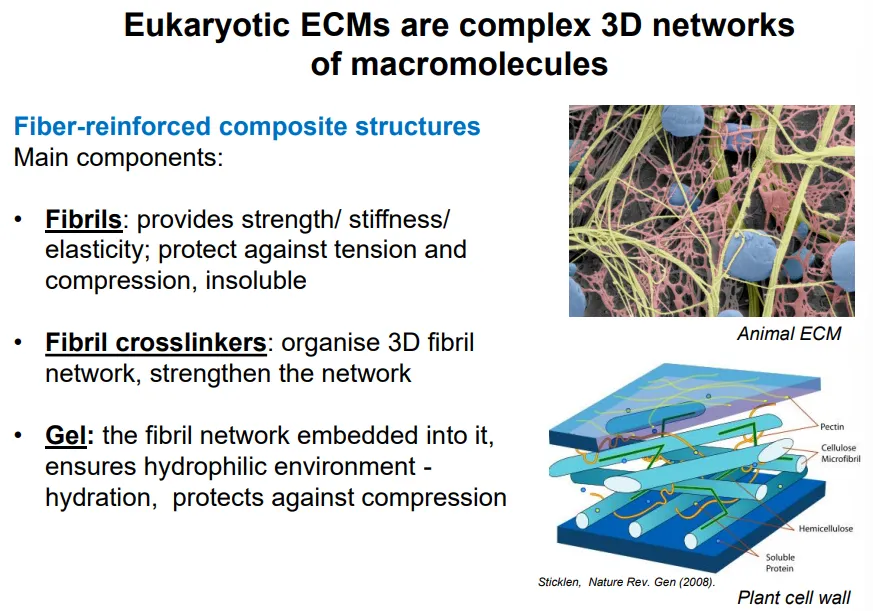
what are the different components of the animal ECM and their functions?
fibrils are made from collagen and elastin proteins:
provide strength/stiffness/elasticity
protect against tension and compression
insoluble
fibril crosslinkers are made from accessory collagen proteins:
organise 3D fibril network
strengthen the network
gel is made from glycosaminoglycans (GAGs) polysaccharide and proteoglycans (GAGs attached to a protein core):
contains the fibril network embedded
creates a hydrophilic environment = hydration
protects against compression
contains protein components (unlike the plant cell wall)
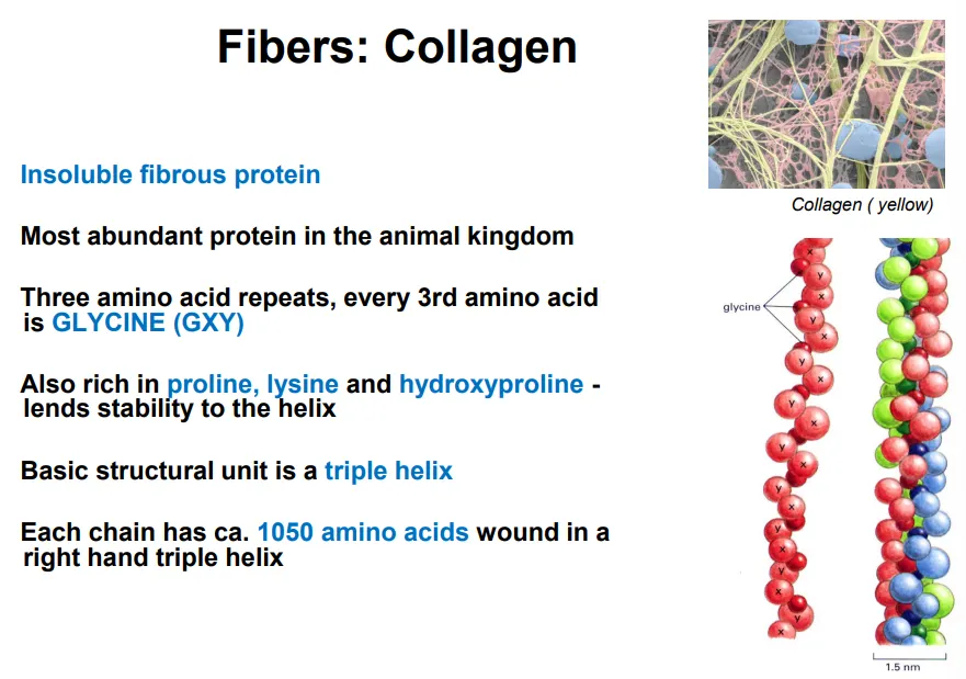
describe the structure of collagen
collagen is an insoluble fibrous protein that makes up the fibrils of the animal ECM
it is a right handed triple helix of three peptide strands made up of three amino acid repeats, every 3rd aa. being glycine
glycine is the smallest amino acid, so when the collagen forms a triple helix, the glycine is at the centre to make the most compact possible shape
rich in proline, lysine and hydroxyproline
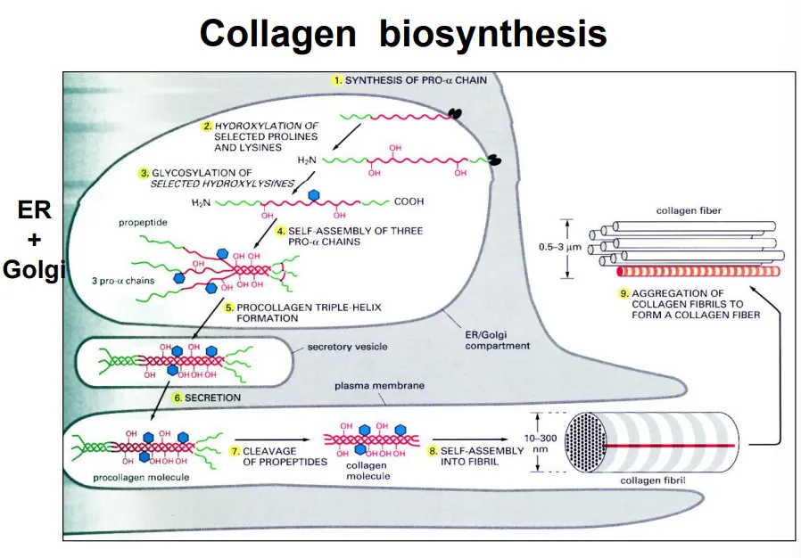
what are the steps of collagen synthesis?
ER: synthesis of pro α-chains (precursors)
ER: proline and lysine hydroxylation- vitamin C is a cofactor
ER: glycosylation of hydroxylysines
ER: self-assembly of the chains into a procollagen triple helix
golgi: N-linked glycan modifications for recognition
secretory vesicles: transfer to plasma membrane
ECM: cleavage of the ends by extracellular proteases
ECM: self-assembly into collagen fibril
ECM: aggregation of fibrils into a fibre
collagen fibres cross-link at specific, regular points, giving them a striated/banded pattern
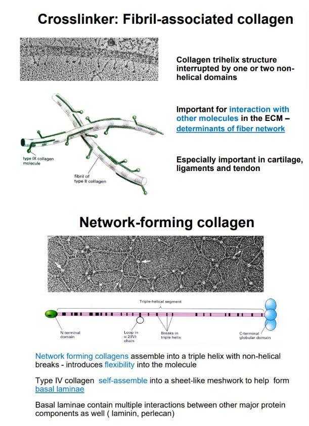
what are the two accessory forms of collagen and their structures?
crosslinker collagen has a trihelix structure interrupted by one or two hinge regions, which allow cross-linking (determines the ECM thickness)
network-forming collagen has more regular non-helical breaks, so they are very flexible and can assemble into a mesh sheet, which can be stacked by interactions between N and C globular domains to form the basal laminae
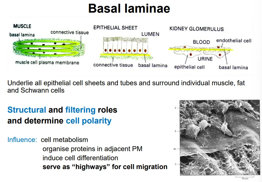
what is the basal laminae and what does it do?
the basal laminae is a specialised form of ECM that lies underneath epithelial cells and surrounds muscle, fat, and schwann cells to facilitate anchorage to connective tissue
this is primarily made up of layers of network-forming collagen sheets, with laminin glycoproteins
this has multiple regulatory roles, eg:
determining cell polarity if needed
filtering intake of molecules
organise proteins in adjacent plasma membrane
induce cell differentiation
serve as “highways” for cell migration
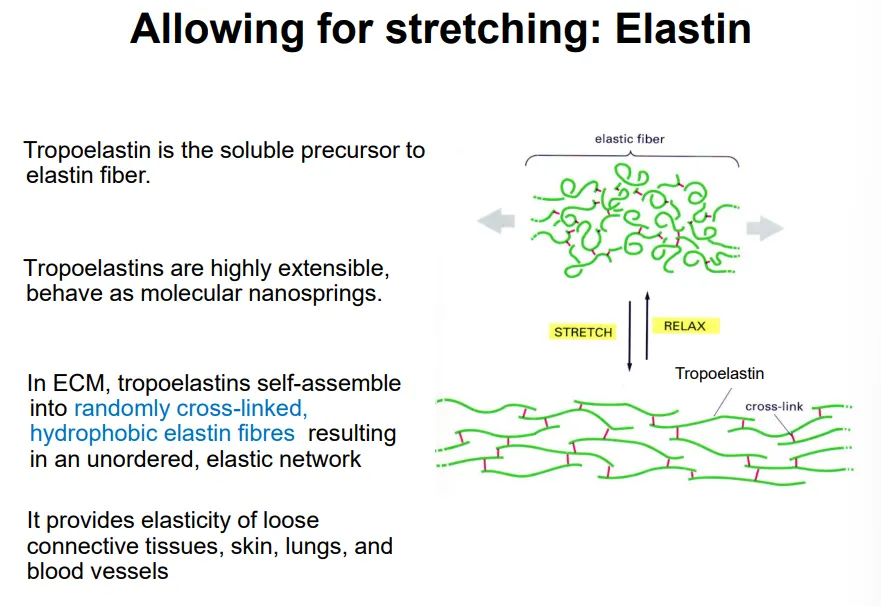
what is the structure of elastin
elastin is an insoluble protein network that forms part of the fibrils of the animal ECM
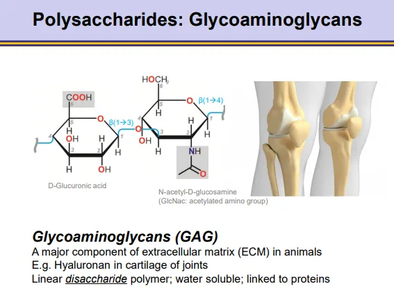
what is the general structure and properties of glycosaminoglycans?
GAGs are polymers of disaccharide monomers, which make up most of the gel in the animal ECM
one monomer is a -uronic acid, the other is an amino sugar
the most abundant is hyaluronic acid, which is glucuronic acid + N-acetylglucosamine
these are strongly hydrophilic and water-soluble
they are negatively charged due to high amounts of sulfur, which attracts sodium- this affects osmosis to give turgor pressure
they are inflexible and have a very low density
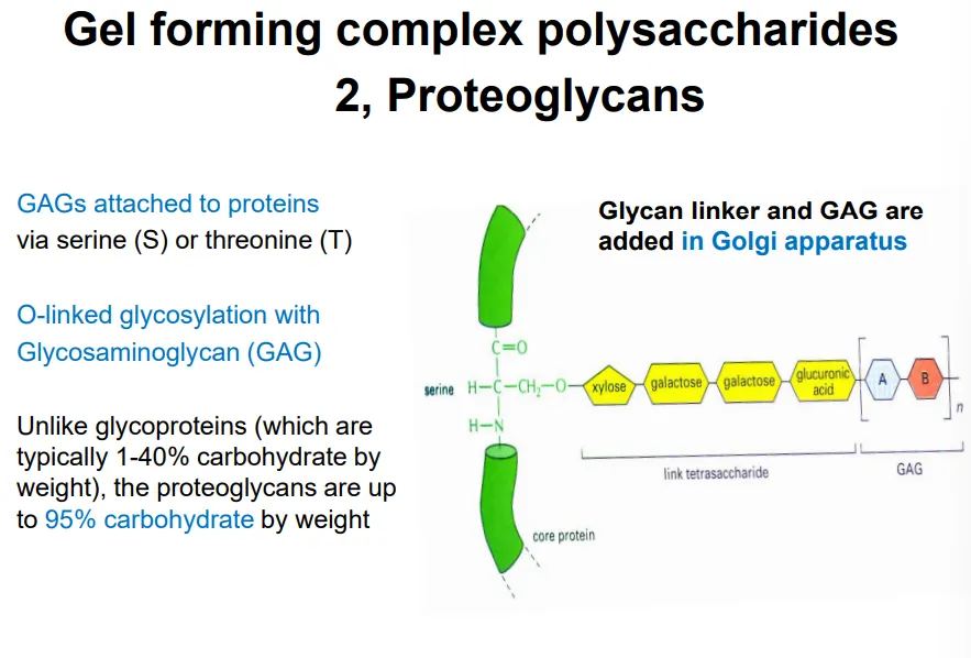
what is the general structure and properties of proteoglycans?
proteoglycans form part of the animal ECM
these are proteins highly modified by O-linked glycosylation, with many glycosaminoglycans (GAGs) attached via serine or threonine residues (in the golgi)
what are the different components of the plant cell wall and their functions?
very little protein content (unlike animal ECM)
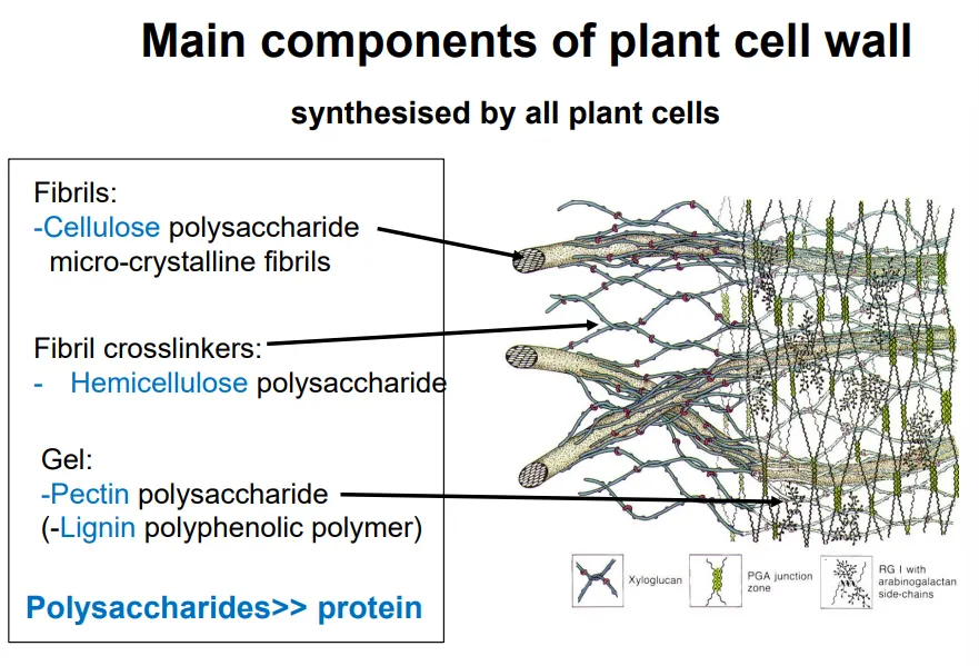
describe the organisation of cellulose fibers in the plant cell wall
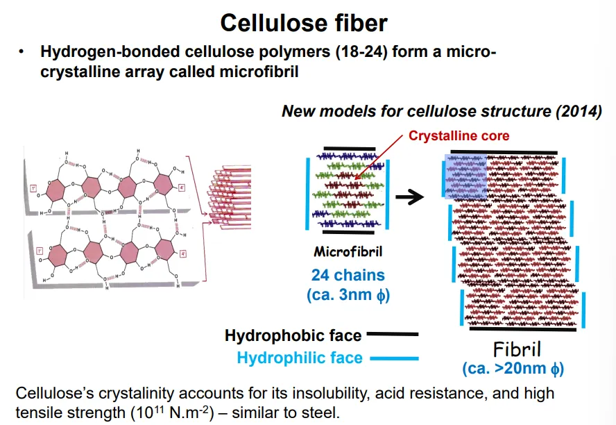
cellulose polymers are produced right next to each other so that they form hydrogen bonds together
this produces a long sheet of adjacent polymer molecules, which is hydrophilic at the edges but hydrophobic on either face
these stack into microfibrils, which have a crystalline core, that can assemble into fibrils due to having a hydrophobic faces on the top and bottom and a hydrophilic faces on either side
how is cellulose deposition in the plant cell wall regulated?
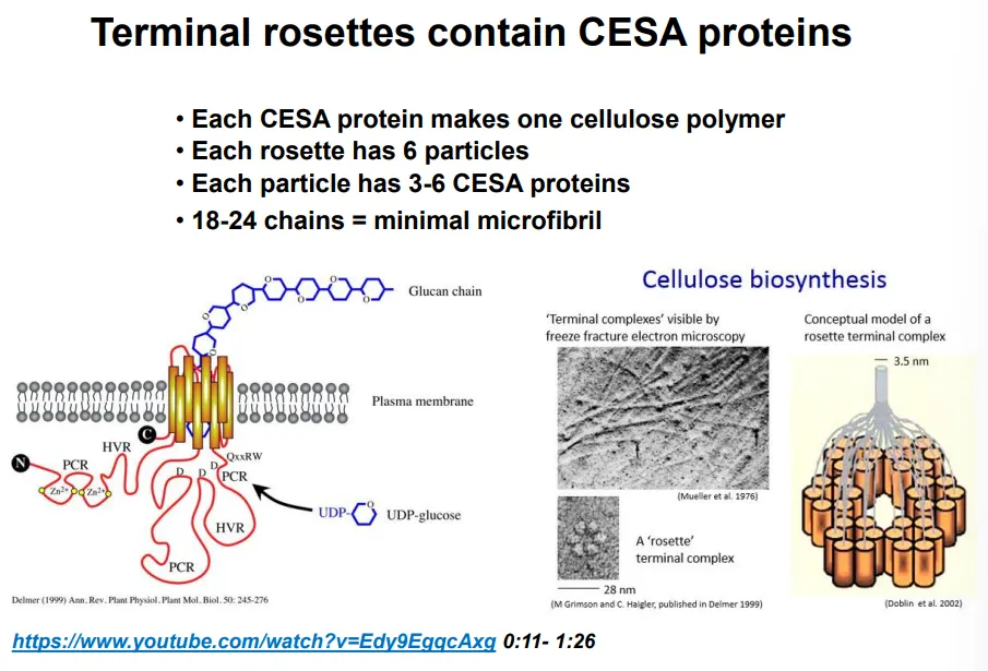
cellulose is synthesised by terminal rosette enzyme complexes in the plasma membrane (acquired by HGT from bacteria)
the terminal rosettes contain 6 particles, each of which contain 3-6 cellulose synthase A (CESA) proteins, which each make one cellulose polymer at a time
this means each rosette makes an 18-24 chain microfibril and twists them together
the cell elongates perpendicular to the orientation of the cellulose fibrils, because new material is deposited in between the fibrils
random cellulose deposition in a cell would mean it could never expand
the deposition of cellulose by terminal rosettes is directed by microtubules to occur in one direction
plant cells stop growing by depositing new layers in different orientations
this deposition is driven by the polymerisation reaction because the polymer is being pushed into an already densely packed matrix, so the terminal rosette is the part that moves instead, along the microtubules
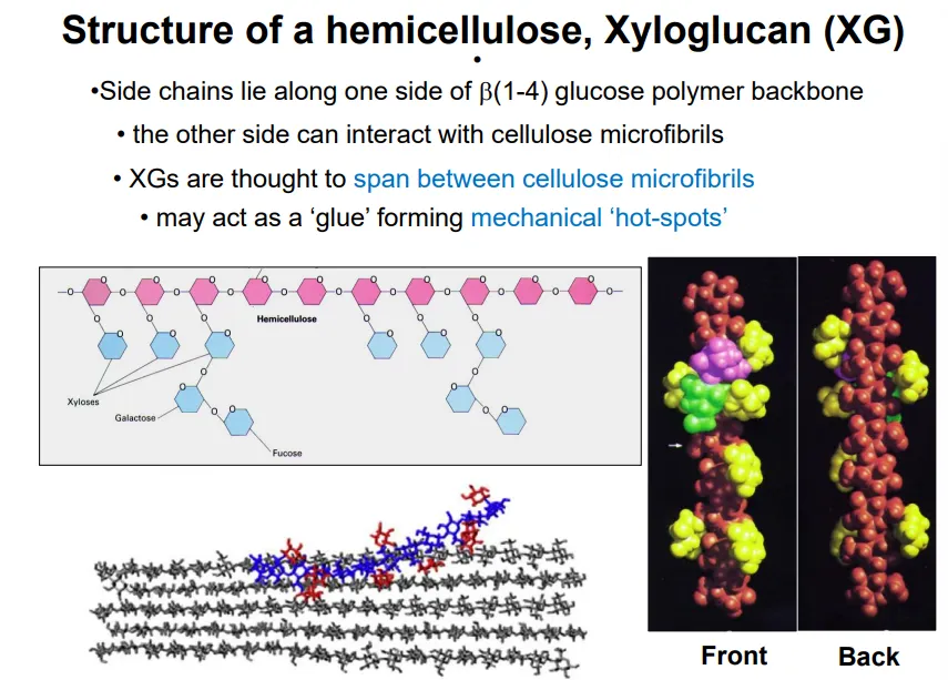
describe the general structure and function of hemicellulose, with an example
hemicellulose cross links the cellulose fibrils in the plant cell membrane
‘half’ cellulose because one side of the chain has many glucan side chains, while the other can interact with cellulose to form mechanical hot spots
eg. xyloglucan
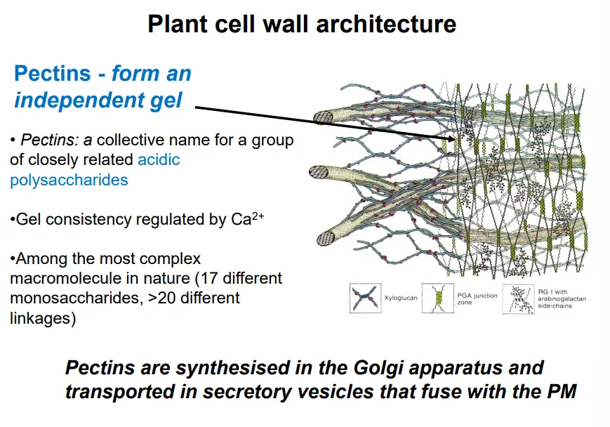
describe the composition and function of the gel in plant cell walls
the gel is mostly made up of pectins (very complex, hydrophilic, acidic polysaccharides), with its consistency regulated by pH and Ca ions
these protect against pathogens and influence the porosity, pH, and ion balance of the cell wall
the gel can also contain lignin (polyphenol), primarily in the secondary cell wall (more rigid, produced by mature cells)
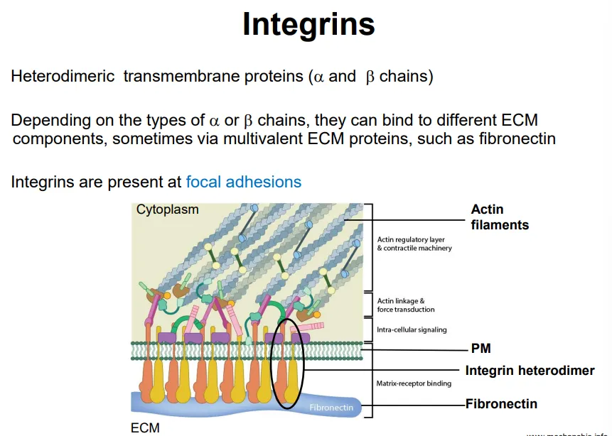
how do animal cells anchor with the ECM?
animal cells have integrins, transmembrane protein receptors that mediate cell adhesion to the ECM
these are heterodimeric with one alpha and one beta chain, which can bind to different ECM components at regions called focal adhesions
these focal adhesions define cell shape, mechanical and chemical signalling and control cell migration
how does the cell modify the cell wall for growth?
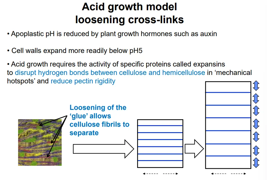
cell growth requires the loosening of the cell wall crosslinking, and the increase of turgor pressure, according to the lockhart equation: R = ɸ(P-Y)
cell expansion happens when P, pressure > Y, the yield threshold
this is determined by the wall extensibility, ɸ (determined by cross linking)
the cross linking is weakened by acidic extracellular pHs (< 5), caused by hormones eg. auxins
this triggers expansin proteins to disrupt H bonds between cellulose and hemicellulose and reduce pectin rigidity
local cell wall loosening at the apical meristem initiates new organ formation
it can also be used to detect abiotic/biotic stress (eg. pectin degradation by pathogens)