sliding filament theory
1/5
There's no tags or description
Looks like no tags are added yet.
Name | Mastery | Learn | Test | Matching | Spaced | Call with Kai |
|---|
No analytics yet
Send a link to your students to track their progress
6 Terms
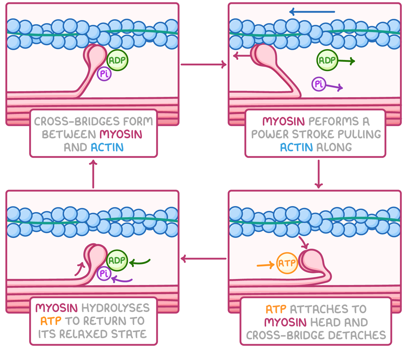
describe the process of muscle contraction according to the sliding filament theory:
action potential travels through T tubules which are in contact w/ sarcoplasmic reticulum which stores Ca2+
action potential causes Ca2+ channels to open on the sarcoplasmic reticulum allowing Ca2+ to diffuse into myofibrils from sarcoplasmic reticulum
Ca2+ causes movement of tropomyosin so myosin binding site no longer blocked on actin filament
myosin heads attach to binding sites on actin, forming a cross bridge
hydrolysis of ATP on myosin heads causes myosin heads to bend
myosin heads change angle and perform power stroke, pulling actin along and releasing ADP + Pi
this causes the sarcomere to shorten, leading to muscle contraction

describe the process of muscle relaxation according to the sliding filament theory:
ATP attaches to myosin head and causes myosin heads to detach, breaking the cross bridge
myosin hydrolyses ATP - catalysed by ATP hydrolase - to return to its original position (relaxation)
myosin head now has a molecule of ADP attached and will reattach itself further along the actin filament
cycle repeats as long as Ca2+ conc remains high (i.e. as long as there is an action potential)
ATP used to actively transport Ca2+ into sarcoplasmic reticulum, stopping the contraction process
what is tropomyosin? what is its significance?
tropomyosin - fibrous protein twisted around 2 actin chains
covers binding site on actin in relaxed myofibril
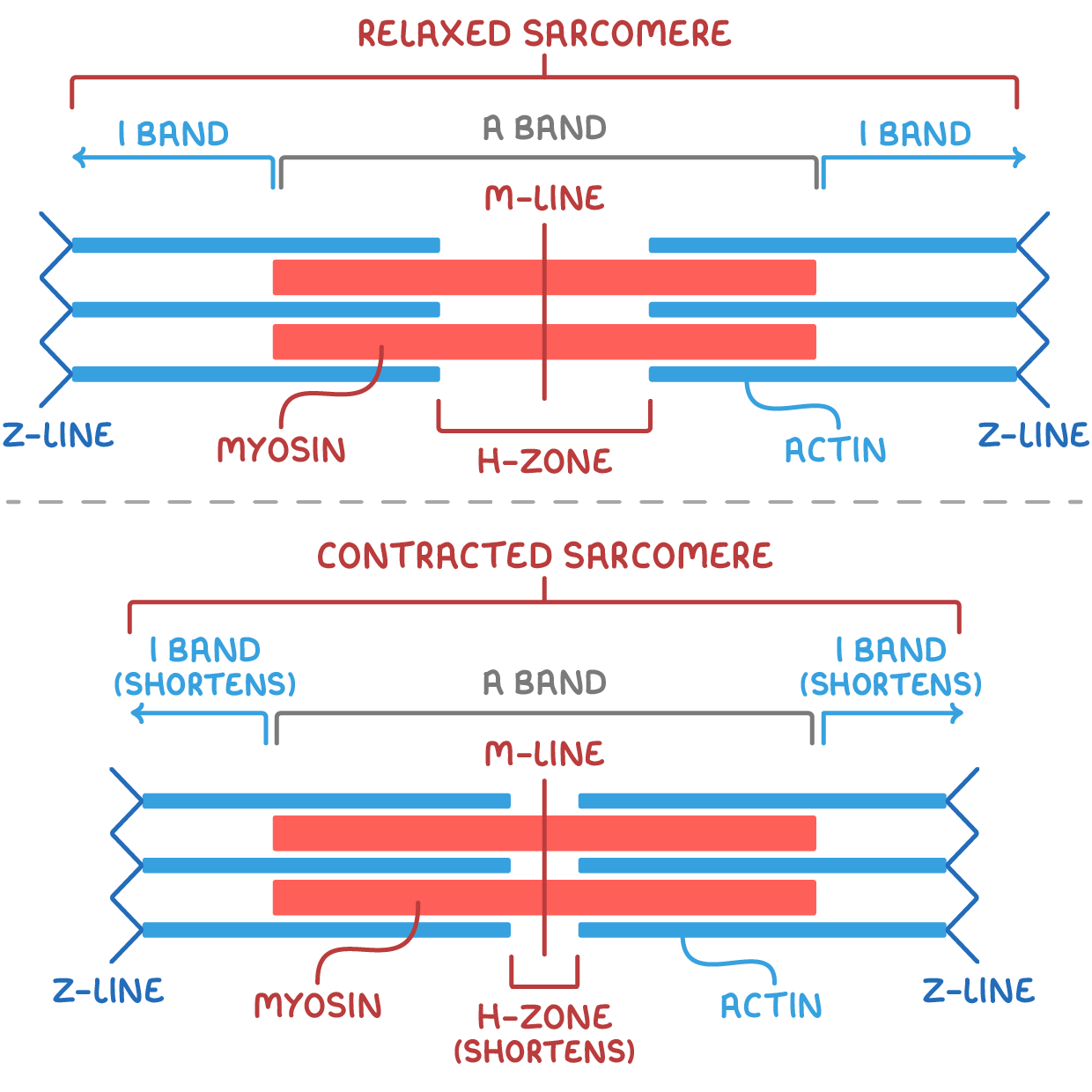
describe how the sarcomere changes during contraction:
sarcomeres shorten, causing a change in the light and dark banding
thin actin filaments slide over thick myosin filaments
Z line pulled closer together
I band shortens
A band remains constant
H zone narrows
during vigorous exercise, the pH of skeletal muscle tissue falls, this fall in pH leads to a reduction in the ability of Ca2+ to stimulate muscle contraction - suggest how (3)
any 3 from:
low pH changes shape of Ca2+ receptor
fewer Ca2+ bind to tropomyosin
fewer tropomyosin molecules move away
fewer actin binding sites revealed
fewer cross bridges can form/fewer myosin heads can bind
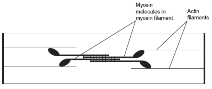
the diagram shows the arrangement of actin and myosin in a sarcomere. one form of muscle disease is caused by a mutated allele of a gene. this leads to production of myosin molecules that are unable to bind to other myosin molecules.
if myosin molecules are unable to bind to other myosin molecules, this prevents muscle contraction - use the diagram and your knowledge of how muscles contract to suggest why (3)
can’t form myosin/thick filaments
can’t pull/move actin
myosin moves
can’t move actin towards each other