BIOC221 Mod 4 - Translation and Protein Degradation
 Call Kai
 Learn
Learn Practice Test
 Spaced Repetition
 Match
Match Flashcards
 Knowt Play
1/25

There's no tags or description
Looks like no tags are added yet.
Name | Mastery | Learn | Test | Matching | Spaced | Call with Kai | Chat |
|---|
No analytics yet
Send a link to your students to track their progress
26 Terms
Describe the features and function of tRNA
tRNA are folded RNA molecules, often in a conformation with a hairpin structure. They are transcribed by RNAP II. Their conformation allows them to bind to a ribosome active site at the same time as a certain codon from an mRNA sequence is being “read” by the ribosome. The tRNA strand is translated by the ribosome to an amino acid corresponding to the tRNA and the codon of the mRNA strand, allowing for amino acid chains and then proteins to be formed.
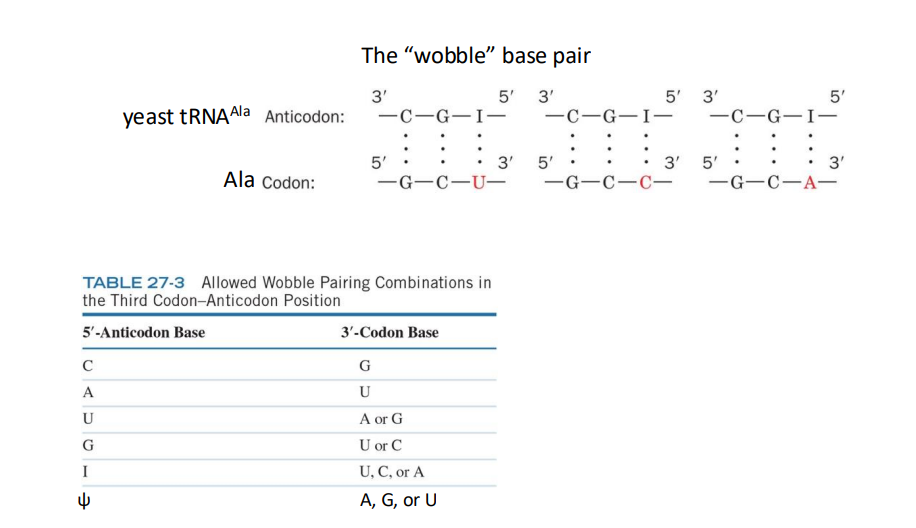
What is another source of redundancy in amino acids that helps maintain the fidelity of the genetic code during protein synthesis
the anticodon on a tRNA binds to mRNA codon, it can recognize >1 codon – the “wobble” base pair. Ensure the fidelity of the genetic code
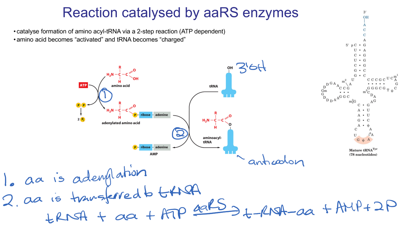
What is the reaction catalysed by aaRS
amino-acyl tRNA Synthetases catalyse two reactions. The first is adenylation of an amino acid (the one that corresponds to this particular tRNA). This reaction requires ATP and is called “activating” the amino acid. This amino-acyl group is then transferred onto one of the corresponding tRNA, “charging” the tRNA.
What are isoaccepting tRNAs and what do they help promote?
Isoacceptor tRNAs: tRNAs recognising different codons for the same amino acid. This is how codon redundancy is enforced, since isoacceptor tRNAs will recognise multiple codons for the same amino acid.
Describe the features of the ribosome
Ribonuclear protein complexes protein = scaffold. This brings together the mRNA strand, centring the codon being read at that moment, and brings in a corresponding tRNA.
rRNA = catalyst, catalysis peptide bond formation.
What are the substrates required to initiate translation?
Requires small ribosomal subunit, capped and polyadenylated mRNA, eIFs (eukaryotic initiation factors), Met-tRNAi Met (iMet-tRNA), GTP.
GTP is required as some of the initiation factors are G proteins.
What roles do the eukaryotic translation initiation factors perform?
Binding to Met-tRNAiMET
Binds to 5’ cap and PABP, RNA unwinding
Binds to A and E sites in 40S subunit
Binds to small subunit
Binds to 43S preinitiation complex
Binds to 60S subunit, displaces other IFs
How is protein fidelity ensured during translation by making sure Met tRNA binds to the correct AUG start codon? (eukaryotes and prokaryotes)
In eukaryotes the correct AUG start codon is flanked by the Kozak sequence which has a higher frequency of certain base pairs. It is not well understood exactly how the kozak sequence helps iMet tRNA bind to the first AUG start codon.
In prokaryotes the relationship is well understood. The Shine Dalgarno sequence is upstream of the first AUG codon and base pairs to a complementary sequence on 16S ribosomal RNA which helps align the iMet tRNA to bind to the correct start codon.
Define Transpeptidation
peptide bond formation
Define Molecular mimicry
when two different molecules can perform similar functions because they have the same shape
Define Peptidyl transfer centre (PTC)
site of peptide bond formation on ribosome
How is translation termination catalysed by eRF complex (and what is eRF)?
eRF is eukaryotic release factor. In Eukaryotes, eRF1 binds to stop codons. Once the stop codon is in the A site, eRF3 (a G protein) binds to eRF1 to help termination, by hydrolysing the ester bond between amino acid and tRNA, releasing the polypeptide chain.
What is the ER import signal for proteins
A hydrophobic Alpha helix on the N terminal of the amino acid chain
How does a protein pass through the ER membrane?
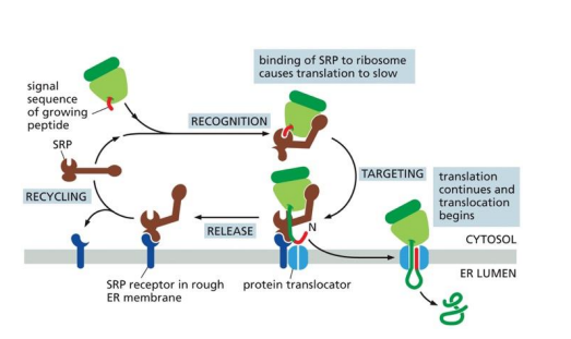
The signal recognition protein (SRP) recognises a hydrophobic Alpha helix at the N terminal of a growing amino acid chain and binds to the amino acid chain and its attached ribosome. The SRP binds to the ribosome A-site, slowing translation. The SRP binds to the ER membrane-bound SRP receptor (SR) via a signal sequence on the SRP and the amino acid chain is fed through the SR into the ER, translation continues and translocation occurs until the full protein is fully translated inside the ER.
While crossing the ER membrane, how is translation resumed after the SRP binds to the SR?
The SRP protein binds to the ribosomal A-site, inhibiting translation. The SRP and SR are both G proteins. The GTP portion of the SR and SRP bind together, activating GTPase activity that converts GTP to GDP, releasing the SRP and GDP portions of the SR from the ribosome-SR complex. This allows translation to continue and the growing amino acid chain is fed through the SR gate into the ER.
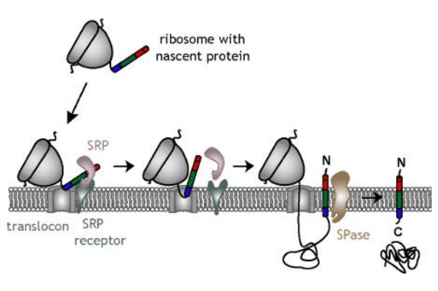
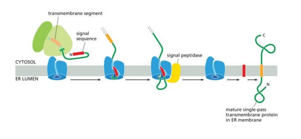
How are transmembrane proteins processed into the ER membrane?
The amino acid chain is fed through the membrane like normal. The transmembrane segement of this chain is stopped when it loops back to go through the membrane again, so the transmembrane sequence stays in the membrane, with the N terminal in the cytosol and the C terminus in the lumen.
How are proteins always N-glycosylated or otherwise glycosylated?
The Oligosacchayltransferase complex (OST) sits right next to translocons in the ER when means that as proteins are translocated into the ER they are actively glycosylated, so every protein in the ER is subject to glycosylation and other PTM’s.
How is glycosylation used to increase protein diversity?
OST complexes can add up to 10 different sugars. This means there are around 7000 combinations of glycosylations that can be done to a single protein. By randomizing this, thousands more different proteins can be created with different jobs, decreasing the DNA storage capacity requirements for that many different proteins.
What PTM is performed on insulin in humans to make it a mature protein?
Proprotein cleavage is carried out by proprotein convertase enzymes, which have specific recognition sequences of amino acids, which they cleave. Proprotein cleavage is performed on premature insulin peptides. Part C is cleaved after parts A and B have formed disulfide bonds and folded into shape.

What are some reasons to degrade proteins?
Controlling cell signalling/regulation
Recycling amino acids
destruction of foreign proteins
misfolded or damaged proteins
What is the role of ubiquitin and what is the role of the Amino Acids on its N and C termini?
Ubiqitination is the process of attaching a ubiquitin protein to a larger one. Ubiqitin is a protein signaller that tells proteases to break down the protein it is attached to. Usually a chain of many ubiqitin are attached to a protein before it is broken down.
The Glycine on the C-terminus is attached to the substrate and the Lysine on the N-terminus is used to bind to the following Ubiquitin in the chain.
What are the steps of ubiqitination
E1 = ubiquitin-activating enzyme: Catalyses attachment of Ub to E1 (Cys residue)
E2 = ubiquitin-conjugating enzyme: Catalyses transfer of Ub from E1 to E2 (Cys residue) Substrate recognition (with E3)
E3 = ubiquitin ligase: Catalyses transfer of Ub from E2 to lysine residue on a substrate (condemned protein) Substrate recognition (with E2)

What determines what kind of signal the ubitination sends to the cell?
The different Lys chains that form peptide bonds in the Ub chain on a protein. Different Lys peptide bond positions between ubiquitin proteins on a ubiqitin chain functions as different cell signals. The one we cover in BIOC221 is homologous Ly48 peptide bonds, which signal proteases to come and lyse the protein that has been ubiquitinised.
Why is ATP required for ubiquitination?
Ubiqitin needs to be activated by binding to E1 which can then promote the binding of this Ub protein to a different protein. This requires ATP for energy and the AMP produced is as an intermediate that binds to Ub before being substituted by E1 itself.
What controls specificity in ubiquitination?
E3 ligases are specific proteins that can recognize a specific protein in solution and bind to add, transferring the Ub bound to it. There is an E3 ligase for every protein.