Biology
1/99
There's no tags or description
Looks like no tags are added yet.
Name | Mastery | Learn | Test | Matching | Spaced | Call with Kai |
|---|
No analytics yet
Send a link to your students to track their progress
100 Terms
macromolecules
polymers are made of monomers
dehydration synthesis- polymerization occurs using polymerase
hydrolysis- breaks up polymers
proteins, carbohydrates, lipids, nucleic acids
proteins
monomer is amino acid, 20 different R groups
dipeptide is two amino acids
different functions: enzymes, hormones, transport, cell structure, receptors, ECM, etc.
carbohydrates
monomer is monosaccharide, CnH2nOn
glucose, fructose, sucrose, ribose, deoxyribose
disaccharides are connected by glycosidic linkage
3 common disaccharides:
1. maltose (glucose + glucose, alpha-1,4) (reducing sugar)
2. sucrose (glucose + fructose, alpha-1,2) (nonreducing sugar)
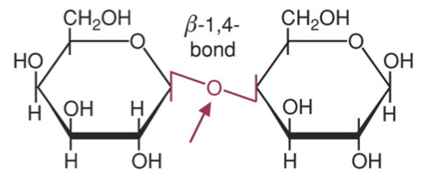
3. lactose (glucose + galactose, beta-1,4)
3 common polysaccharides:
1. glycogen (all glucose, animal storage, alpha-1,4 for linear, alpha-1,6 for branching)
2. starch (all glucose, plant storage, alpha-1,4)
3. cellulose (all glucose, plant structure, beta-1,4)
broken down to CO2 during oxidation, cellular metabolism
lipids
monomer is hydrocarbon
hydrophobic, since C-C and C-H are nonpolar
4 main types:
1. triacylglycerides
2. phospholipids
3. terpenes
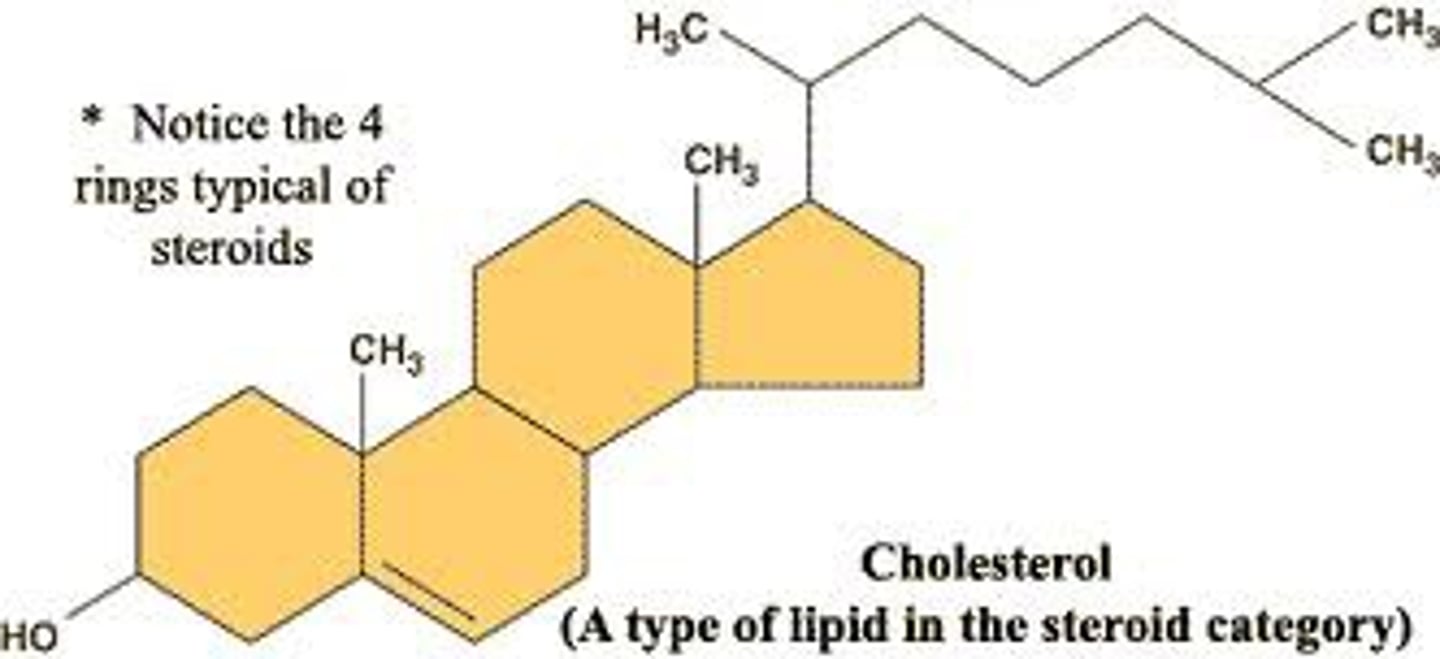
4. cholesterol
5. prostaglandins
fatty acids
alkyl chain that ends with a carboxylic acid
saturated is solid at RT, unsaturated is liquid at RT
all double bonds are cis (z)
only even numbered fatty acids
micelles are formed
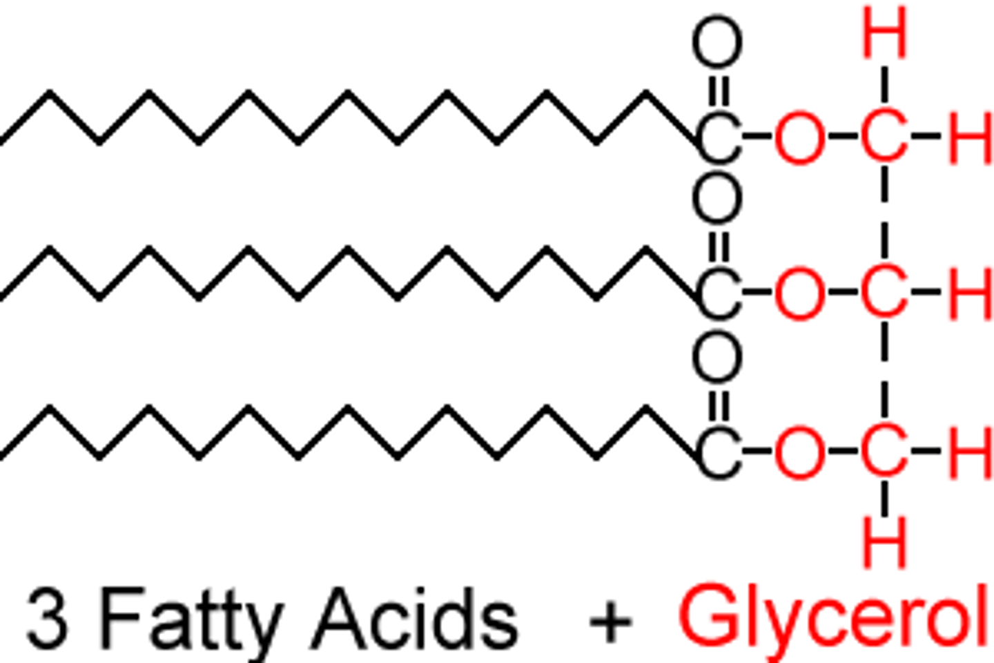
triacylglycerides
fat storage, glycerol with 3 fatty acid chains
lipases hydrolyze fats, hydrophobicity means they pack closer together
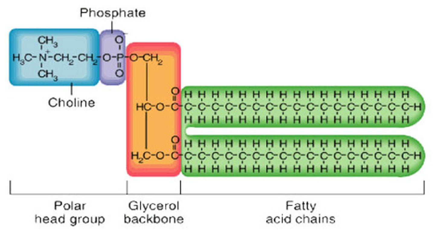
phospholipids
form lipid bilayer membranes, glycerol with 2 fatty acid chains and a phosphate
increase membrane fluidity:
1. unsaturated fatty acids
2. shorter fatty acid tails
3. cholesterol at low temperatures
terpenes
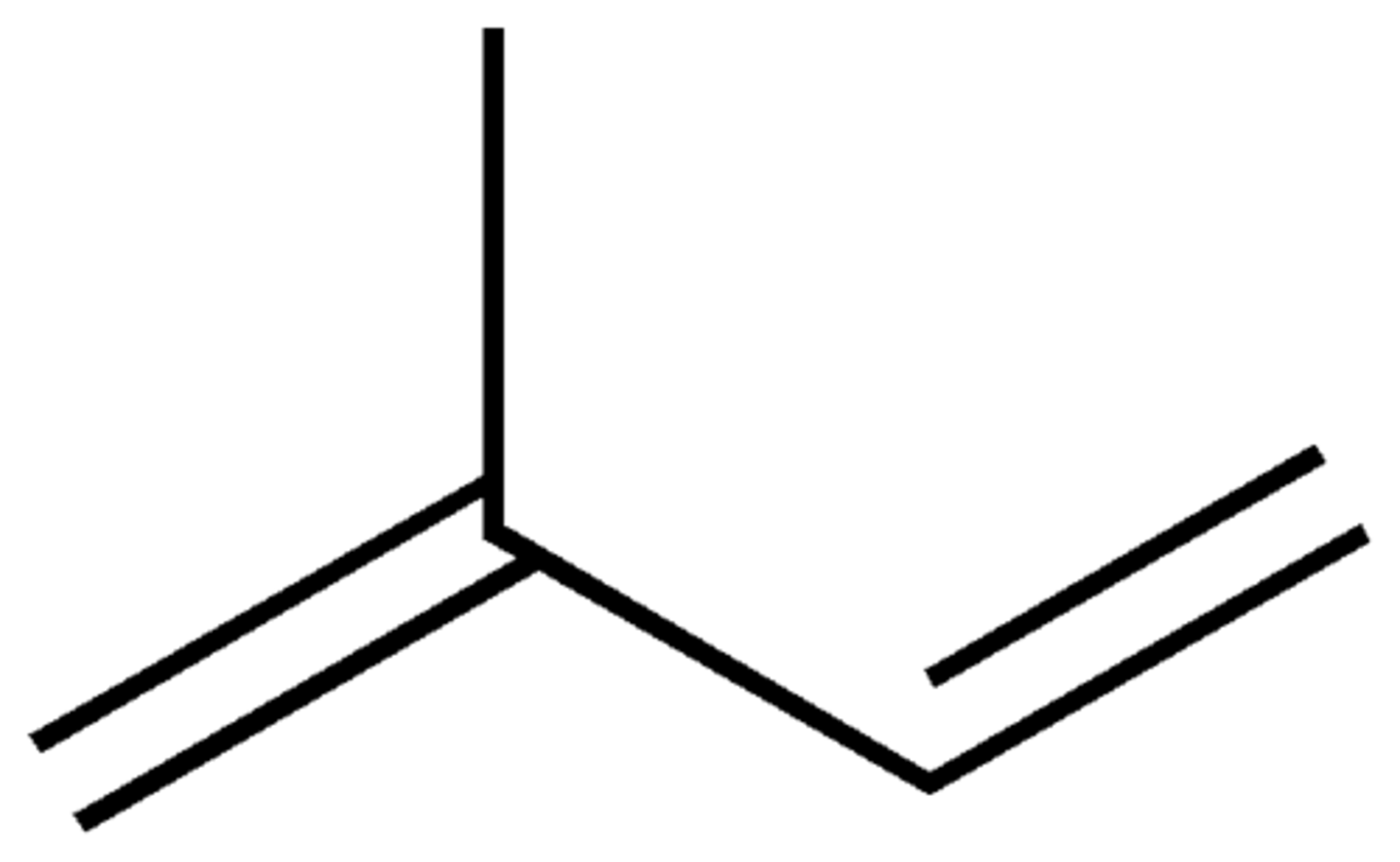
built from multiple isoprene units
monoterpene- 2 isos
squalene- 6 isos (tripene), component of ear wax
terpenoids- functionalized terpenes, cholesterol and steroid hormones, vitamin A
cholesterol
type of sterol, precursor to steroid hormones (testosterone, estradiol), component in all membranes
found concentrated in lipid rafts
only found in eukaryotes
increases fluidity at low temperatures
decreases fluidity at high temperatures
memorize all hormones on pg. 317
prostaglandins
derived from arachidonic acid, have one 5 carbon ring
act as paracrine hormones, signalling only nearby cells
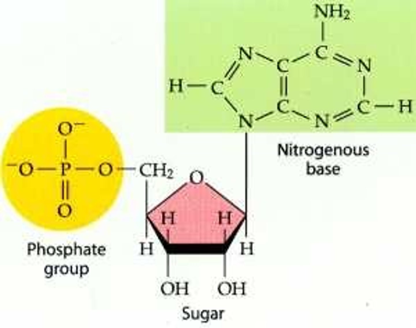
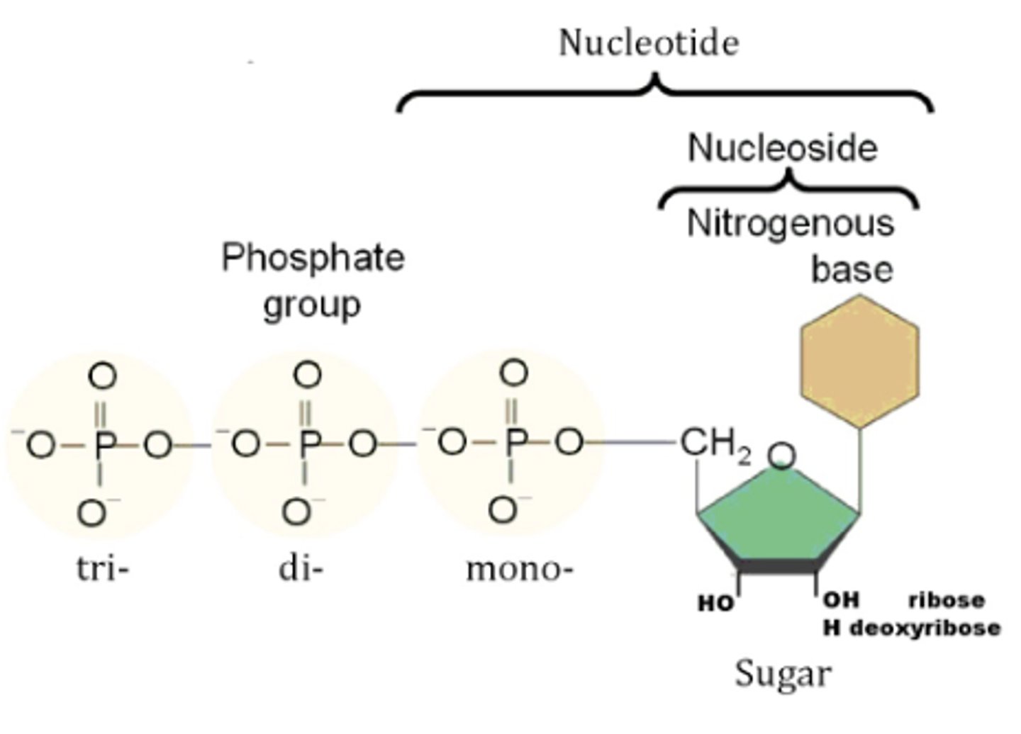
nucleic acids
monomer is nucleotide, includes a sugar, base, and phosphates
5-3 synthesis (refers to carbon number on which bonds form)
nucleotides connected by phosphodiester bonds- OH on bottom of sugar (5 end base) connects to phosphate (3 end base) forming an P-O bond
nucleoside- no phosphate
phosphate anhydride bonds- connect phosphates
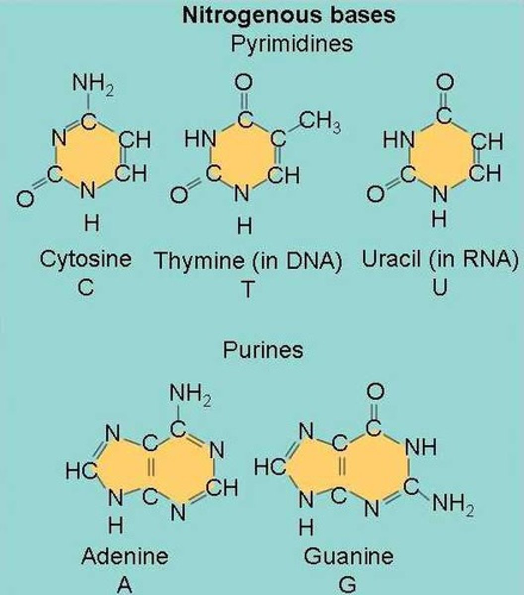
nucleic bases
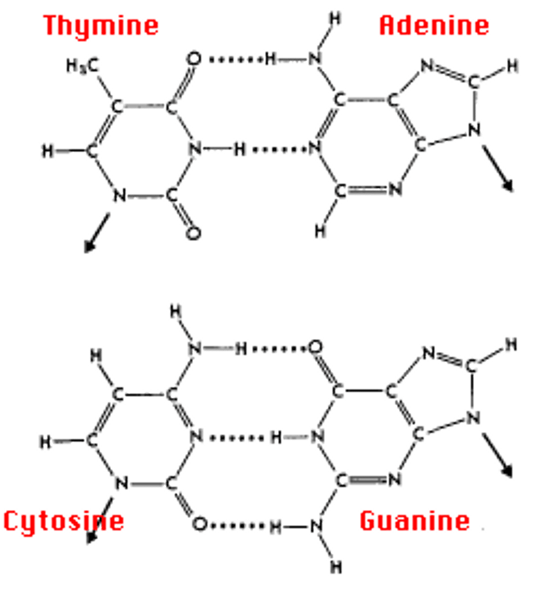
pyrimidine- cytosine, thymine, uracil (CUT, 1 ring)
purine- adenine, guanine (2 rings)
A and T have 2 H bonds
C and G have 3 H bonds
melting temperature- temp at which 1/2 of H bonds can break, eventually will denature
CG bonds are more stable than AT bonds
more bonds and longer strands means higher melting temperature
in dsDNA, A and T found in same quantity, C and G found in same quantity
nucleosides
base attached to ribose (RNA)
adenine- adenosine
guanine- guanosine
cytosine- cytidine
uracil- uridine
thymine- 5-methyluridine
nucleotide functions
genetic information- DNA
transcription/translation- mRNA, tRNA
energy carriers- ATP, cAMP, NADH, FADH2
nucleotide hydrogen bonding
cytosine has 2 H bond donors, 1 H bond acceptors
polymerization vs. hybridization
polymerization- DNA/RNA elongate with phosphodiester bonds to form single-strand
hybridization- DNA/RNA sequences anneal with hydrogen bonds to complementary strand
DNA packaging (eukaryotic/prokaryotic)
prokaryotes- single circular DNA genome
packaging:
1. methylation- protection from restriction enzymes (they chop up viral DNA which is not methylated)
2. supercoiling- in eukaryotes too, most DNA is negatively supercoiled, loops are looped/unlooped using gyrase and topoisomerases
eukaryotes- several linear chromosomes
packaging:
1. histones- proteins with net positive charge, R binds to phosphates on DNA which have negative charge
2. nucleosomes- 8 histones together
3. chromatin- general structure of DNA wrapped around nucleosomes, with linker DNA inbetween
gene silencing/activation
gene silenced by:
1. DNA methylation- nucleotides are methylated
2. histone deacetylation- histones are deacetylated, can be monitored with Western blot
euchromatin- open chromatin, increased expression, genes that are continuously expressed are found here
heterochromatin- closed chromatin, decreased expression, genes here are heavily regulated
epigenetics
study of influences on gene expression that occur without a DNA change
methylation is used as bookmarking during replication for transcriptionally active genes
chromosomes
humans have 46 chromosomes
centromere
center of chromosome connecting sister chromatids after replication
spindle fibers attach when sister chromatids separate
telomere
linear ends of chromosomes in eukaryotes, that loop around to base pair with itself
contain repetitive sequences
shorten after each division, puts a limit on cell divisions
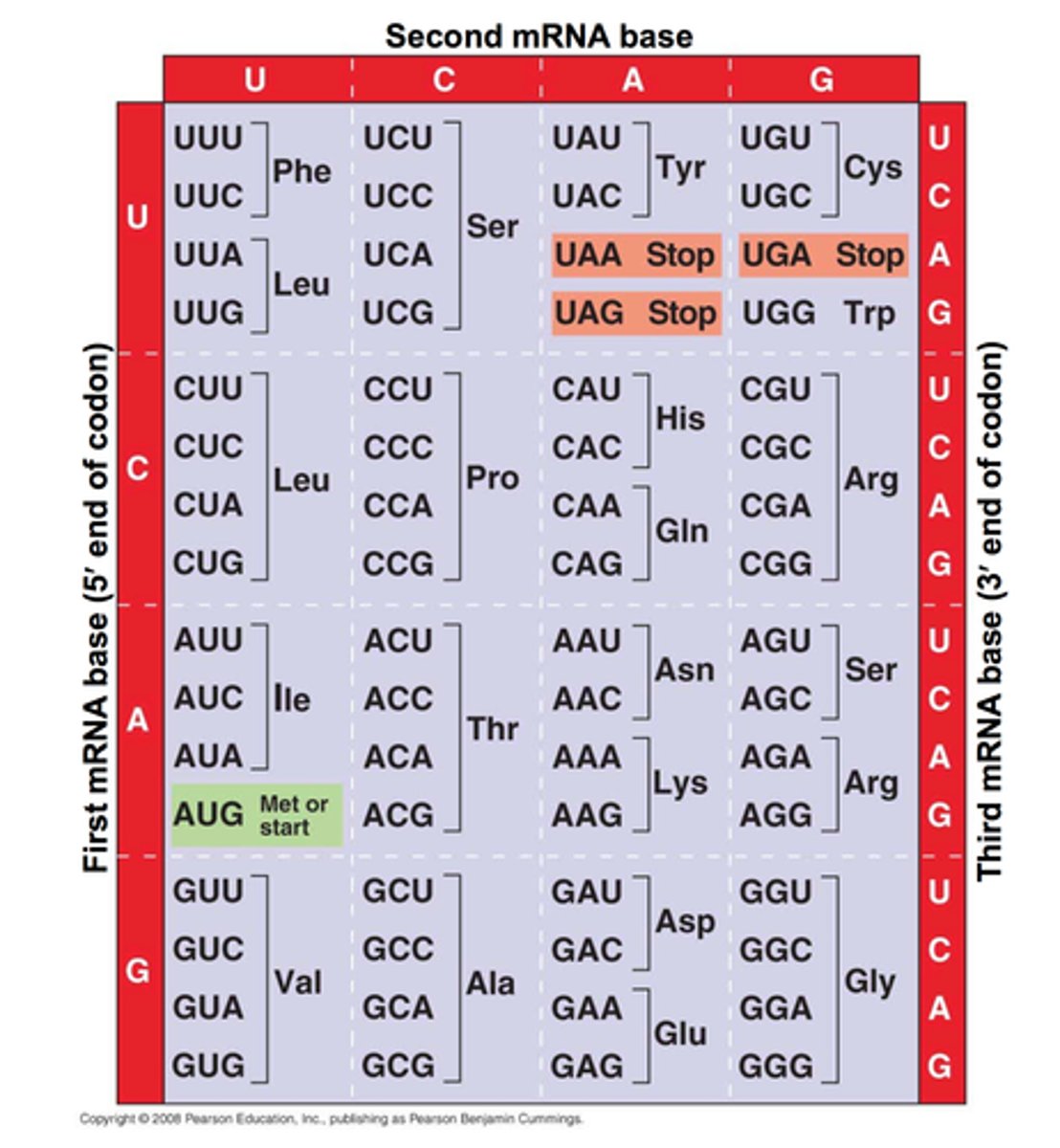
codons
3 nucleotides code for 1 amino acid during translation
other amino acids exist, but no codons for them
start codon- AUG
stop codon- UGA, UAA, UAG
"school starts in august"
"you go away! you are away... you are gone."
mutations (4)
polymerase errors:
1. point mutations- missense (becomes new amino acid), nonsense (becomes stop codon), silent (becomes new codon for same amino acid)
2. small repeats
3. frameshift- insertions and deletions that change reading frame, not a multiple of 3
transition mutation- A <-> G, C <-> T
transversion mutation switched purine/pyrimidine
endogenous damage:
1. reactive oxygen species- oxidize DNA
2. crosslinked bases
3. physical damage
exogenous damage:
1. UV radiation-pyrimidine dimers
2. X rays- double stranded breaks, translocations
3. chemicals- intercalation
transposons:
1. insertions/delections
2. inversions
3. duplications
transposons
transposase- enzyme that cuts and pastes transposon
inverted repeats- points of recognition for transposon
types of transposons:
1. IS element (codes for transposase)
2. complex transposon (carries additional genes)
3. composite transposon (flanked by 2 IS elements)
two transposons:
1. in same direction- DNA folds, deletes the middle gene
2. in opposite directions- DNA folds, inverts middle gene, shouldn't cause any problems if regulatory genes flipped
can cause amplifications, if copy number gets too high that lead to problems
DNA repair
proofreading- during DNA replication, DNA polymerase checks its own work
mismatch repair- after DNA replication, the side with more methyl groups must be the original sequence, so replace sequence on the other side
excision repair- before DNA replication, replace a single base
homologous end joining- after DNA replication, repair double-stranded breaks using sister chromatid as template, homologous crossover
non-homologous end joining- before replication, repair double-stranded breaks, loses some of the sequence
if non-homologous end joining messes up, translocations can occur causing gene fusion
direct reversal- reverse pyrimidine dimer, wants to restore original H bonds
directionality
DNA is read 3 -> 5
DNA is synthesized 5 -> 3
DNA is transcribed/read 3 -> 5
mRNA is synthesized 5 -> 3
mRNA is translated/read 5 -> 3
protein is synthesized N -> C
DNA replication
synthesized 5 to 3, read 3 to 5
semi-conservative, leading strand and lagging strand pass on to separate DNA strands
requires primers, template, and various enzymes:
1. topoisomerase/gyrase- uncoil DNA, creates negative supercoils behind replication
2. helicase- separates DNA strands
3. RNA polymerase- puts in primers, primase
4. DNA polymerase- replicates DNA, proofreads, removes primers
5. ligase- links Okazaki fragments
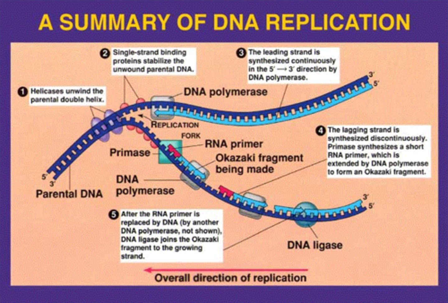
origin of replication
at replication fork where it begins, DNA is split into leading strand (synthesized continuously) lagging strand (Okazaki fragments are synthesized)
location on DNA where replication begins
prokaryotes typically have a single origin of replication
eukaryotes have several on a chromosome
prokaryotic DNA replication
THETA replication- 1 origin, 5 DNA polymerases
DNA polymerase III- fast, main replicating enzyme, can proofread
DNA polymerase I- slow, adds nucleotides at RNA primer until poly III starts, does DNA excision repair
DNA polymerase II- back up for poly III, does DNA repair
DNA polymerase IV, V- error prone, does DNA repair
telomerase
RNA primers add at lagging strand, so when they are removed we get shorter telomeres
prevent damage to ends of linear chromosomes
include their own RNA primer, has reverse transcriptase activity to synthesize DNA for telomere extension
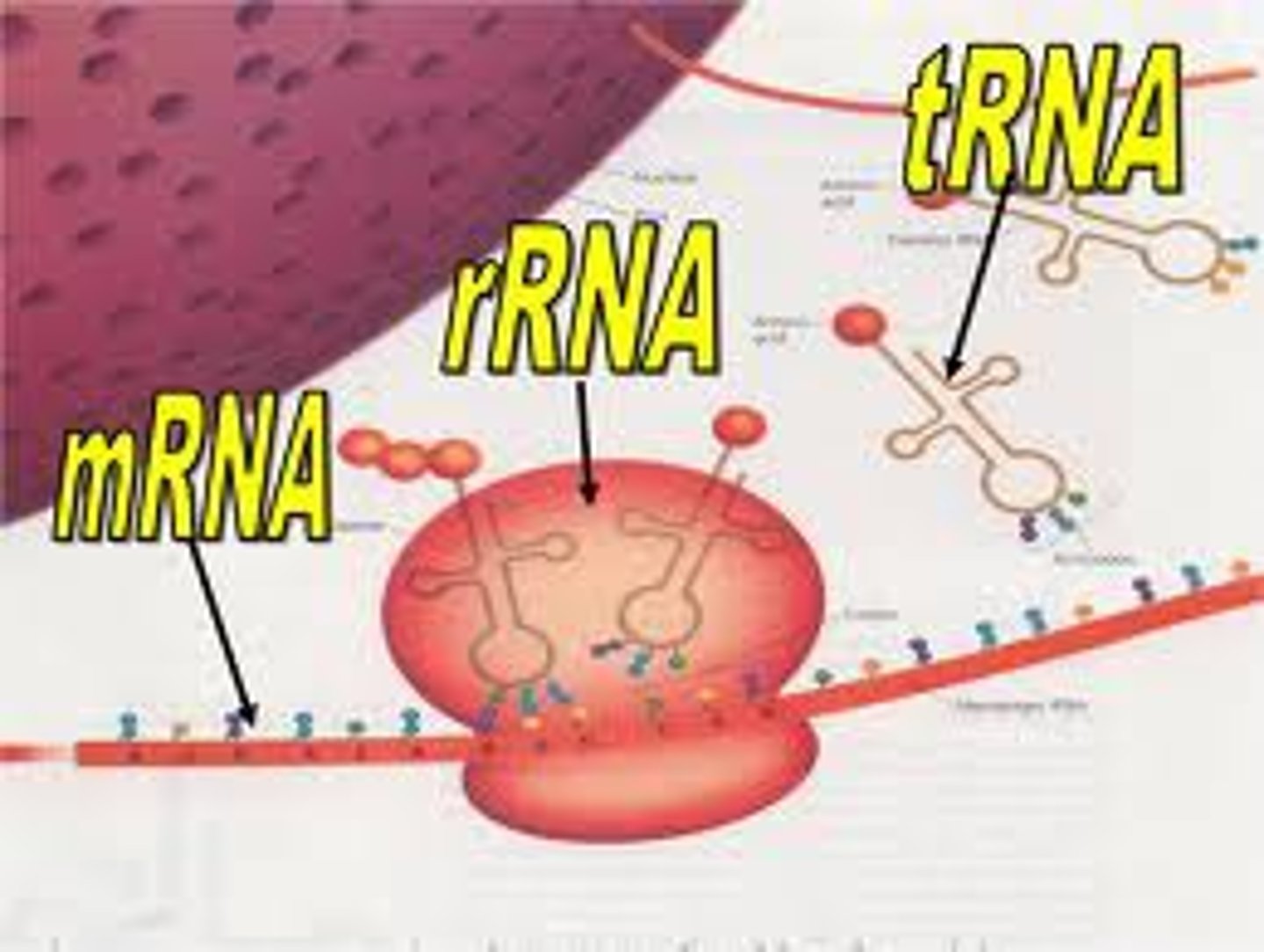
RNA
single stranded, uracil instead of thymine, ribose instead of deoxyribose, has many different shapes
3 types of RNA:
1. rRNA- ribosomal, skeleton of ribosomes
2. mRNA- messenger, feeds into translation
3. tRNA- transfer, carries AA to ribosomes
3 more types of RNA:
1. hnRNA- heterogeneous nuclear, in nucleus, mix of introns and exons
2. miRNA- micro, used for RNA silencing and post transcriptional regulation
3. siRNA- small interfering, synthetic RNA designed to degrade mRNA
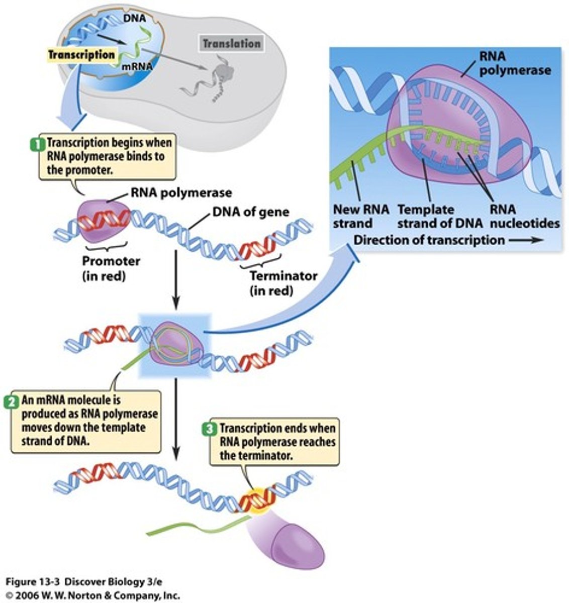
transcription
similarities to replication: has START site, 5 to 3 synthesis, needs DNA template
differences to replication: has STOP site, no primers required, no editing
template strand- anti-sense strand, RNA polymerase attaches
coding strand- sense strand, same code as the resulting mRNA, not transcribed
RNA polymerase attaches to promoter on template strand, which is next to operator and START site
transcription is the primary regulation for translation:
1. promoter- DNA region with affinity for RNA polymerase regulates transcription
2. enhancer- DNA region with affinity for activator transcription factors
3. operator- DNA region with affinity for repressor transcription factors
3. transcription factors- activators and repressors regulate transcription
3. promoter, operator, and enhancer regions are not transcribed to the mRNA
transcription (eukaryotic/prokaryotic)
prokaryotic transcription:
1. transcription and translation happen at same time in the cytoplasm
2. no mRNA processing thus producing human genes in bacteria can be difficult because introns need to be removed
3. polycistronic- 1 mRNA can have many messages
4. 1 RNA polymerase
eukaryotic transcription:
1. transcription and translation happen in different places, nucleus and cytoplasm
2. mRNA processing- 5 G-cap (guanine), 3 poly-A tail (adenine), splicing (remove introns)
3. monocistronic- 1 mRNA has 1 protein for tight regulation
4. 3 RNA polymerases- I (rRNA), II (mRNA), III (tRNA)
gene expression regulation
peptide/steroid hormones, transcription factors, nuclear receptors, other parts of signalling cascades help regulate transcription of genes
different genes do not have to be close to each other or on the same chromosome to be regulated together
environment determines gene expression, ex: addition of new sugar will induce expression of new enzymes to digest that sugar
gene expression regulates all the important processes, like cell differentiation
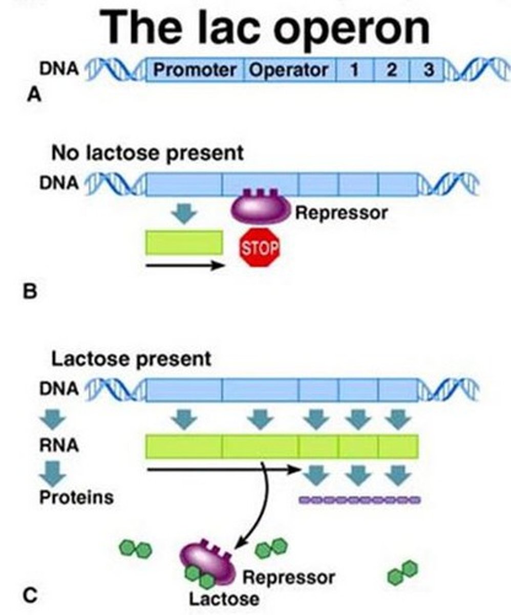
lac operon
only in prokaryotes like E. coli
Jacob-Monod model- describes lac operon
promoter (binds activator) -> operator (binds repressor) -> lac genes, trigger metabolism of lactose
lactose absent- repressor binds
lactose present- repressor leaves
glucose absent- activator binds
glucose present- activator leaves
lac operon only runs when lactose present and glucose absent
splicing
form of post-transcriptional mRNA processing to remove introns and put together exons
spliceosome- contains snRNPs, which contain snRNA, machinery that splices
isoforms- different forms of proteins created through alternative splicing
ribozyme
RNA can perform catalytic functions like enzymes
1. function unit of ribosome is ribozyme
2. splicing can be done with ribozymes
3. proposed that RNA can essentially self-replicate
tRNA
anticodon loop- complementary to codon
acceptor stem- AA binding site
2 ATP needed to load an amino acid
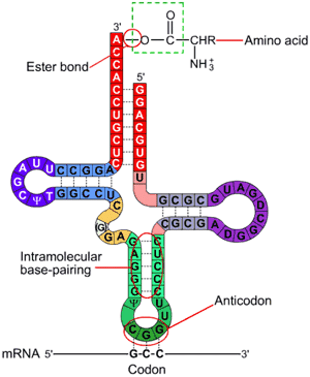
degeneracy
different codons can code for same amino acid
wobble hypothesis
first two codon-anticodon pairs bind, third anticodon is flexible when it is a G/U/I
adenine on tRNA can get converted to inosine
ribosomes
ribosomes are the only organelles that prokaryotes have
eukaryotes and prokaryotes have different ribosomes
ribosomes are synthesized in the nucleolus in eukaryotes
prokaryotes- large subunit (505), small subunit (305)
eukaryotes- large subunit (605), small subunit (405)
Svedberg units measure density, not weight
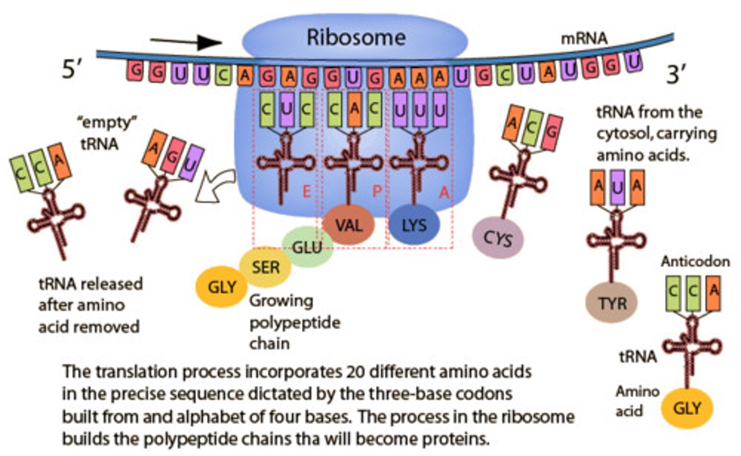
translation
EPA sites:
E site= tRNA exits
P site- growing protein held here
A site- new amino acid added here
1. peptide bond formed between A and P amino acids
2. P amino acid then leaves P tRNA
3. mRNA translocates to move A amino acid to P site
STOP codon- no tRNA, instead binds release factor so final amino acid leaves its tRNA
5/3'-UTR- untranslated regions at each end of mRNA, assist regulation of translation
mRNA is translated 5 -> 3
protein is synthesized N -> C
energy for translation
1. tRNA loading- 2 ATP per AA
2. initiation- 1 ATP
3. A site binding- 1 ATP per AA
4. translocation- 1 ATP per AA
5. termination- 1 ATP
for 50 AA peptide, requires 50 x 4 ATP = 200 ATP
post-translational modifications
protein folding- chaperones assist in finding proper shape
covalent modification- phosphoylation, disulfide bridges
processing- zymogens are inactive enzymes that are activated via cleavage
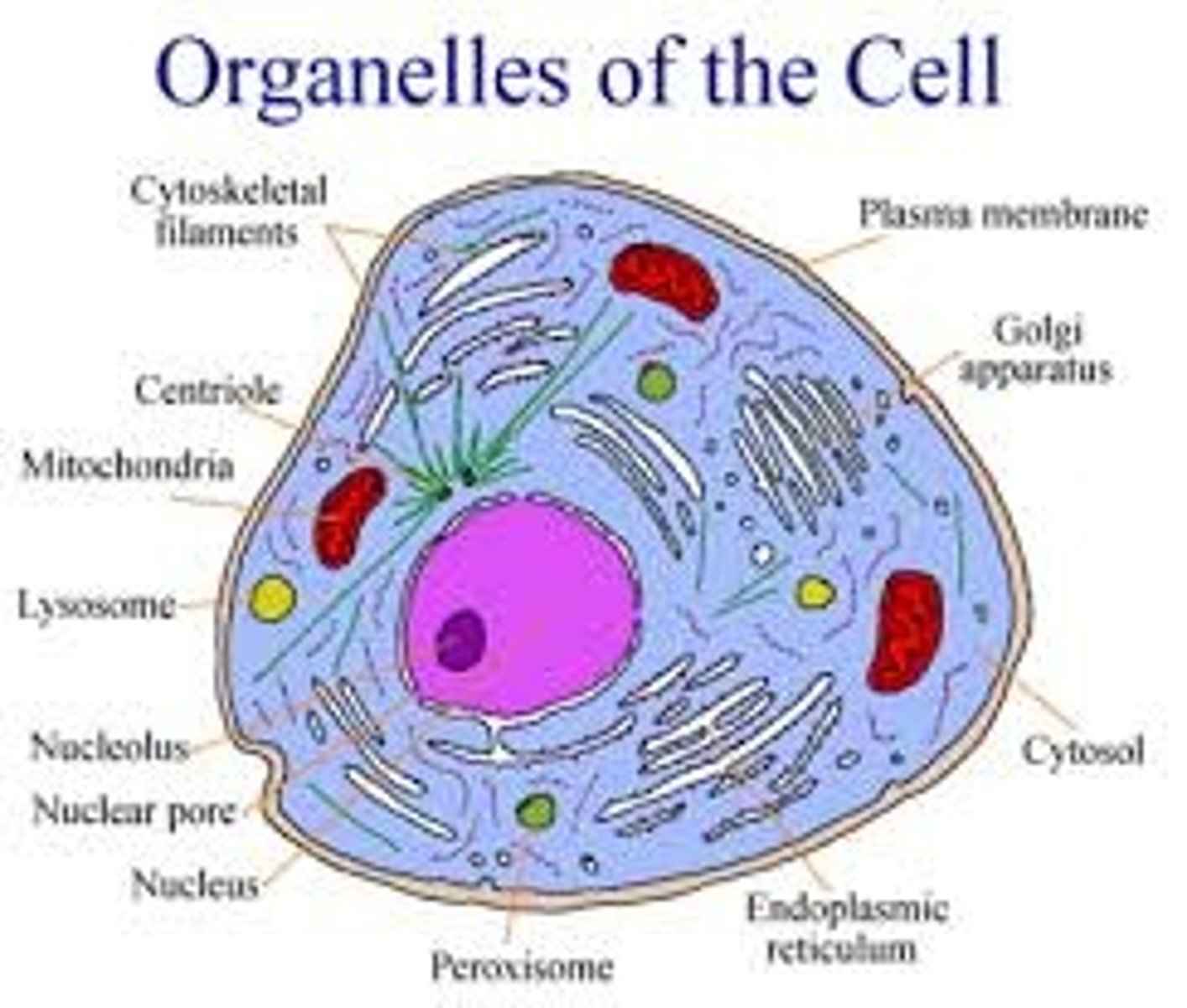
ER
rough ER- proteins (peptide hormones) synthesized by ribosomes here
smooth ER- carbohydrates and lipids (steroid hormones) synthesized here
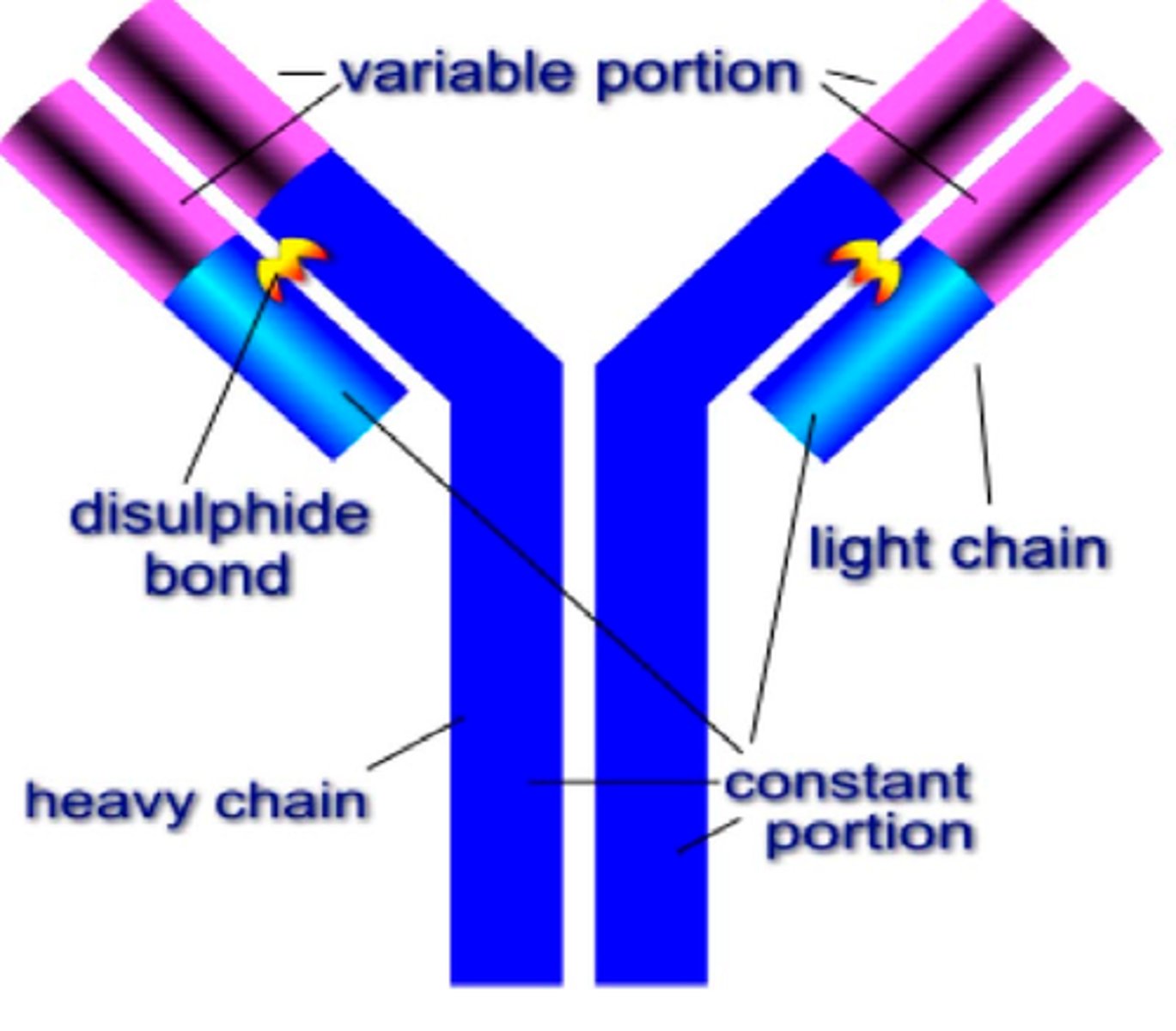
antibodies
immunoglobulin produced by B cells, binds to antigens
variable portion- both tips on top of Y shape, recognizes antigen
constant portion- bottom of Y shape, differs between classes/isotypes
light chain (small piece)
heavy chain (large piece)
viruses
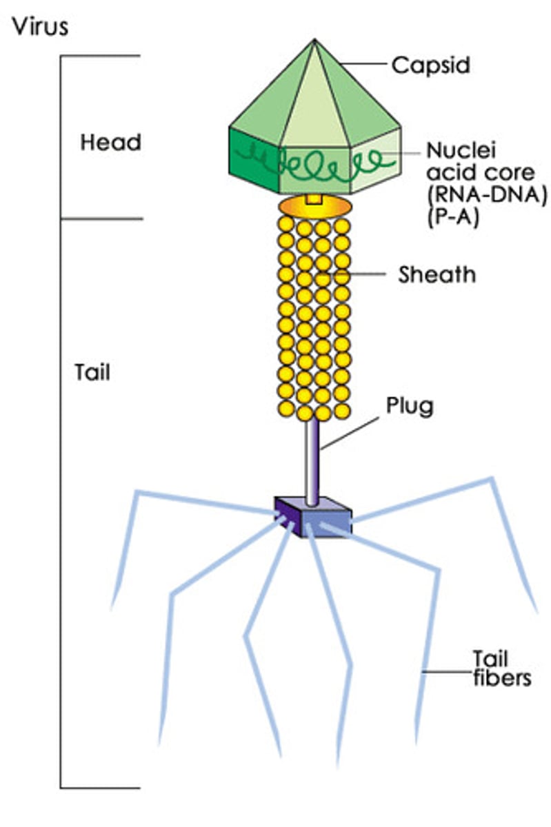
bacteriophage- virus that infects bacteria
made of protein (capsid is usually icosahedral or helical and tail fibers) and nucleic acids (DNA or RNA, ss or ds)
general life cycle:
1. attachment- receptor specificity
2. injection- deliver genome to host cell
some bacteria can enter host cells and lyse them as well
lytic viral life cycle
1. transcribe and translate viral genome- capsid proteins, hydrolase to destroy host cell genome
2. replicate viral genome- automatic assembly into capsid
3. lysis -lysozyme to create holes in cell membrane to release viral particles
virus is actively killing
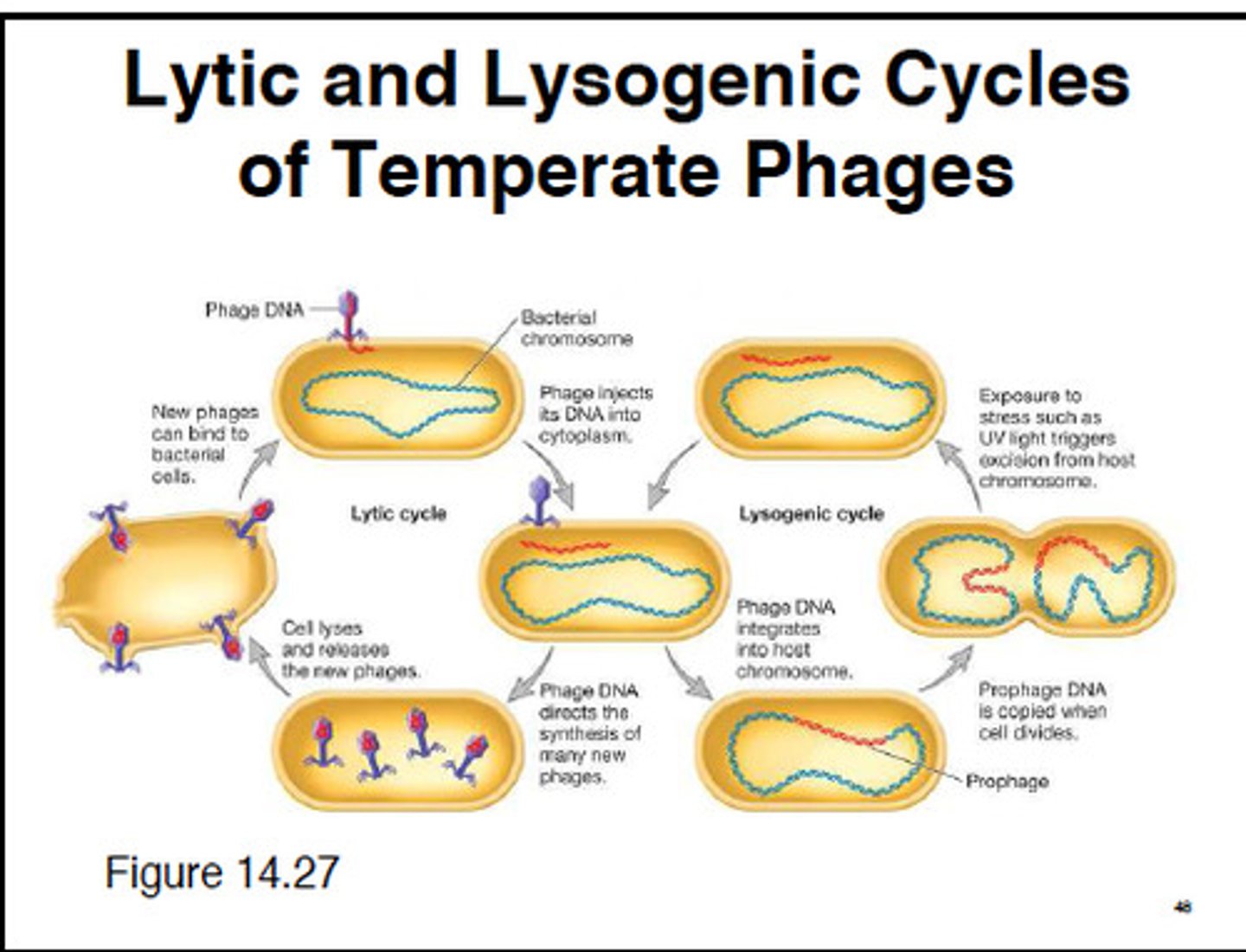
lysogenic viral life cycle
1. provirus- integrate viral genome into host genome, repressor proteins silence host genome
2. reproduction- normal host activity
3. excision and lytic cycle- repressors removed, but some host genome can be removed as well, this diversifies bacterial genome via viral vector
virus is dormant

productive viral life cycle
1. virus released via budding from cell membrane
2. animal cells only
3. produces more virus
4. gets an envelope to protect against immune system and interact with new host cells
virus is smart
viral RNA genomes
(+)RNA:
1. ssRNA is the same as the mRNA
2. directly translated by host cell ribosomes to produce capsids and viral enzymes
3. RNA dependent RNA polymerase creates complementary template to then replicate more of the original ssRNA
(-)RNA:
1. ssRNA is template for mRNA
2. RNA dependent RNA polymerase creates complementary template to replicate more of original ssRNA
3. template is translated by host cell ribosomes to produce capsids and viral enzymes
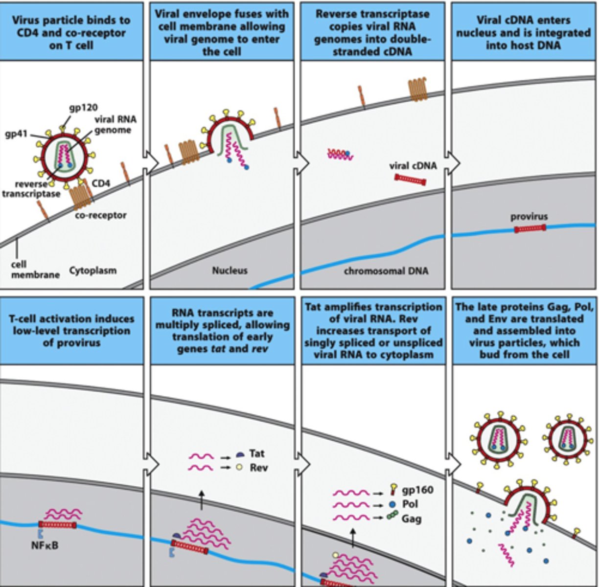
(+)RNA lysogenic (retrovirus like HIV):
1. ssRNA is the same as the mRNA
2. RNA dependent DNA polymerase (reverse transcriptase) converts ssRNA to ssDNA
3. host DNA polymerase converts ssDNA to dsDNA
4. dsDNA inserts into host cell genome, provirus
5. transcribed to replicate original ssRNA and viral enzymes
problems: viral genome is permanently in host genome, rapid mutation due to reverse transcriptase
prions
self-replicating proteins that are very stable
bad prions are produced:
1. spontaneous mutation (mad cow disease)
2. gene can be inherited (fatal familial insomnia)
3. ingestion of diseased tissue (kuru)
bad prions cause cell death:
1. bad prions are produced, accumulating in cell
2. bad prions are ingested, change good prions to bad
viroids
circular RNA with no capsid and can replicate independently
Hepatitis D is a viroid that must coinfect with Hep B, which is caused by a regular virus
one half of circle, (-)RNA, unravels:
1. can reform circle and transcribe other half, (+)RNA
2. can remain unraveled and transcribe other half, (+) RNA
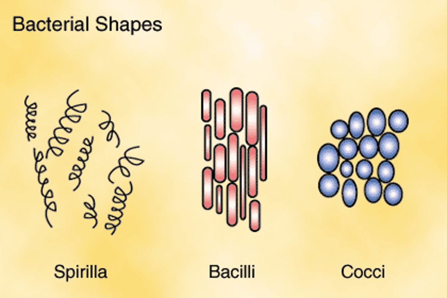
bacteria structure
shapes:
1. round- coccus
2. rod- baccillus
3. spiral- spirillum
cell membranes:
1. gram-positive bacteria- cell wall made of peptidoglycan surrounds cell membrane, stains purple
2. gram-negative bacteria- peptidoglycan cell wall surrounded by inner and outer cell membranes, stains pink, does conjugation
3. flagella- basal structure attached to cell wall/membrane, with a hook that whips around filament
4. no cholesterol
outmost surface has specific proteins that trigger a unique immune response, some foreign cells can vary the proteins presented on the surface to bypass the immune system
bacteria are prokaryotes, lack organelles except ribosomes
bacteria living conditions
temperature:
mesophile- medium temp (0-100C)
termophile- hot temp
psychrophile- cold temp
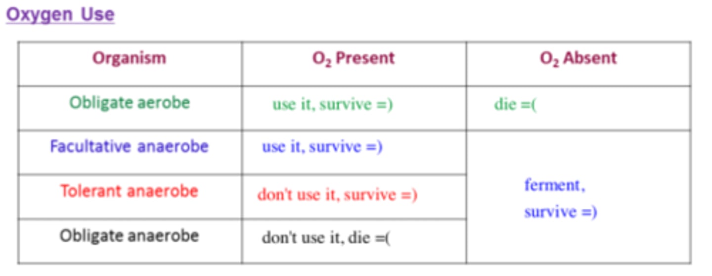
oxygen:
obligate aerobe- needs O2 for cellular respiration
facultative anaerobe- uses O2, can ferment without O2
tolerant anaerobe- can survive with O2/ROS, but ferments without O2
obligate anaerobe- dies with O2/ROS, ferments without O2
bacteria energy sources
ETC happens on membrane
energy source: photo is light, chemo is ATP
carbon source: auto is CO2, hetero is other organisms
1. photoautotrophs- plants
2. chemoheterotrophs- animals
3. photoheterotrophs- carnivorous plants
4. chemoautotrophs- archaebacteria
auxotrophs- cannot synthesize a key molecule to live:
1. arg(-)- can't make arginine
2. leu(-)- can't make leucine
3. lac(-)- can't use lactose
lawn- bacteria growth
plaque- no bacteria growth
chemotaxis
cell movement that occurs in response to chemical stimulus
bacteria reproduction
binary fission- increase cell numbers, but doesn't increase genetic diversity (needs conjugation)
four phases:
1. lag- slow growth
2. log- rapid growth
3. stationary- reaches carrying capacity
4. death- finds new stationary population size
bacteria genetic diversity
transformation- lysogenic phage transfers DNA from environment into bacteria
conjugation- increase genetic diversity, but doesn't increase cell numbers
transduction- viral DNA inserts into bacteria
transfection- transformation into non-bacterial cell
conjugation is a feature of gram-negative bacteria
F factor- circular DNA element that encodes fertility gene, F+ is male, F- is female
conjugation bridge- after male cell produces sex pili and contacts female cell, bridge forms
F factor is replicated in F+ cell and transferred to F- cell
high frequency of recombination (Hfr cell)- F factor integrated into genome, so conjugation can transfer other others of bacterial genome
conjugation mapping- analyze transfer of genes over time into auxotrophic bacteria to map genome
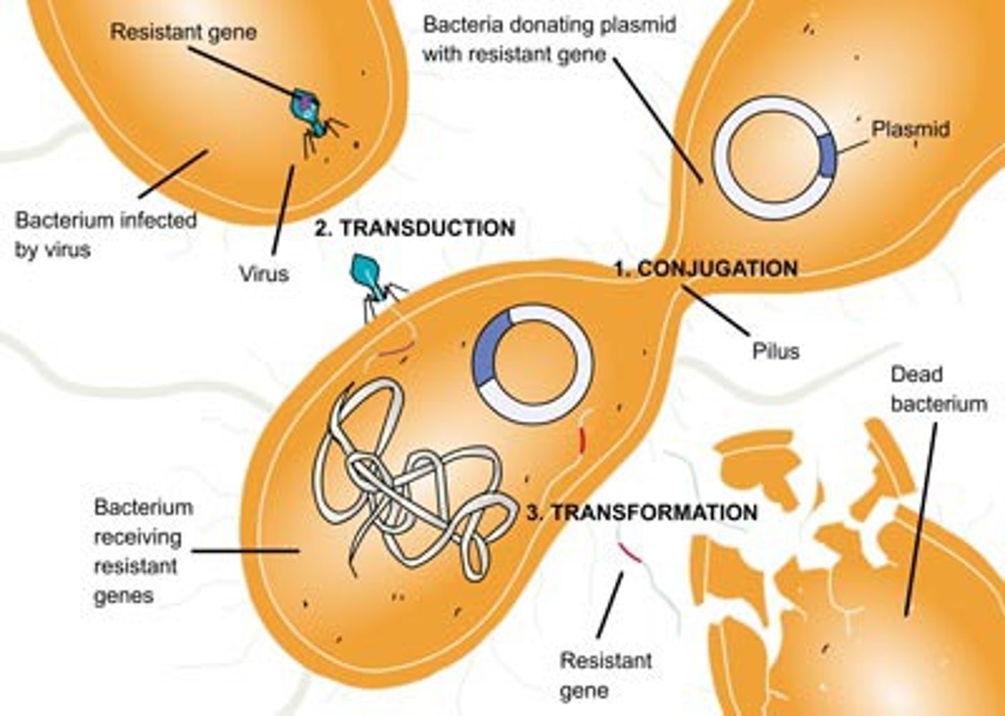
plasmid
a small, circular section of extra DNA that confers one or more traits to a bacterium and can be reproduced separately from the main bacterial genetic code
can be used to mass produce a protein
general types of bacteria
1. eubacteria- typical bacteria
2. archaebacteria- survive in extreme environments
3. parasitic bacteria- can be obligate (replicate inside host cell) or facultative (replicate inside or outside host cell), similar to viruses in that T cells help fight them off
4. symbiotic bacteria- coexist with host like nitrogen-fixing bacteria
organelles
lysosome- hydrolysis/degradation of waste, acidic environment, endocytosis ends here
peroxisome- beta oxidation of fatty acids, produces H2O2 which is broken down to water and CO2
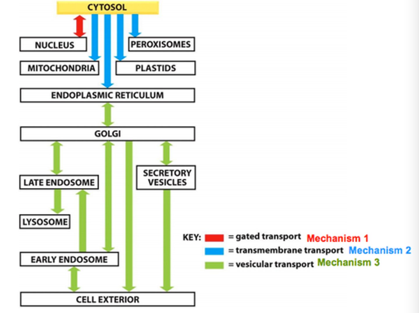
protein transport
transcription occurs in nucleus, translation begins in cytosol
if nuclear/mitochondrial/peroxisomal protein, finish translation in cytosol
if secreted/transmembrane/lysosomal protein (like an antibody), finish translation in rough ER
if transmembrane protein, signal sequence will remain as transmembrane region, otherwise it acts as anchor that is removed later
if nuclear protein, nuclear localization signal
if ER protein, retrograde transport brings it back to ER from Golgi
vesicle buds off ER lumen, bringing the protein to its target
proteasomes
ubiquitin signalling marks proteins for degradation by proteasomes
proteases cut up proteins to smaller peptide pieces
endosomes
created from endocytosis, some viruses enter through endocytosis
cell membrane
4 components:
1. phospholipids- polar head, nonpolar tail, lipid bilayer
2. cholesterol (sterols)- in high temp it stabilizes membrane, in cold temp it keeps membrane fluid (not found in prokaryotic cells)
3. protiens- channels, receptors
4. carbohydrates- attach to proteins, phospholipids in ECM
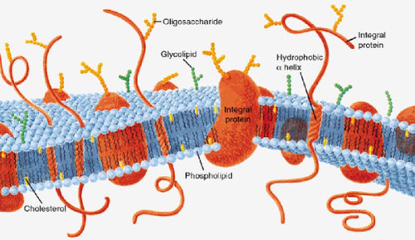
extracellular matrix
collagen, elastin, fibronectin, a lot of water, proteoglycans (ground substance, just carbohydrates attached to proteins)
integrins embedded in membrane connect to fibronectin
electrolytes
Van't Hoff factor (i)- number of ions when one unit dissolves
colligative properties
properties that depend on amount of solute particles but not their identity
four colligative properties:
1. freezing point depression occurs with more ions and higher molality
2. boiling point elevation occurs with more ions and higher molality
3. vapor pressure depression occurs with increased solute
4. osmotic pressure elevation occurs with increased solute, temperature, molarity
bp elevation and fp depression are analogous to cholesterol's temperature dependent role in the plasma membrane
they are also analogous to the role of impurities in a substance (they also increase the ranges of the bp and fp)
osmosis
diffusion- movement of particles down its concentration gradient
osmosis- movement of water down its concentration gradient
osmotic pressure- water moves to high concentration of solute particles, water would move to 1 M NaCl over 1M sucrose
count the number of ions, don't just look at the concentration!
tonicity:
1. hypertonic- more particles than other side of membrane
2. hypotonic- less particles
3. isotonic- equal particles
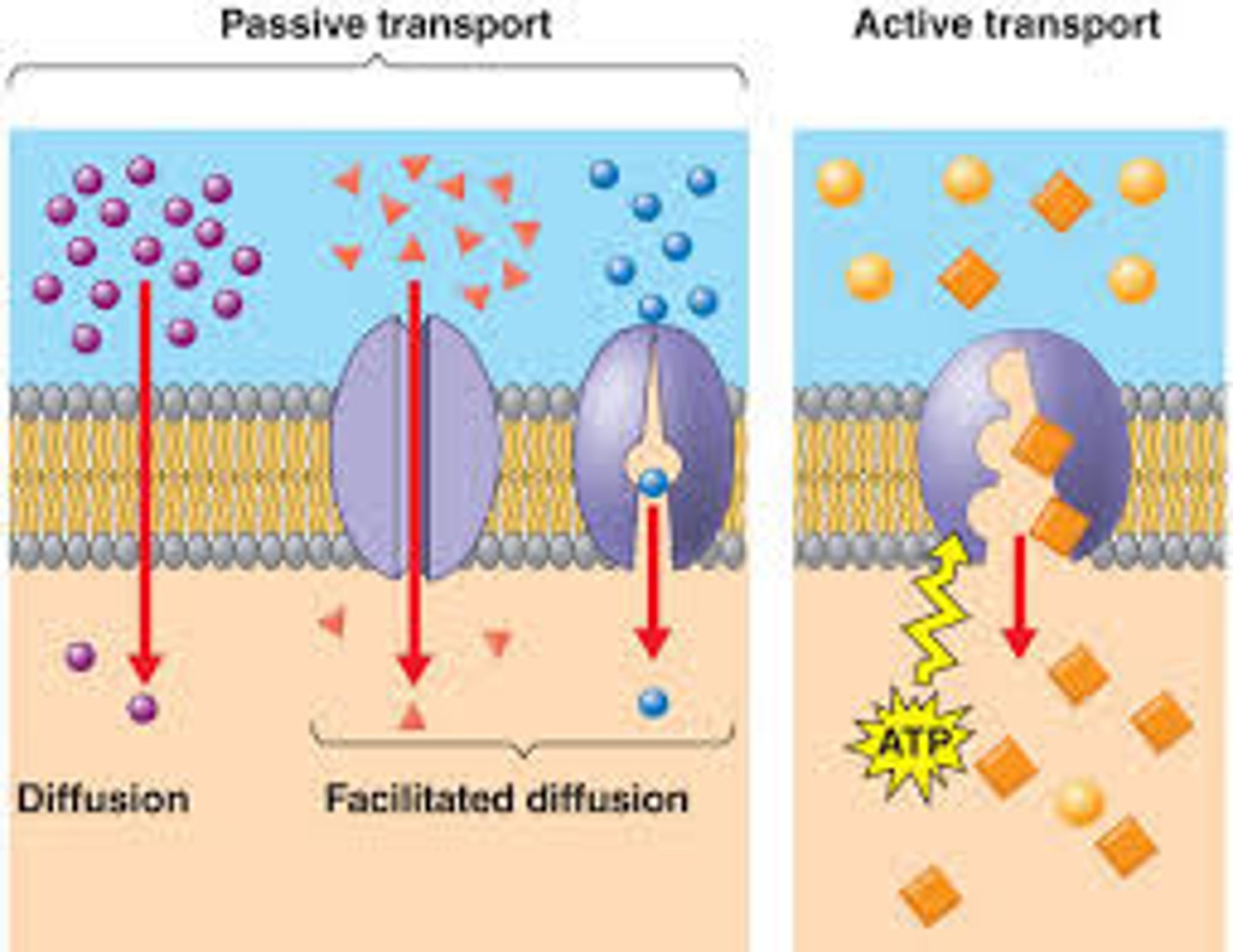
passive transport
no energy required, moving with concentration gradient
simple diffusion:
1. directly cross membrane
2. works well for small hydrophobic molecules like CO2, O2
3. also works for larger planar and hydrophobic molecules
facilitated diffusion- uses helping protein, works well for small hydrophilic molecules like glucose
helper proteins:
1. pores- non-specific, wide opening
2. channels- highly selective, narrow opening
3. cotransport- conformational change to transport, uniports (1 molecule, 1 direction), symports (2 molecules, same direction), antiports (2 molecules, opposing direction)
channel selectivity
aquaporins- select water with hydrophilic amino acids on interior of channel
Na+ is smaller than K+, so a smaller diameter channel will select for Na+ over K+
active transport
energy required, moving against concentration gradient
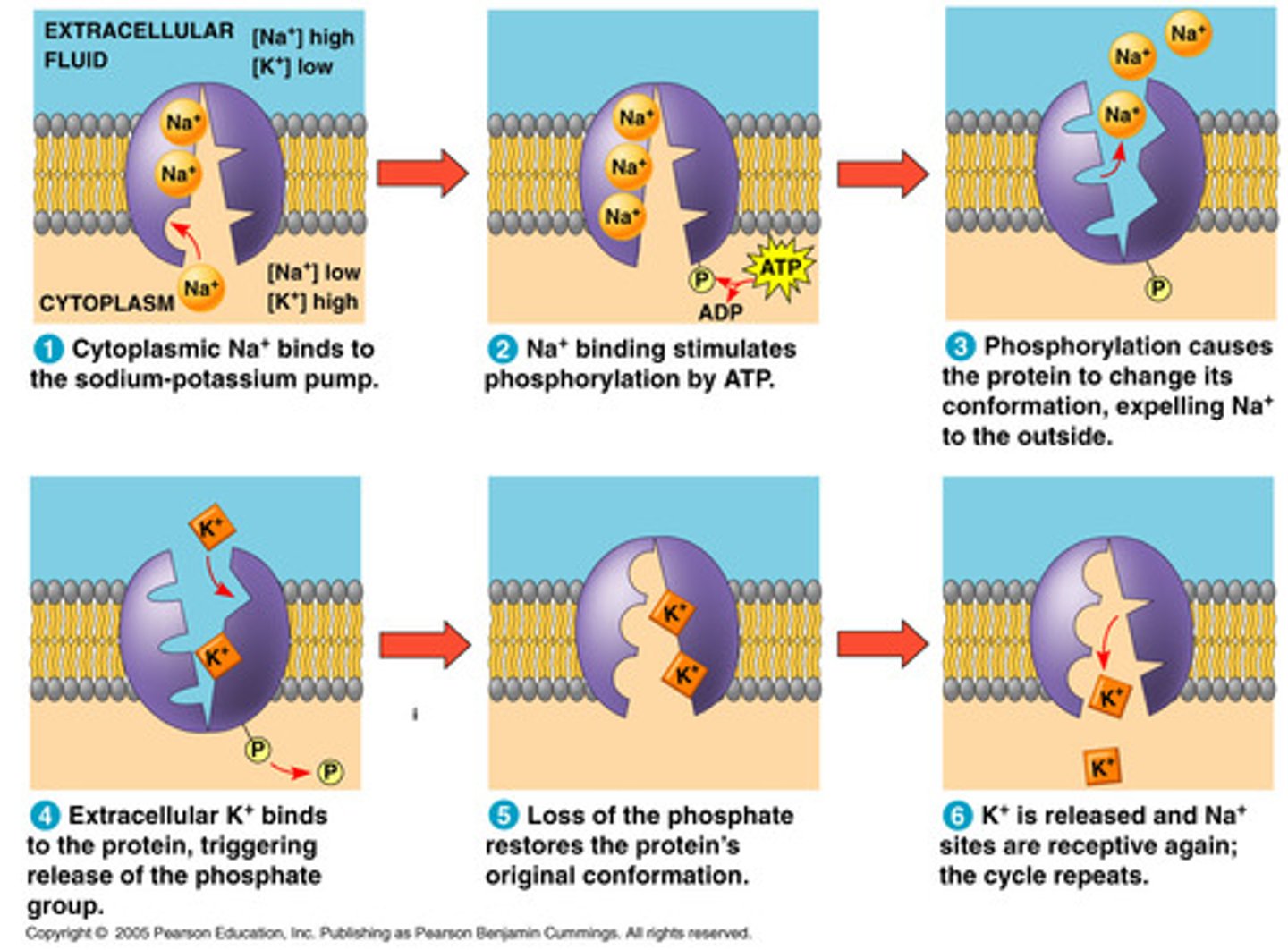
primary- directly use ATP
Na+/K+ ATPase:
1. pumps 3 Na+ out and 2 K+ in, uses 1 ATP
2. maintain osmotic balance
3. establish electrical gradient
4. set up sodium gradient for secondary active transport
secondary- use ATP to establish electrochemical gradient, use gradient to drive transport
Na+/glucose symporter:
1. glucose and Na transported in from lumen
2. powered by Na gradient, created by Na/K ATPase
ABC transporter:
1. uses ATP
2. transports big things
3. can transport drugs out of cancer cells
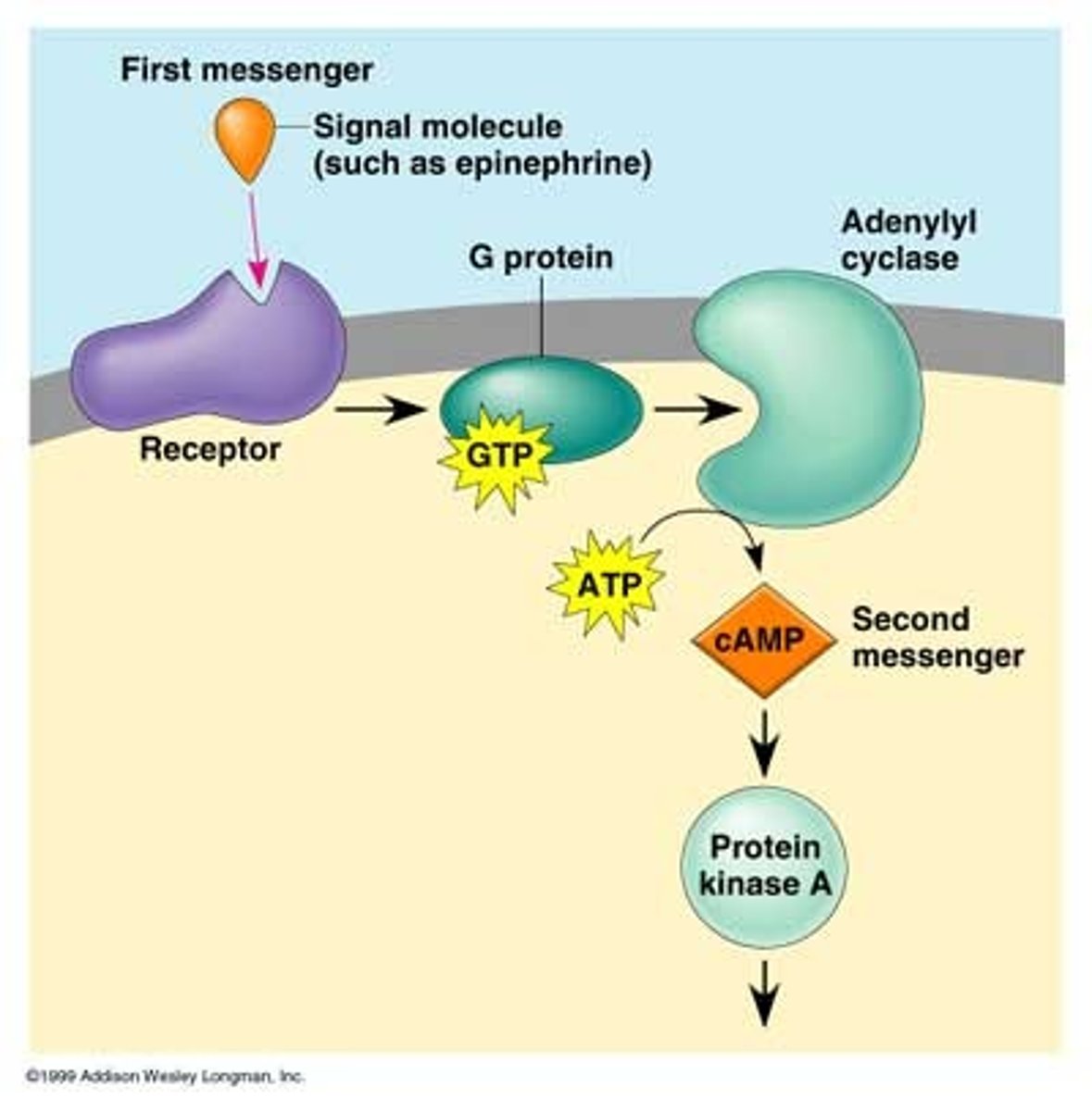
secondary messenger system
G-protein linked receptor activated by ligand, allows GTP to bind to alpha subunit
activates adenylyl cyclase
process ATP into cAMP to amplify effect
cAMP is a secondary messenger, activates cAMP-dependent protein kinases like protein kinase A, which phosphorylate enzymes to activate them
fast but temporary effect
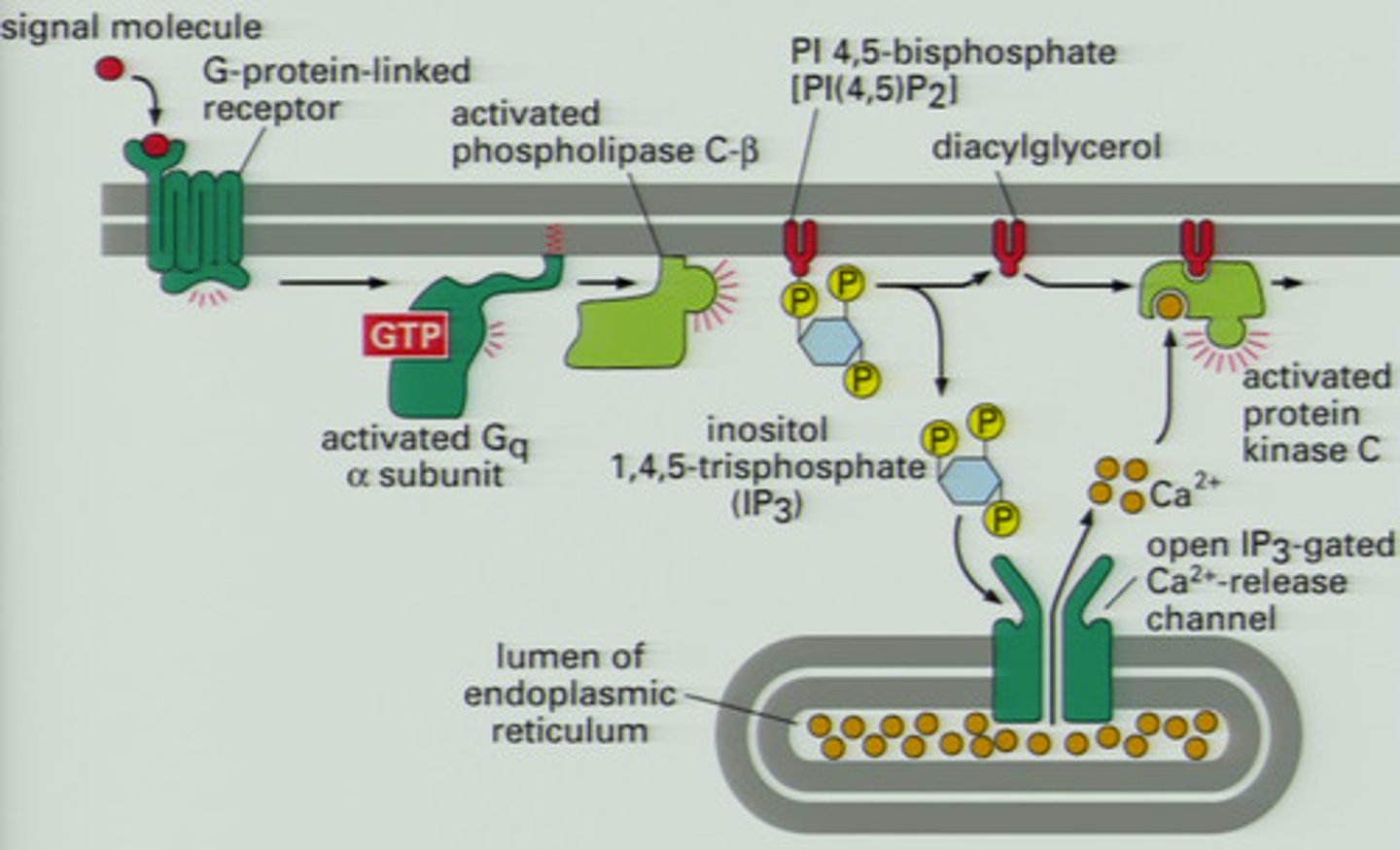
phospholipase C
G-protein linked receptor signals phospholipase C
splits PI into IP3 and DAG:
1. IP3 increases intracellular Ca2+
2. DAG activates kinases (PKC) to activate enzymes
neurotransmitters
GABA, glutamate, dopamine, acetylcholine, serotonin are all neurotransmitters that active receptors on cell membranes
contrast that with peptide and steroid hormones
cytoskeleton
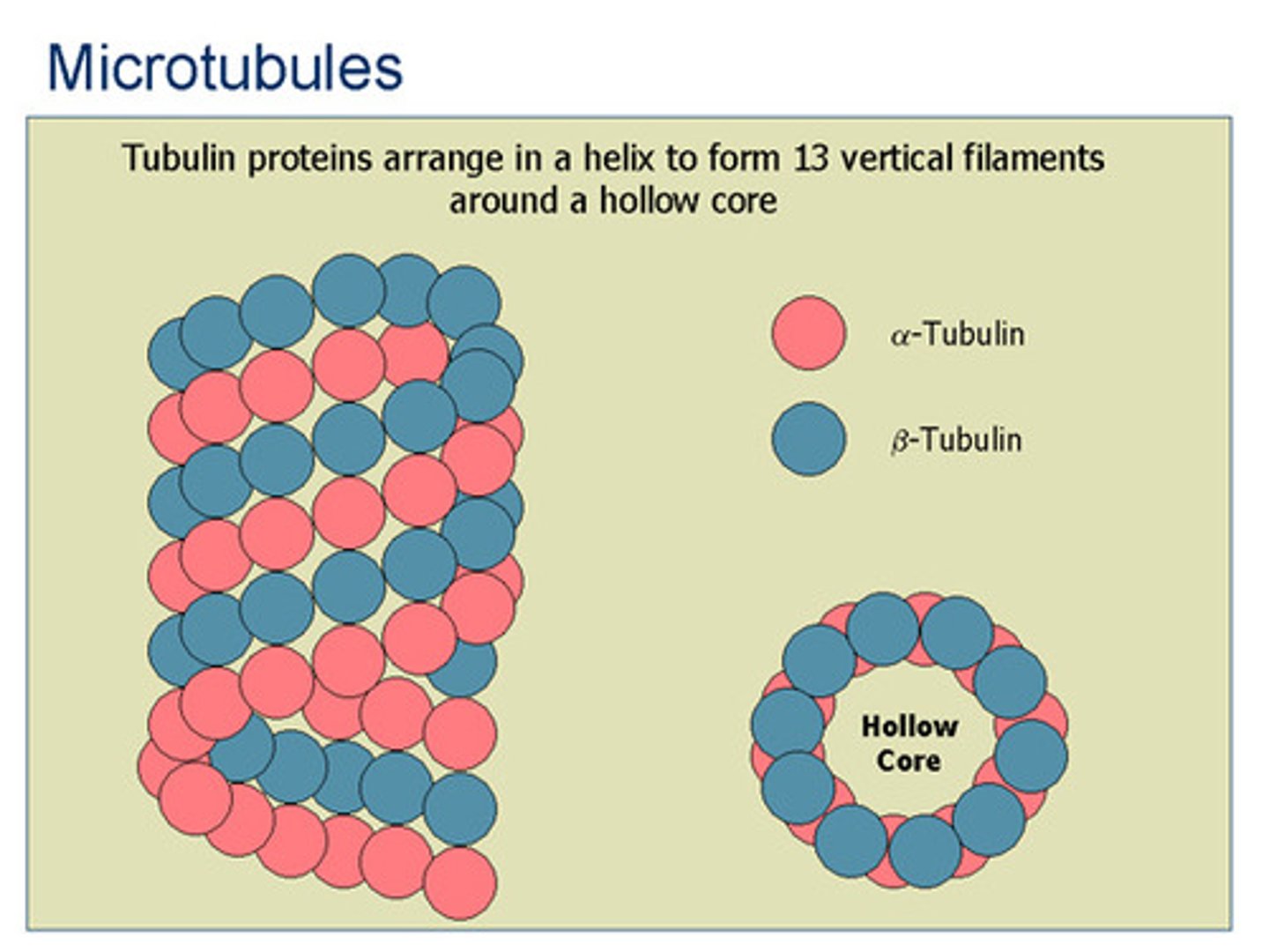
microtubules:
1. alpha and beta tubulin
2. forms dimers, sheets, then LARGE tubes
3. intracellular transport, cilia, flagella, mitotic spindle in mitosis/meiosis
4. can quickly polymerize/depolymerize
5. kinesin and dynein motor proteins
microfilaments:
1. actin, branching from centrosome
2. two actin fibers twist together to form SMALL tubes
3. muscle contraction (myosin, troponin, Ca2+), cytokinesis, adherent and tight junctions
4. some cell mobility
5. myosin motor protein
intermediate filaments:
1. different proteins
2. MEDIUM tubes
3. structure
cilia/flagella have 9 pairs of microtubules on outside, 1 pair in the middle, all connected with dynein to wiggle
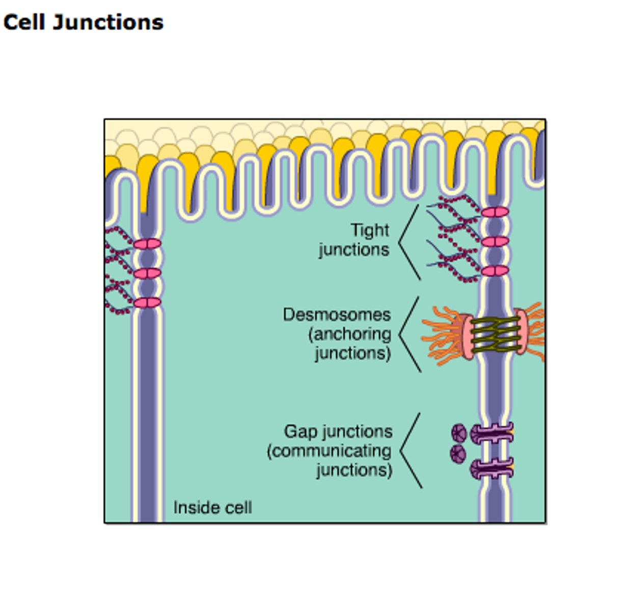
cell junction
adherin junctions- cadherins, actin
desmosomes- intermediate filaments
tight junctions- actin, seal lumen to separate environments (epithelial barriers like blood/lumen in gut)
gap junction- cell-to-cell communication, connexins
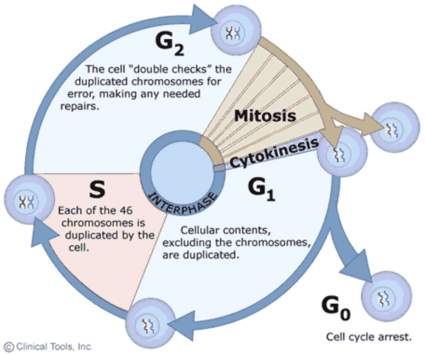
cell cycle and checkpoints
interphase: G1, G0, S, G2
G1- cell activity and growth
G0- non-dividing state at checkpoint, very tightly regulated
S- DNA replication
G2- growth, prepare for mitosis
M- mitosis, PMAT
G1/S checkpoint- tightly regulated, take inventory of nucleotides, enzymes, nutrients for DNA replication, sent to G0 senescence if doesn't pass
G2/M checkpoint- ensure DNA replication complete, check for mutations
cells stuck in at a checkpoint will just continue growing larger
each phase regulated by CDKs responding to cyclin levels and phosphorylation
mitosis
prophase:
1. condense DNA to chromosomes
2. break down nuclear membrane
3. build mitotic spindle, centrioles move to poles
metaphase
1. spindle fibers attach to centromeres at kinetochores
2. align chromosomes at plate
anaphase:
1. sister chromatids separate
2. cytokinesis starts
telophase:
1. decondense DNA
2. reform nucleus
3. break down spindle
4. finish cytokinesis
cytokinesis:
1. actin helps split cell at cleavage furrow
"I piss milk at the cows"
cancer
characteristics:
1. starts with single cell with altered DNA
2. lacks cell cycle control
3. metastasis to surrounding tissue
4. somatic mutation will not be inherited, since it does not affect the germ line!
5. increasing proliferation of cells can lead to cancer
6. different cancer lines have different mutations
7. PET scans with radioactively labeled glucose can help detect cancer cells
cancer genes:
1. oncogenes- a proto-oncogene increases cell cycle activity when active, becomes an oncogene when it mutates to becomes always active
GOF mutation
2. tumor suppressor genes- code for proteins that slow down cell cycle, repair DNA, and trigger apoptosis, p53 is guardian angel
LOF mutation
apoptosis/necrosis
cytochrome C- released from mitochondria
initiator caspase- trigger effectors
effector caspase- trigger deconstruction of cell, cleave proteins
extracellular death signals- surround cell apoptosis
intracellular death signal- tumor suppressor, virus
apoptosis- triggered by internal factors, causes cell shrinkage, doesn't affect other cells
necrosis- triggered by external factors, causes cell swelling, affects neighboring cells
meiosis
S phase:
1. DNA replication
2. sister chromatids are created and attached at centromere
prophase I:
1. homologous chromosomes pair up to from tetrads, connected by synaptonemal complex
2. recombination/crossing-over occurs between homologous pairs
3. chromosomes condense, nuclear envelope breaks down, spindles form
4. longest phase!
metaphase I:
1. tetrads align along metaphase plate
anaphase I:
1. homologous pairs separate, sister chromatids remain together
2. begin cytokinesis
telophase I:
1. chromosomes decondense, nuclear envelope forms, spindles breakdown
2. cytokinesis ends
3. now considered haploid, since each cell has single set
meiosis II:
1. identical to mitosis, but with haploid cells
2. oocyte/spermatocyte is formed, haploid
can form 2^n different gametes, n = haploid number or how many chromosomes
gametes combine to form zygote, diploid
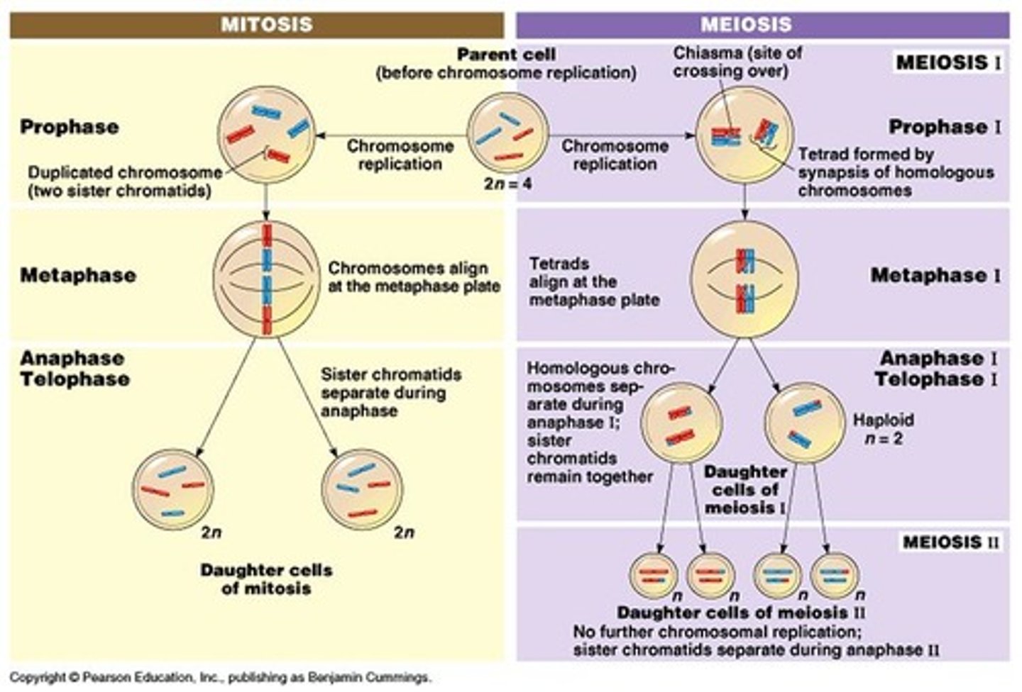
recombination
crossovers- genes swap between homologous pairs, which are connected by the synaptonemal complex
double crossover- crossover at two different places, section in the middle is also exchanged
gives genetic diversity in meiosis that you can't get in mitosis
so how do bacteria achieve genetic diversity?
and how is recombination used for DNA repair?
nondisjunction
either homologous pairs or sister chromatids fail to separate
trisomy- gamete with 3 copies of chromosome, Down syndrome
monosomy- gamete with 1 copy of chromosome
traits
genotype determines phenotype, alleles in a gene determine traits
polymorphic- many different forms of the trait
polygenic- trait determined by many different genes
incomplete dominance
heterozygote displays phenotype that blends alleles
shows with two different uppercase letters, since neither is fully dominant over other, red + white = pink
penetrance vs. expressivity
penetrance- probability of gene being expressed if present, it's either expressed or not
expressivity- how much genotype is expressed as phenotype, incomplete dominance, the extent to which penetrance happens
codominance
full expression of both alleles
ABO blood groups:
each allele codes for a protein on surface of RBCs
IA codes for A protein, IB codes for B protein, i is none
type A: IAIA, IAi
type B: IBIB, IBi
type AB: IAIB
type O: ii
Rh factor
complete blood typing combines ABO blood groups with Rh factor, which is classically dominant
IAiRR is A+, iiRr is O+
universal donor is O-
universal recipient is AB+
epistasis
expression of one gene dependent on expression of another gene
hair shape gene dependent on baldness gene
Mendel's laws
law of segregation- alleles are separated during gamete formation, pair of sister chromatids divide
law of independent assortment- pairs of alleles separate independently of each other
genetic probability
rule of multiplication- probability of both is the overlap
PAB = PA*PB
rule of addition- probability of either is the whole area
PAorB = PA + PB - PAB
linked genes
genes on same chromosome that are close together and might not sort independently
compare expected ratio vs. observed ratio of F2 generation, not F1 generation
homozygous recessive crosses with homozygous dominant will give kids with what appears to be linked genes
dihybrid cross- F1 crosses with F1, both homozygous, generates 9:3:3:1 ratio for F2
observed F2 generation should have less recombinant offspring, more linked offspring
F1 x homozygous recessive generates 1:1:1:1 ratio
recombination frequency- how many recombinant offspring over total offspring, determines how close the genes are together
Hardy-Weinberg equilibrium
study population genetics by assuming allele frequencies don't change over time
p = dominant allele frequency
q = recessive allele frequency
p + q = 1 (allele frequency)
p^2 + 2pq + q^2 = 1 (genotype frequency)
conditions for which equation holds:
1. no mutation- but they do happen
2 no natural selection- but evolution happens
3. no migration- but they move
4. no random drift- but one allele might randomly dominate
5. random mating- but they have preferences
it takes 1 generation to reach new equilibrium if old one is disturbed
genetic drift
random changes in allele frequencies that occurs in small populations
ex: a well-adapted Y chromosomes may not be passed down if a man only has daughters, causes degeneration of Y chromosome
natural selection
modes of natural selection:
1. directional selection- one end of bell curve is advantageous
2. divergent selection- both ends of bell curve are advantageous, so middle dies
3. stabilizing selection- middle of bell curve is advantageous, so both ends die
4. artificial selection- humans intervene
5. sexual selection- mating ritual or display is advantageous
6. kin selection- social sacrifices are advantageous
bottleneck
a change in allele frequency following a dramatic reduction in the size of a population
differential reproduction
survival of those who live to make babies