Cells and Their Internal Structures
1/12
There's no tags or description
Looks like no tags are added yet.
Name | Mastery | Learn | Test | Matching | Spaced | Call with Kai |
|---|
No analytics yet
Send a link to your students to track their progress
13 Terms
Define magnification, resolution, and contrast.
Three key terms for microscopy:
Magnification — how much bigger the image appears compared to the actual object
Resolution — ability to distinguish two close objects as separate. Higher resolution = sharper, more detailed image
Contrast — difference in brightness between the object and background. Stains are often used to increase contrast
Differentiate between transmission electron microscopes (TEM) and scanning electron microscopes (SEM).
TEM | SEM | |
|---|---|---|
Full name | Transmission Electron Microscope | Scanning Electron Microscope |
How it works | Electrons pass through the specimen | Electrons scan the surface of the specimen |
Image type | Flat 2D internal view | 3D surface view |
Best for | Internal organelle structure | Surface texture and shape |
Identify the best type of microscope to use for different samples.
Situation | Best Microscope |
|---|---|
Living cells or tissues | Light microscope |
Internal organelle detail | TEM |
3D surface of a virus or cell | SEM |
Quick cheap observation | Light microscope |
Highest possible resolution | Electron microscope |
List and describe the structures that all cells have in common.
All living things are made of cells
The cell is the basic unit of life
All cells come from pre-existing cells
Cell membrane — controls what enters and exits the cell
DNA — genetic information that runs the cell
Ribosomes — make proteins
Explain the relationship between surface area and volume of a cell and how this explains why most cells are small.
As a cell gets bigger its volume grows faster than its surface area
This is a problem because nutrients and waste enter/exit through the surface (membrane)
If the cell gets too big, the membrane can't keep up with the demands of the volume inside
Define and differentiate between the genome and proteome.
Genome — the complete set of DNA in a cell. All of your genetic instructions. Every cell in your body has the same genome.
Proteome — the complete set of proteins a cell produces. Different cells express different proteins even though they have the same genome.
Define cytosol and its function.
Cytosol — the liquid component inside the cell that surrounds the organelles
It's mostly water but also contains salts, sugars, proteins, and other molecules
Together cytosol + organelles = cytoplasm
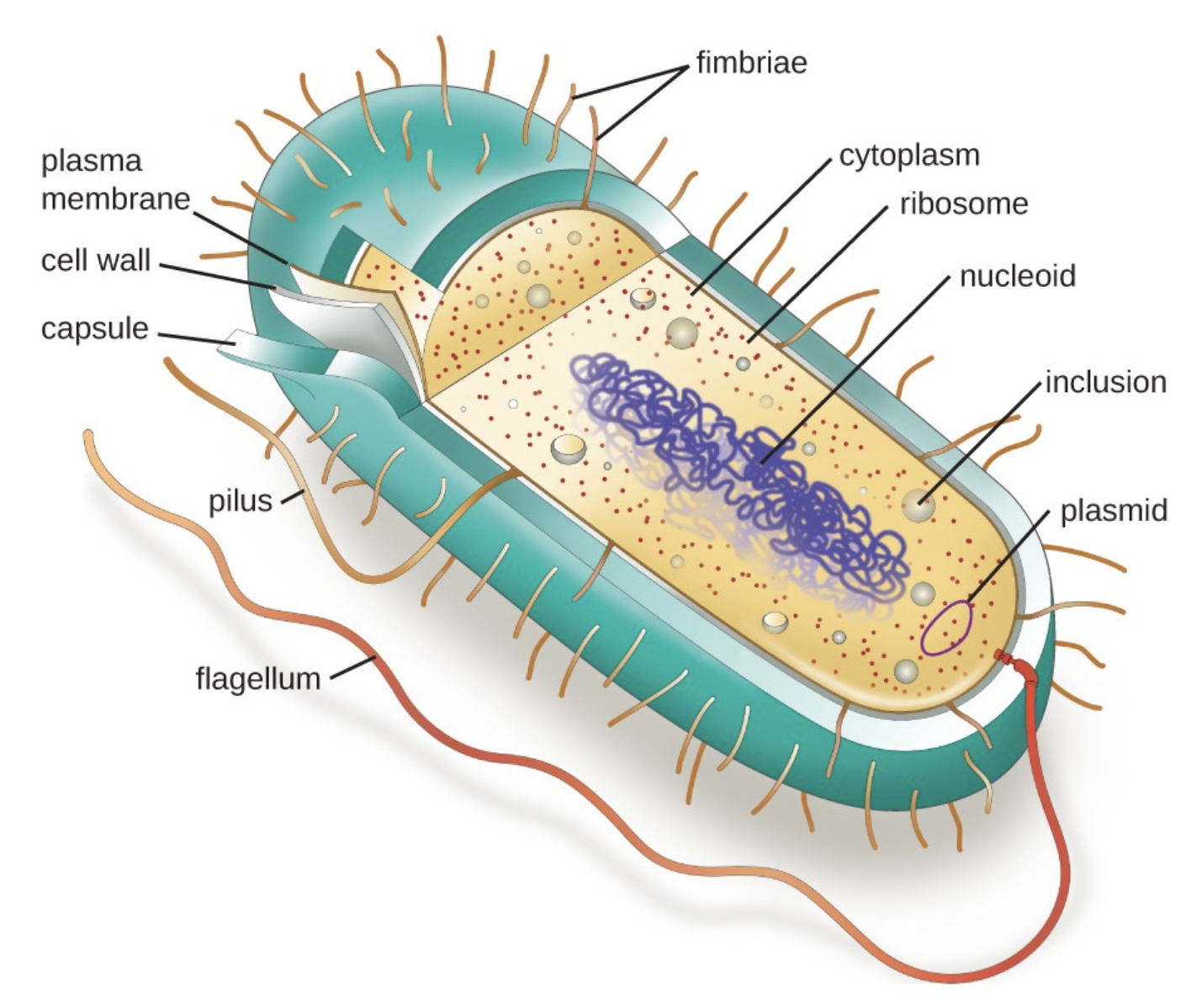
Diagram a bacterial cell illustrating the location and functions of the main structural components including the DNA, the nucleoid, ribosomes, plasmids, cell membrane, cell wall, capsule/glycocalyx, pili, and flagella.
Structure | Function |
|---|---|
DNA/Nucleoid | Region where DNA is located — no membrane around it |
Ribosomes | Make proteins |
Plasmids | Small circular DNA separate from main DNA — often carry antibiotic resistance genes |
Cell membrane | Controls what enters and exits |
Cell wall | Rigid outer layer that gives shape and protection |
Capsule/Glycocalyx | Sticky outer coat — helps bacteria attach to surfaces and evade immune system |
Pili | Short hair-like projections — help bacteria attach to surfaces or other cells |
Flagella | Long whip-like tail — used for movement |
List the components of the cytoskeleton and explain their general functions.
Component | Size | Function |
|---|---|---|
Microfilaments | Thinnest | Made of actin. Give cell shape, help with cell movement and division |
Intermediate filaments | Medium | Provide mechanical strength and anchor organelles |
Microtubules | Thickest | Made of tubulin. Cell shape, move organelles, form spindle fibers during cell division. Also make up cilia and flagella |
List and describe the major eukaryotic organelles and explain the function of each.
Organelle | Function |
|---|---|
Nucleus | Controls cell activity, stores DNA |
Nucleolus | Inside nucleus, makes ribosomes |
Ribosomes | Make proteins |
Rough ER | Has ribosomes, makes and processes proteins |
Smooth ER | No ribosomes, makes lipids, detoxifies drugs/alcohol |
Golgi Apparatus | Packages and ships proteins to their destination |
Mitochondria | Makes ATP (energy) — powerhouse of the cell |
Lysosomes | Digest waste, old organelles, and foreign invaders |
Vacuoles | Storage — large in plant cells, small in animal cells |
Chloroplasts | Photosynthesis — only in plant cells |
Cell wall | Rigid support — only in plant cells |
Centrioles | Help organize cell division — only in animal cells |
Differentiate between a prokaryotic cell and a eukaryotic cell based on structural/organelle differences.
Feature | Prokaryote | Eukaryote |
|---|---|---|
Nucleus | No — DNA floats in nucleoid | Yes — DNA enclosed in nucleus |
Membrane bound organelles | No | Yes |
Size | Smaller | Larger |
DNA shape | Circular | Linear chromosomes |
Ribosomes | Yes (smaller) | Yes (larger) |
Examples | Bacteria, archaea | Animals, plants, fungi, protists |
Differentiate between a plant cell and animal cell based on structural/organelle differences.
Feature | Plant Cell | Animal Cell |
|---|---|---|
Cell wall | Yes | No |
Chloroplasts | Yes | No |
Large central vacuole | Yes | No (small vacuoles) |
Centrioles | No | Yes |
Shape | Rigid, rectangular | Flexible, irregular |
Diagram the process of making a protein such as insulin for secretion.
The endomembrane system is a series of organelles that work together to make, process, and ship proteins.
Organelles involved:
Nucleus → Rough ER → Smooth ER → Golgi → Vesicle → Cell membrane
The pathway using insulin as an example:
Step | What happens |
|---|---|
Nucleus | DNA instructions for insulin are read and sent out as mRNA |
Rough ER | Ribosomes on rough ER read mRNA and build the insulin protein |
Smooth ER | Protein gets some initial processing |
Golgi Apparatus | Insulin gets packaged, modified, and labeled for shipping |
Vesicle | Insulin is wrapped in a membrane bubble for transport |
Cell membrane | Vesicle fuses with membrane, insulin is released outside cell (exocytosis) |