Medical Anatomy: Lecture 2
1/86
There's no tags or description
Looks like no tags are added yet.
Name | Mastery | Learn | Test | Matching | Spaced | Call with Kai |
|---|
No analytics yet
Send a link to your students to track their progress
87 Terms
name the different regions of the vertebral column
cervical
thoracic
lumbar
sacral
coccygeal
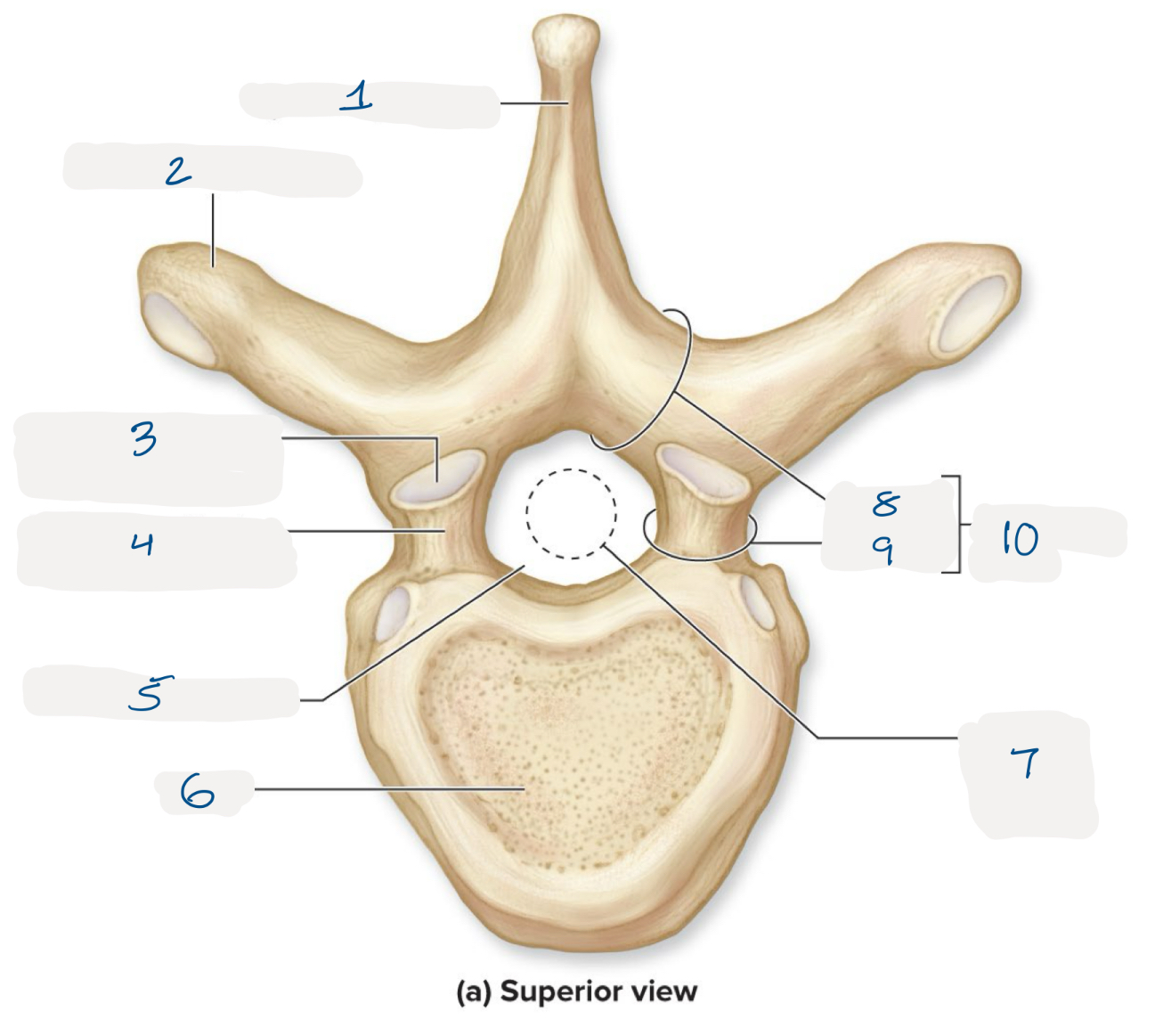
name the numbered regions of the vertebrae
spinous process
transverse process
superior articular facet
superior articular process
vertebral foramen
body
spinal cord location
lamina
pedicle
vertebral arch
where do spinal nerves pass through
intervertebral foramen
what are the first 2 cervical vertebrae called and what makes them unique
C1 ( atlas)
large superior articulating facet for joint with cranium
C2 (axis)
additional posterior process called “dens” which allows head to swivel
atlanto-axial joint movement
features of cervical vertebrae
small size
spinous process has bifid tip
transverse foramen (space for vertebral arteries)
features of thoracic vertebrae
medium size
elongated spinous process
costal facets for rib joints
features of lumbar vertebrae
large size
rounded spinous process
sacrum
5 fused vertebrae
sacral promontory
auricular surface (articulates with pelvis)
coccyx
3-4 fused tiny vertebrae
lumbosacral joint
intervertebral disc between L5 and sacrum
facet joint between L5 inferior articulating facets and sacral superior articulating facets
sacroiliac (SI) joint
sacral auricular surface and auricular surface of ilium
intervertebral discs; articulation, structural type, functional type, & movements
vertebral bodies of adjacent vertebrae
cartilaginous
amphiarthrosis
extension, flexion, lateral flexion of vertebral column
facet joints; articulation, structural type, functional type, & movements
superior & inferior articulating processes of adjacent vertebrae
synovial
diarthrosis
rotation, extension, flextion, lateral flexion of the vertebral column
atlanto-occipital joint; articulation, structural type, functional type, & movements
superior articulating facets of atlas and occipital condyles of occipital bone
synovial
diathrosis
extension, flexion or the head, slight lateral flexion
atlantoaxial; articulation, structural type, functional type, & movements
anterior arch of atlas and dens of axis
synovial
diarthrosis
head rotation
sacroiliac; articulation, structural type, functional type, & movements
auricular surface of ilium to auricular surface of sacrum
synovial
diarthrosis
nutation (sacral flexion) & counternutation (sacral extension)
nervous system
neural communication by electrical and chemical signals that are rapid and cause immediate responses
responsible for voluntary, involuntary movements, reflexes, learning, memory, & emotions
central nervous system (CNS)
brain
spinal cord
peripheral nervous system (PNS)
nerves & receptors outside of the brain and spinal cord
name the 2 nervous systems
sensory and motor
sensory nervous system
detects stimuli and transmits infromation from receptor to the CNS
somatic sensory
sensory input form the receptors of the 5 sense and proprioceptors
visceral sensory
sensory input from receptors of internal organs and blood vessels
motor nervous system
initiates and transmits information from the CNS to effectors
somatic motor
motor output to skeletal muscle
autonomic motor
motor output to cardiac muscle, smooth muscle, and glands
autonomic
organ, gland regulation
sympathetic
parasympathetic
neurons
electrical cells that transmit the signals through the nervous system
directional flow of electrical activity
action potentials are the long distance signals
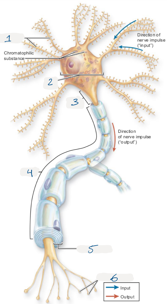
name the numbered structures of the neuron
dendrites
cell body
axon hillock
axon
myelin sheath
synaptic knobs
synapses
connection points between 2 neurons or between a neuron and a muscle, gland, or other cell
glia
supportive cells within the central and peripheral nervous system
myelin sheath
external wrapping around the axon that insulates and increases the speed of electrical transmission
myelin makes the axon appear white
white matter
myelinated axons
tracts
bundles of axons in the CNS
nerves
bundles of axons in the PNS
tracts with white matter
bundles of axons in the CNS
nerves with white matter
bundles of axons in the PNS
gray mater
non-myelinated axons
nuclei
cell bodies in the CNS
ganglia
cell bodies in the PNS
nuclei with gray matter
cell bodies in the CNS
ganglia with gray matter
cell bodies in the PNS
nerve
bundles of axons in the peripheral nervous system traveling to/from the CNS
reflex
simplest, fastest pathways
input, integration, output
sensory pathways
inputs processed within multiple CNS regions
motor pathways
outputs regulated by multiple CNS regions
afferent
brings sensory information from the outside world into the body
receptors
sense the stimulus
sensory neurons
transmit the signals into the nervous system
sensory ganglion
group of sensory neuron cell bodies outside of the CNS
sensory fibers
bundle of axons within a nerve that carry sensory information toward the CNS
somatic sensory pathway
dorsal horn
dorsal root
dorsal root ganglion
spinal nerve
somatic sensory receptors (tactile receptors, proprioceptors)
visceral sensory pathway
dorsal horn & lateral horn
dorsal root
dorsal root ganglion
spinal nerve
visceral sensory receptors (baroreceptor, chemoreceptor)
somatic effectors
anterior horn
anterior root
spinal nerve
somatic effector (skeletal muscle)
autonomic effector
anterior & lateral horn
anterior root
spinal nerve
preganglion synapse on postganglion
postganglion reach the effector organs (cardiac muscle, smooth muscle, gland)
efferent
motor control is efferent output from the nervous system
motor commands are sent to move muscles and related structures
motor fibers
bundle of axons within a nerve that carry motor information from CNS toward skeletal muscle
motor neurons
transmit the motor commands from the CNS to skeletal muscle
what do autonomic pathways do
regulate the organs
central nervous system control
2 major divisions of the CNS
sympathetic
fight or flight
parasympathetic
rest and digest
two-neuron chains
autonomic pathways have 2 neuron chain in the PNS
pre-ganglionic neuron exits the CNS
post-ganglionic neuron signals directly to the organ
ganglion
a group of autonomic cell bodies outside of the CNS
parasympathetic nervous system
cranial nerves (C3, C7, C9, C10)
pelvic splanchnic nerves (S2, S3, S4)
long pre-ganglionic axons
terminal ganglia within organs
sympathetic nervous system
lateral horns T1-L2 to sympathetic trunk
cervical ganglia
thoracic, lumbar, sacral splanchnic nerves
abdominal ganglia
short pre-ganglionic axons
prominent ganglia near the spine
spinal cord
protected by the vertebrae within the spinal canal
part of the CNS
foramen magnum ——> L1/L2
anchored laterally by denticulate ligaments and inferiorly by filum terminale
name the spinal cord meninges and other spaces in order of out to in
wall of vertebrae
epidural space
dura mater
arachnoid mater
subarachnoid space (filled with CSF)
pia mater
what space does a lumbar puncture needle reach?
needle is inserted into the subarachnoid space to get a sample of CSF fluid
what space does an epidural needle reach?
epidural needle stays in the epidural space before the dura matter
name the gross features of the spinal cord
cervical
thoracic
lumbar
sacral
coccygeal
conus medullaris
cauda equina
filum terminale
cauda equina
the spinal cord ends at L1/L2
lumbar and sacral parts of the spinal cord end much higher than their vertebrae send nerves below through cauda equina
sectional anatomy of the spinal cord
white matter:
posterior, lateral, anterior funiculus
gray matter:
posterior, lateral, anterior horns
central canal
posterior median sulcus
anterior median fissure
spinal nerves
combined sensory, motor, and autonomic nerve roots that exit the spinal canal via the intervertebral foramen
rami
spinal nerve branches
form nerve plexuses
posterior ramus
innervate deep muscles and skin of the back
anterior ramus
innervate anterior and lateral portions of the trunk, upper limbs, and lower limbs
rami communicantes
between the spinal nerves and the sympathetic trunk ganglia of the sympathetic trunk
how to number spinal nerves
cervical spinal nerves exit above corresponding cervical vertebrae
C8 spinal nerve exits above T1
thoracic, lumbar, sacral, coccygeal spinal nerves exit below corresponding vertebrae
cervical plexus
spinal nerves C1-C4
innervate anterior neck muscles, skin of the neck, and parts of the head and shoulders
phrenic nerves C3, C4, C5
innervate the diaphragm
brachial plexus
spinal nerves C5-T1
innervate the pectoral girdle and the entire upper limb of one side
roots, trunks, divisions, cords, branches (richard tucker drinks cold beer)
thoracic nerves
no plexus
T1-T11 anterior rami form intercostal nerves (between adjacent ribs)
T12 isubcostal nerve
lumbosacral plexus: lumbar
spinal nerves L1-L4
anterior division
includes obturator nerve
posterior division
includes femoral nerve
lumbosacral plexus: sacral
spinal nerves L4/5, S1-S4
includes sciatic nerve
deep back muscles of the vertebral column
action:
vertebral extension, lateral flexion of the spine
innervation:
dorsal rami of spinal nerves
erector spinae muscles
lateral to medial
iliocostalis group
longissimus group
spinalis group
transversospinalis muscles
multifidus
semispinalis
rotatores
action:
vertebral extension
lateral flexion or rotation of the spine
quadratus lumborum
origin:
12th rib; transverse process of lumbar vertebrae
insertion:
iliac crest
innervation:
T12-L4 spinal nerves
action:
spinal extension, lateral flexion, depresses rib 12
serratus posterior
serratus posterior superior
serratus posterior inferior
both extend, laterally rotate the spine, and stabilize posture
trapezius
action:
elevation, superior rotation of the scapula (superior), retraction of the scapula (middle), depression of the scapula (inferior)
origin:
occipital bone, ligamentum nuchae, spinous processes of C7-T12
insertion:
clavicle, acromion and spine of scapula
innervation:
CN XI accessory nerve
test: “shrug your shoulders”
latissimus dorsi
action:
extension, adduction, medial rotation of the arm
origin:
spinous processes of T7-T12, ribs 8 to 12, iliac crest, thoracolumbar fascia
insertion:
intertubercular sulcus of the humerus
innervation:
thoracodorsal nerve (C6-C8)
vertebral landmarks
vertebra prominens: C7
iliac crest (intercristal line): palpated near L3/L4 and imaging at L4/L5
what muscles border the triangle of ausculatation
trapezius
latissimus dorsi
rhomboid major