Cardiovascular System Part 1
1/46
There's no tags or description
Looks like no tags are added yet.
Name | Mastery | Learn | Test | Matching | Spaced | Call with Kai |
|---|
No analytics yet
Send a link to your students to track their progress
47 Terms
Cardiovascular System
A closed system of the heart and blood vessels.
Transportation
Function of the Cardiovascular System
To deliver oxygen, nutrients, hormones, and other substances to different parts of the body.
To remove carbon dioxide and other waste products.
Fist, cone-shaped (pinecone)
What is the size and shape of the heart?
Less than a pound
How much does the heart weigh?
Thoracic cavity
The heart is in the Medial section of the ______, in between the lungs.
Inferior mediastinum
The heart is enclosed within the ______?
Apex
This rests on the diaphragm and is pointed toward the left hip.
Base
This lies beneath the second rib and is pointed at the right shoulder.
This is where the great vessels emerge
Midsternal Line
1
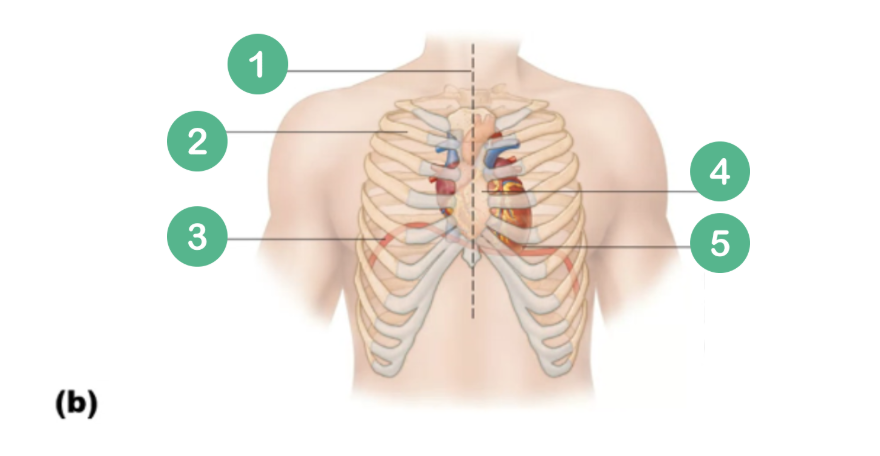
2nd Rib
2

Diaphragm
3

Sternum
4

Point of Maximal Intensity
5

Pericardium
A double-walled sac that covers and protects the heart.
Fibrous pericardium and Serous pericardium
What are the two main layers of the pericardium?
Fibrous pericardium
This layer of the Pericardium is outer, loose, and superficial.
Serous pericardium
This layer of the Pericardium is inner, deep to fibrous.
Has two layers: Parietal pericardium and Visceral pericardium
Parietal pericardium and Visceral pericardium
What are the two layers of the Serous Pericardium?
Parietal pericardium
Layer of the Serous pericardium (1/2)
What is the outside layer that lines the inner surface of the fibrous pericardium?
Visceral Pericardium
Layer of the Serous pericardium (2/2)
Also known as Epicardium
This layer is directly attached to the heart muscle.
Between the parietal pericardium and the visceral pericardium.
Where is the pericardial cavity located?
Serous fluid
What fills the pericardial cavity?
Fibrous Pericardium
Parietal Layer (of Serous Pericardium)
Pericardial Cavity (with Serous Fluid)
Visceral Layer (Epicardium)
List the layers of the heart covering from Superficial to Deep.
Epicardium (outer), Myocardium (middle), and Endocardium (inner).
What are the three layers of the heart wall?
Epicardium
Outside layer; the visceral pericardium.
Myocardium
The middle layer of the heart, composed primarily of cardiac muscle.
Endocardium
The inner layer of the heart wall, also known as endothelium.
Atria (Right & Left)
What are the RECEIVING chambers of the heart?
Receiving chambers
Help fill ventricles
Low pressure
Ventricles
What are the DISCHARGING chambers of the heart?
Discharging/Pumping chambers
Thick-walled
Propel blood into circulation
Interatrial Septum
This separates the two ATRIA longitudinally.
Interventricular Septum
This separates the two VENTRICLES longitudinally.
Fossa ovalis
An oval-shaped depression in the interatrial septum, remnant of the fetal foramen ovale.
Foramen ovale
An opening in the fetal heart (in the interatrial septum) that allows blood to flow from the right to left atrium.
Right Side
Which side is the pulmonary circuit pump (pumps blood to the lungs)
Left side
Which side is the Systemic circuit pump (pumps blood to the rest of the body)
Arteries
Carry blood Away from the heart.
Veins
Carry blood toward the heart
Pulmonary circulation
To transport oxygen-poor blood from the right side of the heart to the lungs, and return oxygen-rich blood back to the left side of the heart.
Right Heart, Pulmonary Trunk, Pulmonary Arteries, Lungs, Pulmonary Veins, Left Heart
Describe the path of blood in pulmonary circulation.
Oxygen-poor blood away from the heart (to the lungs).
What type of blood do the pulmonary arteries carry, and in which direction?
Oxygen-rich blood towards the heart (from the lungs)
What type of blood do the pulmonary veins carry, and in which direction?
AV (Atrioventricular) valves
Prevent backflow into the atria during ventricular contraction.
Anchored to the ventricular walls by chordae tendineae ("heartstrings") to prevent eversion.
Open during relaxation (diastole), closed during contraction (systole)
Semilunar valves
Prevent backflow into the ventricles during relaxation.
Closed during heart relaxation (diastole), open during ventricular contraction (systole)
Pressure changes in the heart chambers.
What dictates the opening and closing of heart valves?
AV Valves: Open (passively filling ventricles).
Semilunar Valves: Closed
During ventricular relaxation (diastole), what is the status of the AV and Semilunar valves?
AV Valves: Closed (preventing backflow).
Semilunar Valves: Open (blood ejected into arteries)
During ventricular contraction (systole), what is the status of the AV and Semilunar valves?
Chordae tendineae
This anchors AV valve cusps to the ventricular walls, preventing them from being forced backward into the atria during contraction