124 WBC Weekend Lab
1/52
There's no tags or description
Looks like no tags are added yet.
Name | Mastery | Learn | Test | Matching | Spaced | Call with Kai |
|---|
No analytics yet
Send a link to your students to track their progress
53 Terms
low power scan (10x)
eval overall stain quality
eval cell distribution
determine optimal exam area
high power scan (40x)
wbc estimation
oil immersion (100x)
eval RBC morphology
platelet estimation
wbc differential
optimal exam area of a blood smear
monolayer of cells
method for wbc differential on blood smear
battlement pattern
neutrophil band
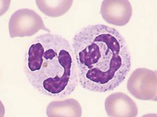
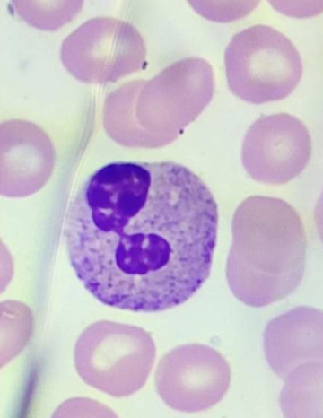
segmented neutrophil
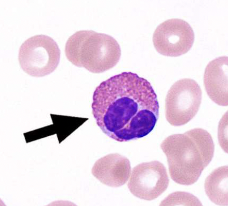
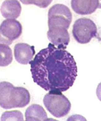
segmented eosinophil

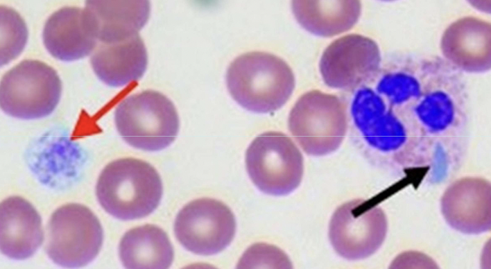
segmented basophil
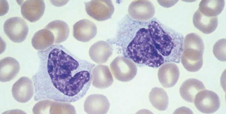
monocyte
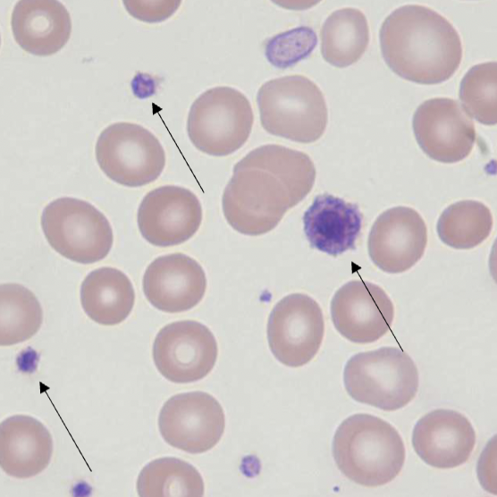
thrombocyte (platelet)
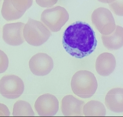
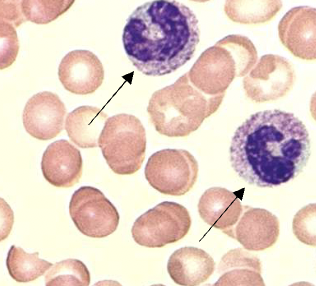
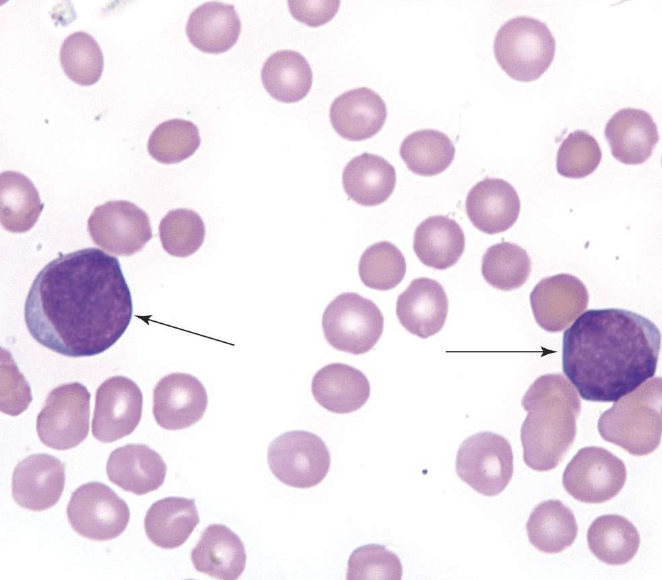
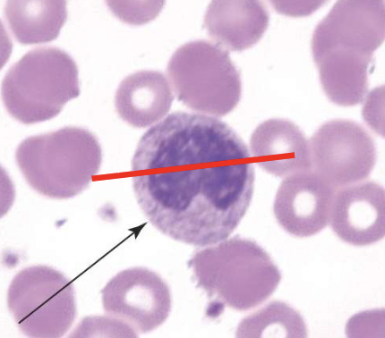
lymphocyte
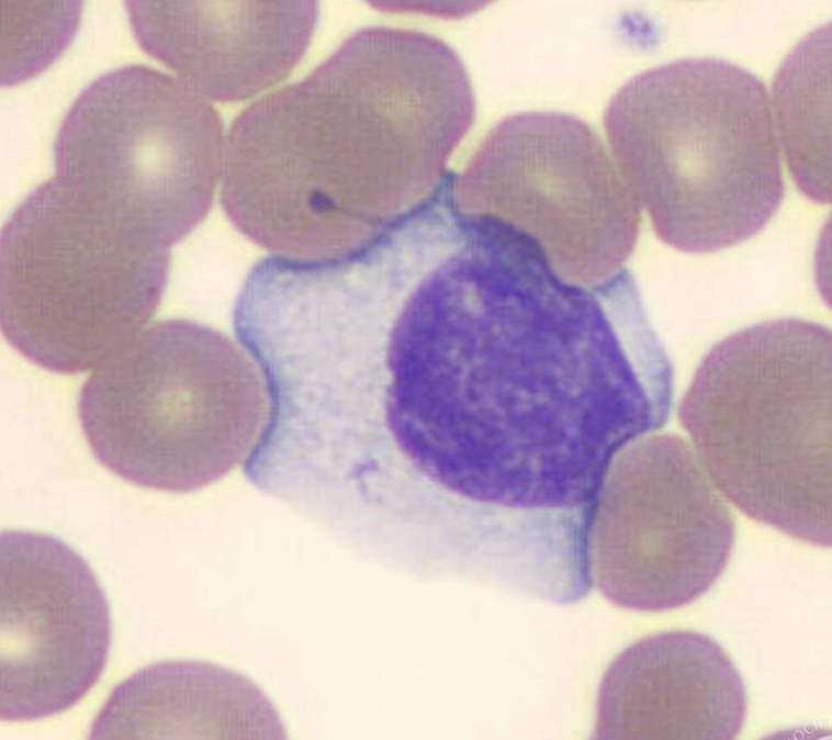
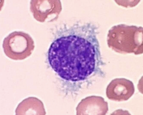
reactive lymphocyte
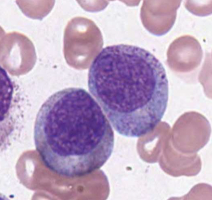
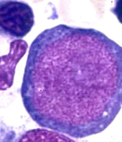
myeloblast
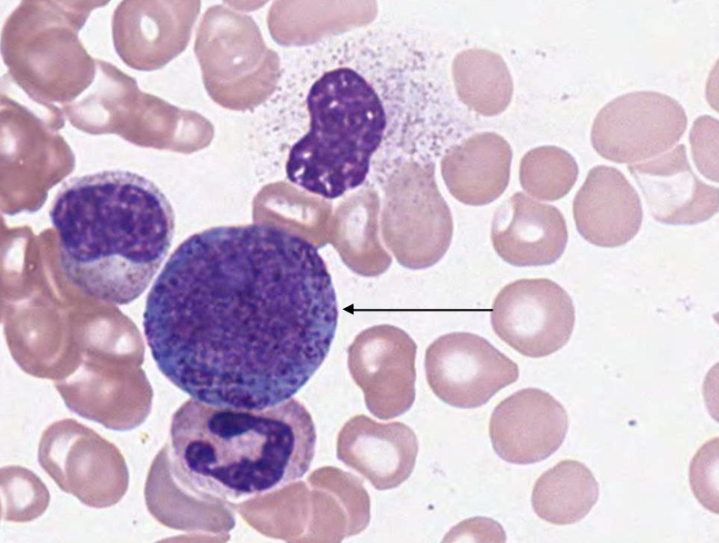
promyelocyte
neutrophilic myelocyte
neutrophilic metamyelocyte
neutrophil maturation (myelopoiesis)
myeloblast
promyelocyte
myelocyte
metamyelocyte
neutrophilic band
segmented neutrophil
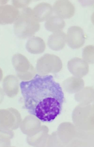
plasma cell
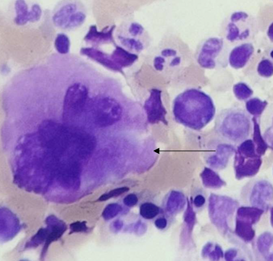
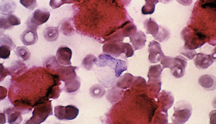
megakaryocyte
rubriblast (rbc)
wright stain steps
methanol - fixes cells to slide
eosin - has affinity for basic components (stains red)
methylene blue - has affinity for acidic components (stains blue)
wbc count estimation calculation
avg. # of wbc/HPF
count in 10 areas then divide by 10 to get avg.
wbc count estimation
2-4 WBC → 4-7k/uL
4-6 WBC → 7k10k/uL
6-10 WBC → 10-13k/uL
10-20 WBC → 13-18k/uL
platelet count estimation
count # of platelets in 10 HPF areas
multiply avg. by 2000 = plt/uL
thrombocytosis
> 20 plt/uL
thrombocytopenia
< 20 plt/uL
wbc differential
identify 100 wbcs in hpf
bone marrow differential
identify ~500 NUCLEATED cells
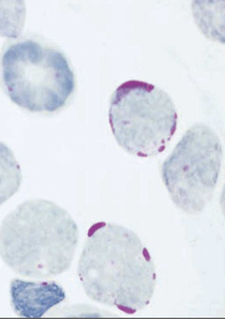
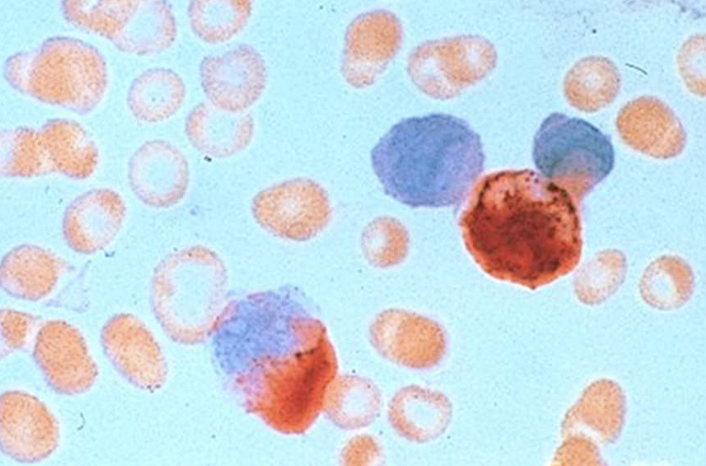
sickledex procedure
4 mL phosphate buffer to tube containing sodium hydrosulfite
50 uL patient sample to tube
cap & shake
incubate at room temp. for 10-20 min.
read tube against black lines
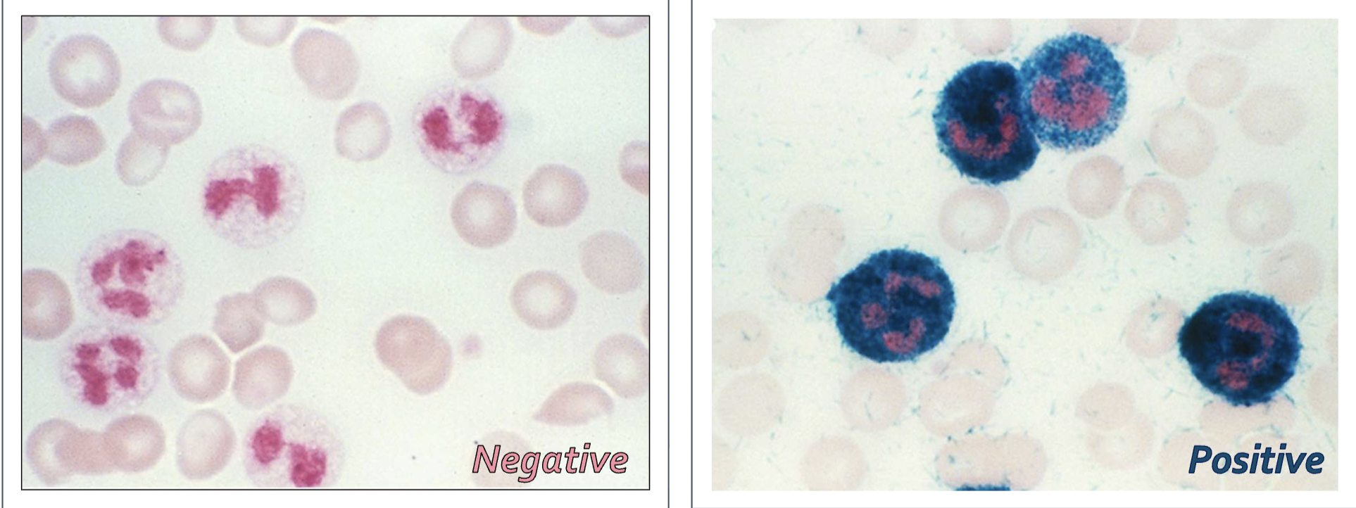
monospot assay
detects heterophile antibodies
monospot procedure
1 drop patient sample
1 drop diluent
place strip in tube w/ sample
wait 5 min.
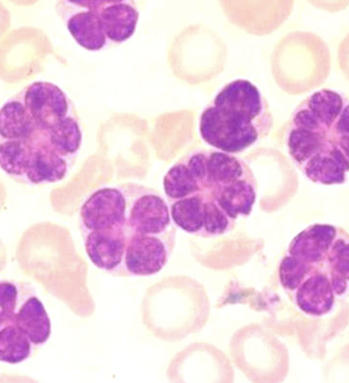
pelger-huet anomaly
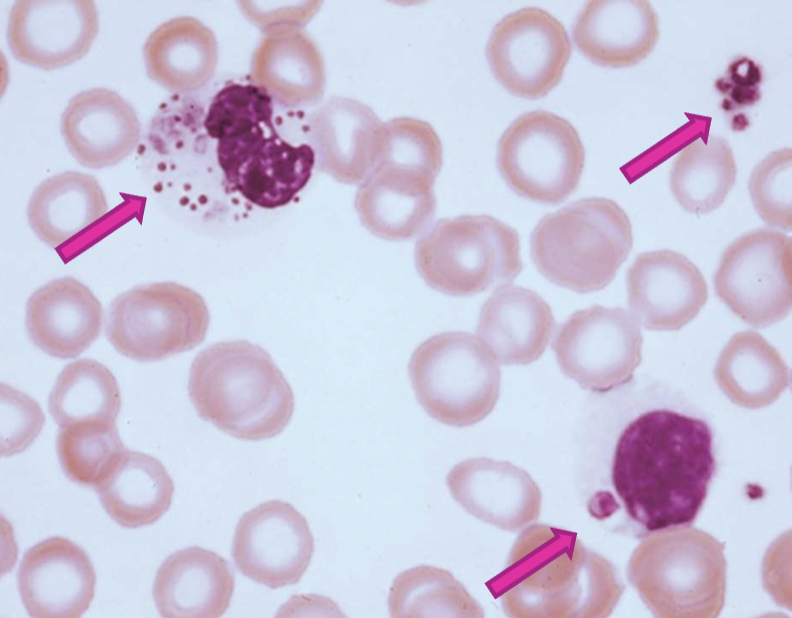
alder’s anomaly
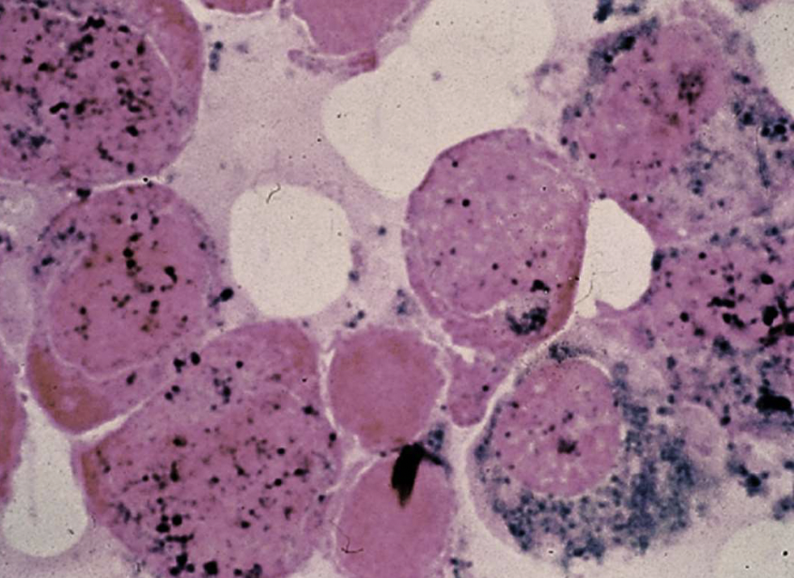
chediak-higashi syndrome
may-hegglin anomaly
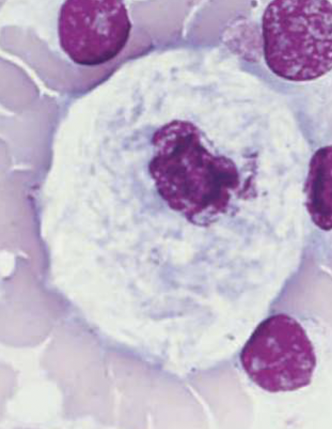
gaucher cell
niemann-pick cell
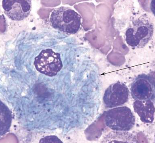
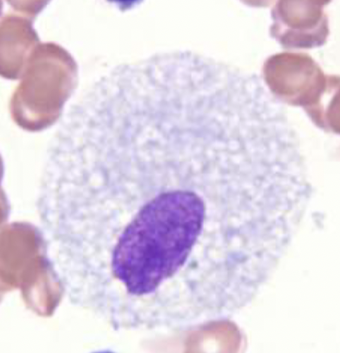
sea-blue histiocyte
MPO stain
SBB stain
PAS stain
CAE stain
NSE stain
LAP stain
TRAP stain
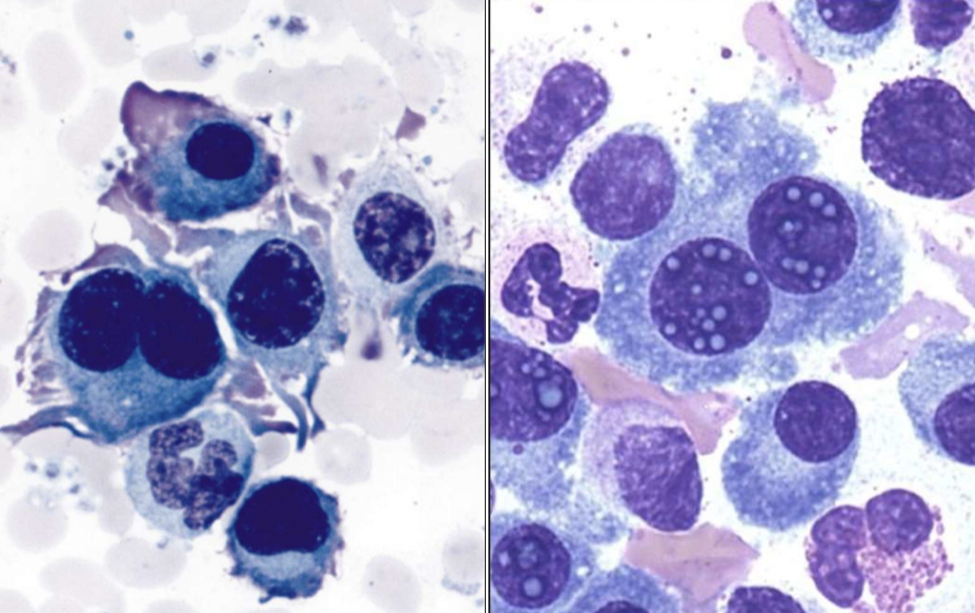
waldenstrom’s macroglobulinemia
flame cells
dutcher bodies
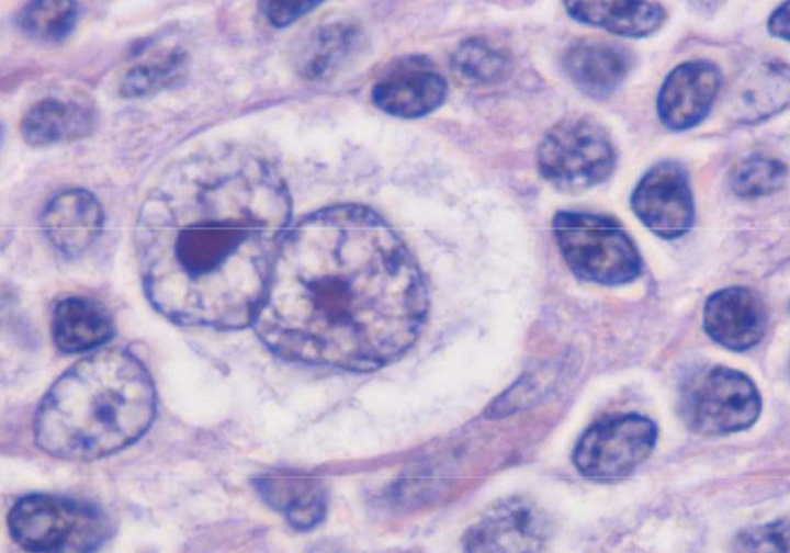
reed-sternberg cell in hodgkin’s lymphoma
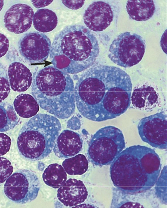
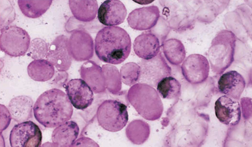
multiple myeloma (excess production of plasma cells)
sezary cell
burkitt’s lymphoma
chronic leukemia
acute leukemia