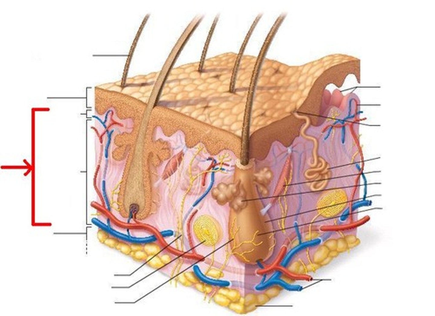
Unit 1.1 Integumentary System
1/34
There's no tags or description
Looks like no tags are added yet.
Name | Mastery | Learn | Test | Matching | Spaced | Call with Kai |
|---|
No analytics yet
Send a link to your students to track their progress
35 Terms
What are the three layers of the integumentary system?
Epidermis, dermis, and subcutaneous
What are the functions of the epidermis?
- Provides a physical and chemical barrier
- Regulates fluid
- Provides light touch sensation
- Assists with excretion
- Critical to endogenous vitamin D production
- Contributes to cosmesis/appearance
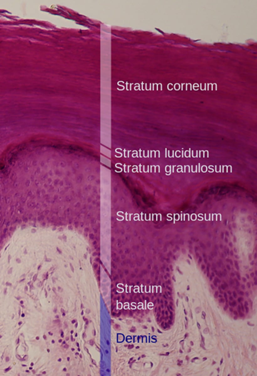
What are the layers of the epidermis?
Stratum corneum
Stratum lucidum
Stratum granulosum
Stratum spinosum
Stratum Basale
What are the characteristics of the stratum corneum layer?
- Contains multiple layers of flattened, dead, interlocking keratinocytes
- typically dry
- water resistant
- permits slow water loss by insensible perspiration
What are the characteristics of the stratum lucidum layer?
appears as a glassy layer in thick skin only
What are the characteristics of the stratum granulosum layer?
- keratinocytes produce keratohyalin and keratin
- keratin fibers develop as cells become thinner and flatter
- Gradually, the cell membranes thicken, the organelles disintegrate, and the cells die
What are the characteristics of the stratum spinosum layer?
- Keratinocytes are bound together by maculae adherens attached to tonofibrils of the cytoskeleton
- Some keratinocytes divide in this layer
- Langerhans cells and melanocytes are often present
What are the characteristics of the stratum basale layer?
- Is the deepest, basal layer
- Attachment to basal lamina
- Contains epidermal basal (stem) cells, melanocytes, and Merkel cells
What is the function of the keratinocyte cell?
These are continuously dividing cells that produce keratin
What keratins function?
a protective protein that creates a tough, water-resistant physical barrier to protect the body from trauma and infection
What is the function of melanocyte cells?
They produce the pigment melanin
What is the function of melanin?
which gives skin its color and provides protection against the harmful effects of ultraviolet (UV) radiation
What is the function of Langerhans cells?
They help fight infection by attacking and engulfing (phagocytizing) foreign material that enters the skin
What is the function of Merkel cells?
These are specialized mechanoreceptors that provide information regarding light touch sensation
What are the functions of the dermis?
- Supports and nourishes the epidermis
- Houses epidermal appendages
- Assists with infection control
- Assists with thermoregulation
- Provides sensation
What are the two layers of the dermis?
papillary layer and reticular layer
What structures are in the papillary layer?
Dermal papillae, pain receptors, capillary loops, and extensions of skin appendages
What structures are in the reticular layer?
Blood vessels, appendages: sebaceous and sweat glands, hair follicles, and nerve receptors: pressure
What do fibroblast cells do?
They are the primary cells of the dermis and are responsible for producing collagen and elastin fibers, which give the skin its structural strength and flexibility
Where are the Meissner's Corpuscles located in the dermis?
papillary layer
What's the function of the Meissner's corpuscle?
Specifically known for detecting sensitive light touch
Where are the Pacinian Corpuscles located in the dermis?
reticular layer
What's the function of the Pacinian corpuscle?
Specifically known for detecting deep pressure and rapid vibrations
Where does erosion take place?
Epidermis
What are the clinical signs of erosion (superficial partial thickness)?
Characterized primarily by erythema (redness) . Because the epidermis is avascular, there is minimal to no bleeding
What's the appearance of erosion (superficial partial thickness)?
These wounds look like a reddened area or a scrape where the very top layer of skin has been removed, but the integrity of the deeper, blood-vessel-containing dermis remains intact.
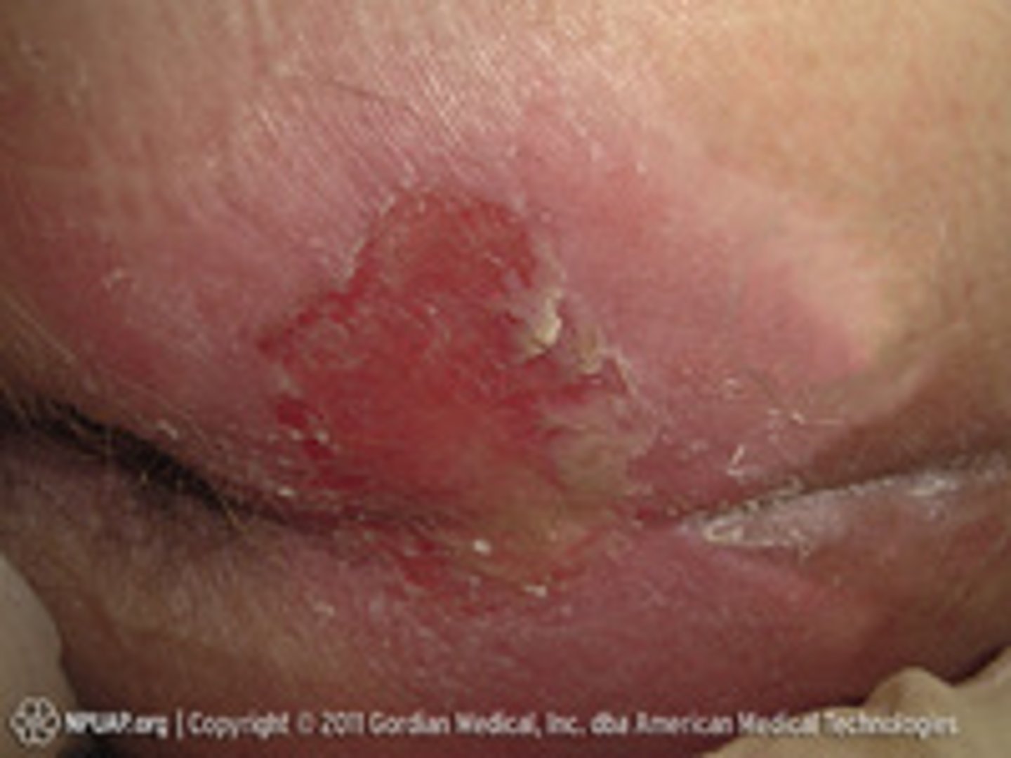
What's an example of erosion (superficial partial thickness)?
A superficial (first-degree) burn or a simple abrasion
Where does partial-thickness skin loss occur?
Dermis
What are the clinical signs of partial-thickness skin loss?
Unlike superficial wounds, partial-thickness wounds involve the highly vascular dermis, leading to bleeding.
What's the appearance of partial-thickness skin loss?
They often appear as a blister or a raw, moist wound bed. Because they do not penetrate the entire dermis, they do not typically show subcutaneous fat or deeper structures.
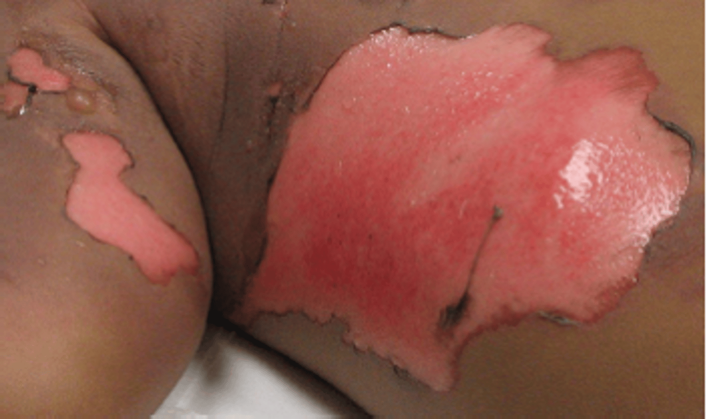
What's an example of partial-thickness skin loss?
Skin tears, Stage 2 pressure injuries, and superficial or deep second-degree burns
Where does full-thickness skin loss occur?
epidermis, dermis, and subcutaneous
What are the clinical signs of full-thickness skin loss?
These wounds may exhibit the exposure of bone, tendon, ligament, or muscle .
What's the appearance of full-thickness skin loss?
often looks like a crater or "pothole". You can differentiate the depth by the color of the exposed tissue:
o Subcutaneous (Adipose) Tissue: Glistening white to pale yellow
o Muscle: Bright/dark red and bleeds readily.
o Tendon/Ligament: Glistening white and fibrous.
o Bone: Shiny, smooth, and milky white.
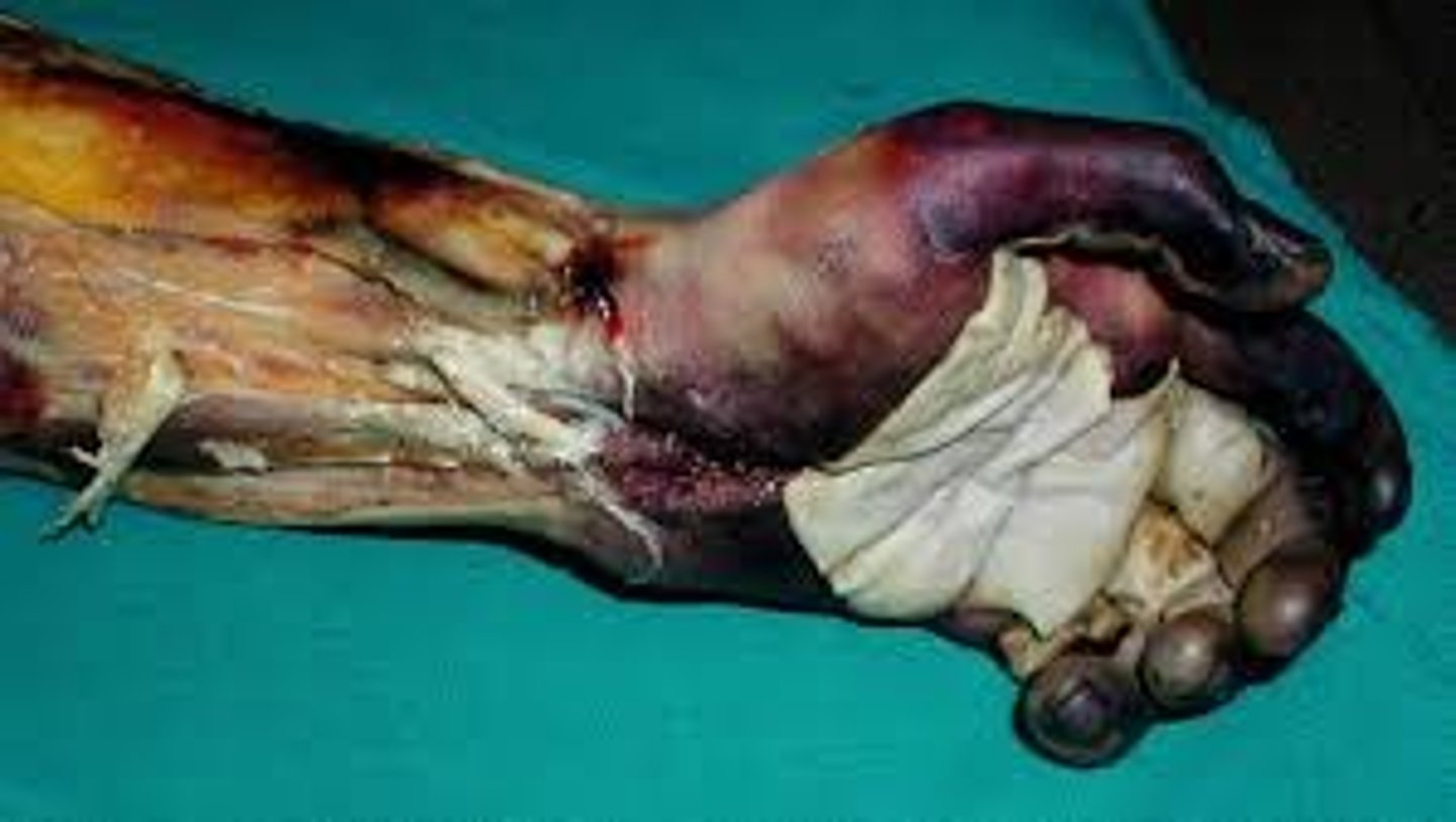
What's an example of a full-thickness skin loss?
Surgical incisions, "third-degree" burns, and Stage 3 or 4 pressure injuries