NURS 222 Exam 3
0.0(0)
Studied by 1 person Call Kai
 Learn
Learn Practice Test
 Spaced Repetition
 Match
Match Flashcards
 Knowt Play
Card Sorting
1/74

Earn XP
Description and Tags
Last updated 12:19 AM on 11/7/22
Name | Mastery | Learn | Test | Matching | Spaced | Call with Kai | Chat |
|---|
No analytics yet
Send a link to your students to track their progress
75 Terms
1
New cards
What does Confrontation Test test?
Tests Visual Field
2
New cards
How to perform Confrontation test
Stand 2 feet away. Both of you cover opposing eye. Wiggle finger and advance from periphery; several directions. You and patient should see object at same time.
3
New cards
What does Corneal Light Reflex test?
Assesses parallel alignment of eye axes And inspects extraocular muscle function
4
New cards
How to perform Corneal Light Reflex test
Shine light at bridge of nose, 12 inches away. Patient stares ahead. The light should reflect in exactly the same spot on each cornea.
5
New cards
What does the Cover test test?
Only done if corneal light reflex is asymmetric. Inspect Extraocular Muscle Function.
6
New cards
How to perform Cover test
Stare ahead, cover one eye with card-it should be a steady gaze. Next, uncover eye and look for steady gaze. Repeat. Should observe steady gaze in opposite eye as you cover. Should observe steady gaze in tested eye as you uncover it.
7
New cards
What does Diagnostic Positions test?
CN III, IV, VI. Inspect extraocular muscle function.
8
New cards
How to perform Diagnostic Positions test
Have patient keep both eyes open. Keep target 12 inches away from patient. “Follow my pen”. Move yo the 6 positions, hold, then back to center (2, 3, 4, 8, 9, 10 o’clock positions)
Should see parallel tracking or “OU” (= in both eyes)
Should see parallel tracking or “OU” (= in both eyes)
9
New cards
Inspect External Eyeball structures (General)
Observe that patient can move easily around room without groping.
10
New cards
Inspect external Eyeball structures (Eyebrows)
Observe that eyebrows are present. Eyebrows move symmetrically, no lesions.
11
New cards
Inspect external Eyeball structures (Eyelids and lashes)
Observe that upper lid can overlap superior iris. Closes completely. Lashes are even and curve outwards.
12
New cards
Inspect external Eyeball structures (Eyeballs)
Observe that eyeballs are aligned. No sunken or protrusion appearance.
13
New cards
Inspect external Eyeball structures (Conjunctiva and sclera)
Using thumbs, slide lower lid down. Eyeball should be moist and glossy. Conjunctivae should be clear revealing color below (should be pink over lower lids and white over sclera)
14
New cards
Inspect external Eyeball structures (Lacrimal apparatus)
Feel along upper lid for swollen gland. Press finger against sac, not nose. NO swelling should be present on upper lid. No liquid should be regurgitated from puncta.
15
New cards
Inspect Anterior Eyeball Structures (Cornea and lens)
Shine light from side and check for smoothness and clarity of cornea, anterior chamber, and lens. No cloudiness or irregular ridges should be found.
16
New cards
Inspect Anterior Eyeball Structures (iris and pupil)
Iris should be flat, round with even color throughout.
Pupils should be round, regular, and equal in size (~ 3-5 mm). Measure them
Pupils should be round, regular, and equal in size (~ 3-5 mm). Measure them
17
New cards
Inspect Anterior Eyeball Structures (Test pupillary light reflex)
Darken the room. Have patient gaze into distance. Advance penlight from side. Pupils should constrict (= direct light reflex)
Simulataneous constriction of other pupil (consensual) Record R 3/1= 3/1 L
Simulataneous constriction of other pupil (consensual) Record R 3/1= 3/1 L
18
New cards
Inspect Anterior Eyeball Structures (Accommodation)
Have patient focus on a distant object. “Shift your gaze to my finger” (~ 3 inches away). Pupils should constrict. Convergence of axes of eyes.
19
New cards
snellen test
a test of visual acuity using a Snellen chart
20
New cards
snellen test (results)
Patient stands 20 feet away to read chart. Numerator is 20, or however far away the patient stand to read the chart. Note smallest line where patient can read and record visual acuity for that line. This is the denominator. The larger the denominator, the poorer the patients vision is. 20/20 is normal.
21
New cards
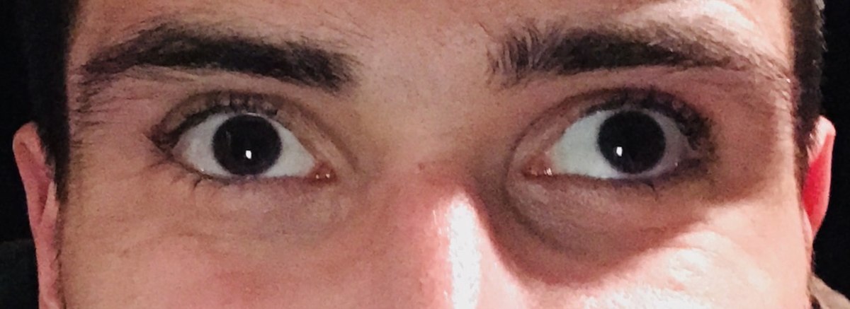
Strabismus
Is a congenital condition where both eyes do not focus on an object simultaneously; these eyes appear crossed. Impairment of the extraocular muscles or their nerve supply causes strabismus.
22
New cards
presbyopia
Impaired near vision in middle age and older adults caused by loss of elasticity of the lens and associated with the aging process.
23
New cards
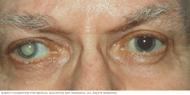
cataracts
increased opacity of the lens, which blocks light rays from entering the eye. Cataracts develop slowly and progressively after age 35 or suddenly after trauma.
24
New cards
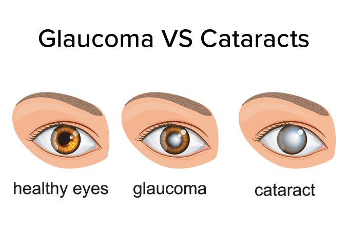
glaucoma
Intraocular structural damage resulting from elevated intraocular pressure. Obstruction of the outflow of aqueous humor causes this. Without treatment the disorder leads to blindness.
25
New cards
photophobia
A morbid fear of light. Tears, headaches, bright colored spots in vision
26
New cards
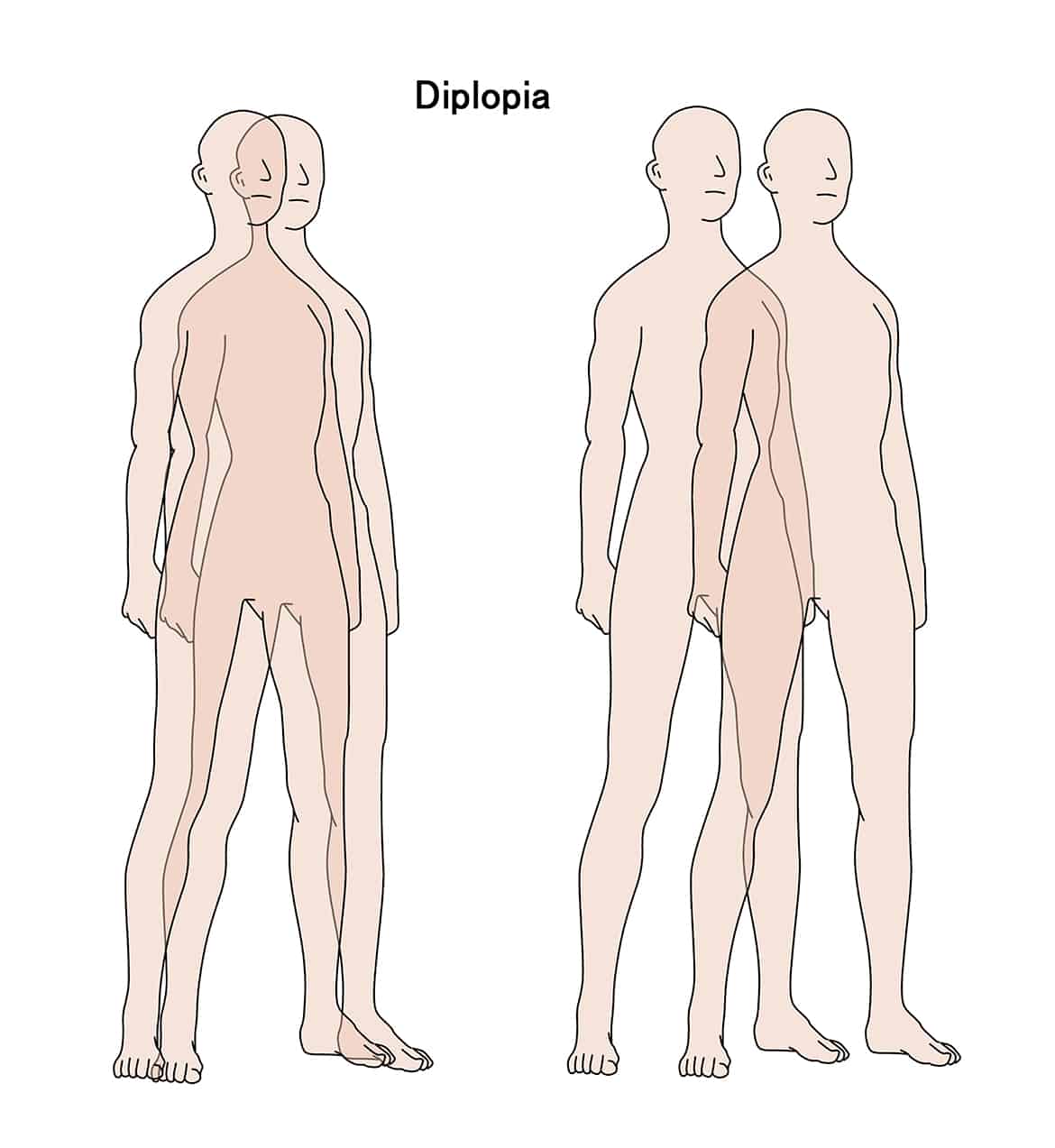
Diplopia
visual impairment in which an object is seen as two objects. Double vision.
27
New cards
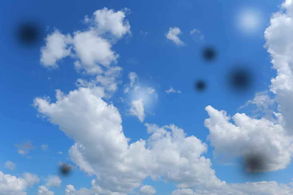
Floaters
spots in vision that look like black or gray specks or strings that drift across the eyes.
28
New cards
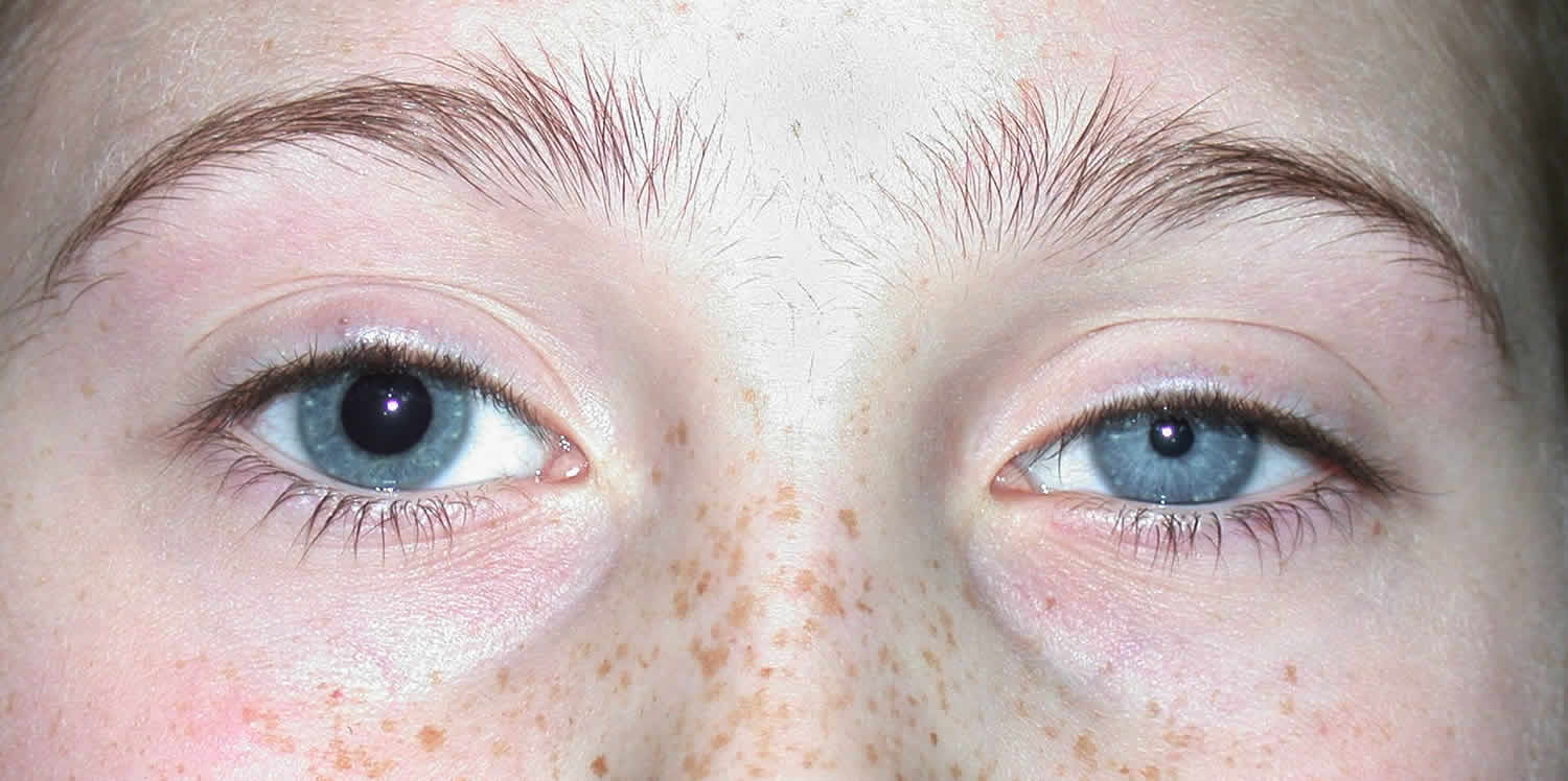
aniscoria
Unequal pupil sizes
29
New cards
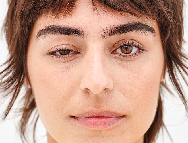
Ptosis
drooping of the upper eyelid caused by muscle paralysis and weakness
30
New cards
ear exam steps
1. Health History
Earaches/ear pain? Trauma to head? Any ear infections? Now or in past? Discharge from ears? Any hearing loss currently? Any loud noises at home or job? Any ringing/buzzing in ears? Taking any medications? Ever felt vertigo (subjective and objective? How do you clean your ears? Use ear protection from loud noises or while swimming? Last hearing test?
2. Inspect external ear
Size/Shape of auricle, Position and alignment on head, skin condition (color, lumps, lesions,), Check for movement of auricle and tragues for tenderness, evaluate external auditory meatus (size, swelling, redness, discharge, cerumen, lesions, foreign bodies)
3. Otoscopic examination
External Canal, cerumen, discharge, foreign bodies, lesions, redness or swelling of canal wall.
4. Inspect tympanic membrane
Should be pearly grey, shiny, translucent, flat. Membrane should be intact.
5. Test Hearing Acuity
CN VIII, whispered voice test
Earaches/ear pain? Trauma to head? Any ear infections? Now or in past? Discharge from ears? Any hearing loss currently? Any loud noises at home or job? Any ringing/buzzing in ears? Taking any medications? Ever felt vertigo (subjective and objective? How do you clean your ears? Use ear protection from loud noises or while swimming? Last hearing test?
2. Inspect external ear
Size/Shape of auricle, Position and alignment on head, skin condition (color, lumps, lesions,), Check for movement of auricle and tragues for tenderness, evaluate external auditory meatus (size, swelling, redness, discharge, cerumen, lesions, foreign bodies)
3. Otoscopic examination
External Canal, cerumen, discharge, foreign bodies, lesions, redness or swelling of canal wall.
4. Inspect tympanic membrane
Should be pearly grey, shiny, translucent, flat. Membrane should be intact.
5. Test Hearing Acuity
CN VIII, whispered voice test
31
New cards
presbycusis
age related hearing loss
32
New cards
Otosclerosis
Abnormal bone growth within the ear. Causes hearing loss due to the ear’s inability to amplify sound.
33
New cards
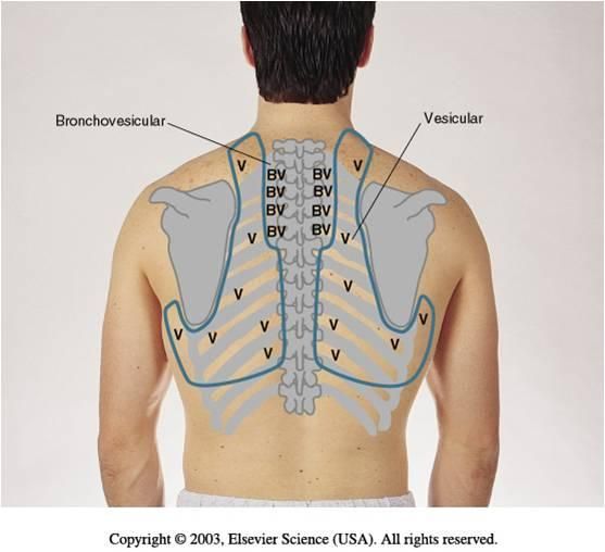
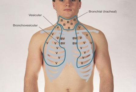
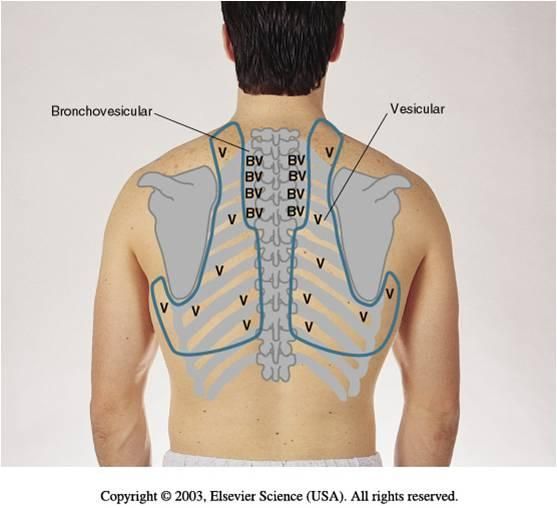
Vesicular breath sounds
Heard in peripheral and lateral lung fields as well as over clavicles.
Longer inspiration and shorter exhalation
Longer inspiration and shorter exhalation
34
New cards
Bronchial breath sounds
Heard over and around the trachea. Harsh. Shorter inspiration and longer exhalation.
35
New cards
Bronchovesicular
Heard over main bronchi anteriorly and posteriorly. Equal inspiration and exhalation
36
New cards
bruit
Audible vascular sound associated with turbulent blood flow.
37
New cards
Murmurs
Due to structural defects in valves
Blowing and swooshing sound
Velocity of blood increasing
Viscosity of blood decreasing
Blowing and swooshing sound
Velocity of blood increasing
Viscosity of blood decreasing
38
New cards
regurgitation
backflow of blood through a defective heart valve
39
New cards
tachycardia
abnormally rapid heartbeat
40
New cards
bradycardia
abnormally slow heartbeat
41
New cards
orthopnea
form of dyspnea in which the person can breathe comfortably only when standing or sitting erect
42
New cards
tachypnea
Fast breathing; > 20 breaths per minute
43
New cards
Bradypnea
Slow breathing; <12 breaths per minute
44
New cards
eupnea
normal relaxed breathing
45
New cards
Cheyne stokes respirations
Cycles of alternating apnea and hyperventilation associated with a critical condition
46
New cards
hyperpnea
energetic respiration that occurs normally after exercise or abnormally with fever or various disorders
47
New cards
apnea
Stopped respiration w/o breathing. Temporary cessation of breathing.
48
New cards
arteriosclerosis
Thickening and hardening of arterial walls
49
New cards
dyspnea
difficult or labored respiration
50
New cards
layers of heart wall (outside to inside)
Pericardium (surrounds and protects heart)
Myocardium (muscle wall, responsible for pumping and contracting)
Endocardium (lines inner surfaces of heart)
Myocardium (muscle wall, responsible for pumping and contracting)
Endocardium (lines inner surfaces of heart)
51
New cards
heave
move or cause to move in a specified way, direction, or position
52
New cards
pulmonary ventilation
process of air flowing into the lungs through inspiration and out of the lungs through expiration
53
New cards
Right sided heart failure
Usually happens due to a respiratory problem. Blood backs up into the body. Blood cannot go forward. Leads to peripheral edema.
Key signs: JVD, peripheral edema, enlarged spleen/liver
Key signs: JVD, peripheral edema, enlarged spleen/liver
54
New cards
left sided heart failue
Blood backs up to the lungs, blood cannot go forward to the body. Drowning in blood. Leads to crackles, dyspnea, orthopnea, confusion, memory loss
55
New cards
Path of blood flow from Vena Cava to heart
Vena Cava -> R. Atrium -> Tricuspid Valve -> R. Ventricle -> Pulmonary valve -> Pulmonary Artery -> Lungs -> Pulmonary Vein -> L. Atrium -> Mitral Valve -> L. Ventricle -> Aortic Valve -> Aorta
56
New cards
Conduction system of heart
1. SA Node (pacemaker)
2. AV node (delays signal to allow atria time to contract before ventricles stimulated
3. Bundle of His
4. R/L Bundle Branches (septum)
5. Purkinje Fibers: cause ventricles to contract (apex)
2. AV node (delays signal to allow atria time to contract before ventricles stimulated
3. Bundle of His
4. R/L Bundle Branches (septum)
5. Purkinje Fibers: cause ventricles to contract (apex)
57
New cards
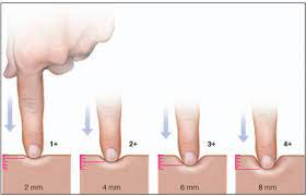
Peripheral Pulses
Brachial, radial, femoral, popliteal, dorsalis pedis, posterior tibial
Graded with +1, +2, +3, +4, +5 (+2 is normal)
Graded with +1, +2, +3, +4, +5 (+2 is normal)
58
New cards
Pretibial edema
caused by poor circulation or fluid retention.
59
New cards
Crackles
Popping/crackling in the lungs
60
New cards
wheezing
Musical-ish sounds
61
New cards
pleural friction rub
Leather rubbing together
62
New cards
Ronchi
Snoring sound
63
New cards
stridor
a whistling sound when breathing; indicates obstruction of the trachea or larynx
64
New cards
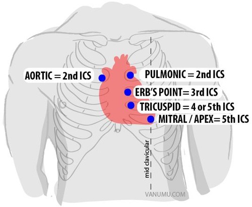
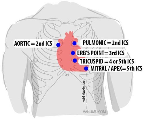
Aortic Area
Located 2nd intercostal space, right sternal border, S2 louder than S1
65
New cards
Pulmonic Area
Located 2nd intercostal space, Left sternal border, S2 louder than S1
66
New cards
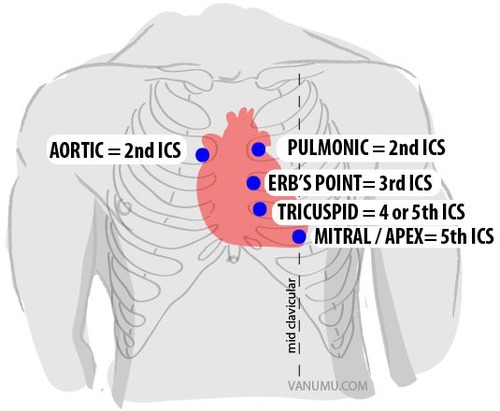
Erb’s Point
Located 3rd intercostal space, left sternal border, S1=S2
67
New cards
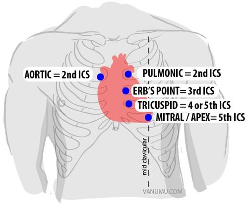
Tricuspid Area
Located 4th Intercostal space, left sternal border, S1 louder than S2
68
New cards
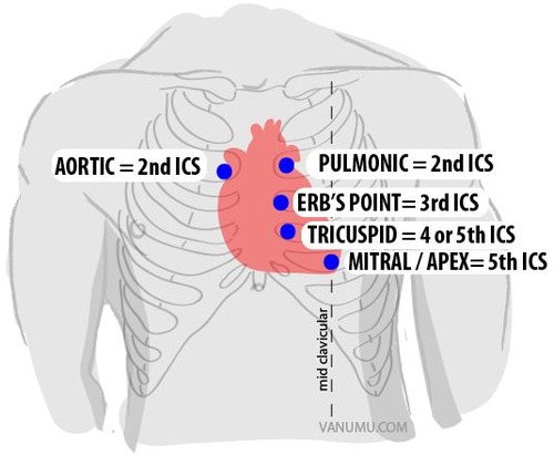
Mitral (apical) Area
Located 5th intercostal space, left midclavicular line, S1 louder than S2
69
New cards
Respiratory Assessment
1. Health History
Cough? Any shortness of breath? Any chest pain with breathing? Past history of lung disease? Cigarettes, age started, how many per day, for how long, have you tried to quit? Any living or work conditions that affect your breathing? Last Tb skin test, chest X-ray? Last flu vaccine?
2. Inspection
Any deformities of the thoracic cage? Respirations-rate, rhythm, accessory muscle use. Skin color and condition. Nails (clubbing). Person’s position. Facial expression. Level of consciousness (person, place, time, situation)
3. Palpation
Confirm symmetric expansion. Tactile Freitas (hands vertical on either side of spine, have patient speak). Skin temperature and moisture. Detect any lumps, masses, tenderness. Trachea.
4. Auscultation
Listen to posterior, lateral, anterior. Note any abnormalities.
Cough? Any shortness of breath? Any chest pain with breathing? Past history of lung disease? Cigarettes, age started, how many per day, for how long, have you tried to quit? Any living or work conditions that affect your breathing? Last Tb skin test, chest X-ray? Last flu vaccine?
2. Inspection
Any deformities of the thoracic cage? Respirations-rate, rhythm, accessory muscle use. Skin color and condition. Nails (clubbing). Person’s position. Facial expression. Level of consciousness (person, place, time, situation)
3. Palpation
Confirm symmetric expansion. Tactile Freitas (hands vertical on either side of spine, have patient speak). Skin temperature and moisture. Detect any lumps, masses, tenderness. Trachea.
4. Auscultation
Listen to posterior, lateral, anterior. Note any abnormalities.
70
New cards
Heart Assessment
1. Health History
Chest Pain/Tightness, shortness of breath, use more than one pillow to sleep, do you have a cough, tire easily, facial skin ever turn blue or ashen, swelling of feet/legs, awaken at night to urinate, past history of heart disease, change in usual/daily activities, medications? Smoking? Cardiac risk factors (diabetes, hypertension, high cholesterol, obesity, sedentary lifestyle, age)
2. Neck
Carotid Arteries: observe and palpate the R and L; grade, equality
3. Precordium
Skin color and condition. Chest wall pulsation. Location of apical impulse if able to feel. Can you see the impulse. Apical pulse, rate, rhythm.
4. Auscultation
Identify an atomic area to listen. identify S1 and S2, note any variation.
Chest Pain/Tightness, shortness of breath, use more than one pillow to sleep, do you have a cough, tire easily, facial skin ever turn blue or ashen, swelling of feet/legs, awaken at night to urinate, past history of heart disease, change in usual/daily activities, medications? Smoking? Cardiac risk factors (diabetes, hypertension, high cholesterol, obesity, sedentary lifestyle, age)
2. Neck
Carotid Arteries: observe and palpate the R and L; grade, equality
3. Precordium
Skin color and condition. Chest wall pulsation. Location of apical impulse if able to feel. Can you see the impulse. Apical pulse, rate, rhythm.
4. Auscultation
Identify an atomic area to listen. identify S1 and S2, note any variation.
71
New cards
Valves of heart
AV: Triscuspid (Right A/V) and Mitral (Left A/V)
Semilunar: Pulmonic and Aortic
Semilunar: Pulmonic and Aortic
72
New cards
Peripheral vertigo
impaired balance with the ability to walk
73
New cards
Central vertigo
more severe instability, cannot walk or stand without falling
74
New cards
Grading tonsils
+1: Visible
+2: Halfway between tonsillar pillars and uvula
+3: touching uvula
+4: touching each other
+2: Halfway between tonsillar pillars and uvula
+3: touching uvula
+4: touching each other
75
New cards
tongue normal findings
Should be pink, moist, roughened with papillae