Quiz 4 LO's: Infectious Diseases
1/110
There's no tags or description
Looks like no tags are added yet.
Name | Mastery | Learn | Test | Matching | Spaced | Call with Kai |
|---|
No analytics yet
Send a link to your students to track their progress
111 Terms
LO1: Define opportunistic infections
infections caused by organisms that are usually nonpathogenic, but something has changed so they can now cause disease
LO1: What are some things that allow an opportunistic infection to develop?
decrease in salivary flow
antibiotic administration
immune system alterations
LO2: What is the inflammatory response to the immune system like?
nonspecific, rapid first response to injury
includes cells of the innate immune system like neutrophils and macrophages
results in edema and accumulation of a large number of WBCs at the injury site
LO2: What is the immune response to the immune system like?
specific, humoral and cell-mediated, work to prevent injury and defend against m/os
has a memory
m/os are antigens in which specific antibodies are created to fight the specific antigens
LO3: What are some examples of opportunistic infections that can occur in the oral cavity?
Oral Candidiasis --> overgrowth of candida albicans
includes:
- pseudomembrane candidiasis
- erythematous candidiasis
- denture stomatitis
- chronic hyperplastic candidiasis
- angular cheilitiis
- chronic mucocutaneous candidiasis
- median rhomboid glossitis
LO4: What organism causes tuberculosis? Where does it go in the body?
infectious chronic granulomatous disease caused by mycobacterium tuberculosis
primary infection of the lungs or other areas like head, neck, larnyx, and cervical lymph node
LO4: What is the route of transmission for tuberculosis?
inhaled droplets of the bacteria lodge in the alveoli of the lungs --> cause fever, fatigue, cough, weight loss
after undergoing phagocytosis, they become resistant and multiply in the macrophages
they then flow into the blood stream and spread through the body via the vascular system
can be transmitted through dental health care personnel - avoid dental work with active TB
LO4: How is a diagnosis made for tuberculosis?
Oral lesions are identified by biopsy
Granulomas as the characteristic histopathological lesions of TB --> composed of areas of necrosis surrounded by macrophages, multinucleated giant cells, and lymphocytes
Skin tests with injection of purified protein derivative - if the body have a Type 4 hypersensitivity reaction, then the body knows the bacteria and chest radiographs are taken to determine if TB is active
How do we treat tuberculosis?
treat primary disease
antituberculosis agents --> isoniazid, rifampin, rifapentine
LO4: What organism causes actinomycosis?
actinomyces israelii
LO4: What is the route of transmission for actinomycosis?
infection often follows trauma, nonvital tooth, periodontal infection, tooth extraction, or abrasion of mucosa
LO4: How is a diagnosis made for actinomycosis?
known for the formation of abscess that drain by the formation of sinus tracts
organisms appear in the pus as tiny, yellow grains and are called sulfur granules
diagnosis is made by identifying the colonies in the tissue from a culture of the lesion
How do you treat actinomycosis?
long-term high doses of antibiotics --> amoxicillin or tetracycline
LO4: What organism causes syphillis?
treponema pallidum
LO4: What is the route of transmission for syphilis?
through direct contact of people, sexual contact, transfusion of infected blood, transfer from placenta to fetus
bacteria can penetrate mucous membranes, but need a break in the skin to invade there
LO4: Describe the primary stage of syphilis
lesion is called chancre --> highly infectious and forms at the site where bacteria enters the body, typically the lips (males = upper lip, women = lower lip)
Chancre, small size in soft tissue only
regional bilateral lymphadenopathy occurs
lesion heals spontaneously and enters a latent period
can be treated at this stage
LO4: Describe the secondary stage of syphilis
occurs 6 weeks after initial lesion
diffuse eruptions of skin and mucous membranes occur
skin lesions can appear as a rash & sore throat, fever, and lymphadenopathy also occur
oral lesions are mucous patches --> painless, grey plaques covering ulcerated mucosa and MOST INFECTIOUS
lesions heal spontaneously, but can recur for months or years
disease may be latent for years before entering stage 3
LO4: Describe the tertiary stage of syphilis
occurs years after initial infection if it has not been treated
involves problems with the CV, CNS, and vascular system, organ complications
lesion is called a gumma and is noninfectious --> commonly appears on the tongue and palate as a firm mass that becomes an ulcer
gumma can be destructive to the palatal bone
gumma can cause interstitial glossitis on the tongue
What is congenital syphilis? What does it cause in the fetus?
syphilis that is passed from mother to fetus
facial and dental abnormalities --> abnormally shaped teeth, deafness, saddle nose, frontal bossing, vaulted palate, ocular keratosis
LO4: How is a diagnosis made for syphilis?
SKIN: dark field examination to identify spirochetes
ORAL: Venereal Disease Research Lab test or fluorescent treponemal antibody absorption test
**must have the correct antibodies formed in the body for the test to be correct
How do we treat symphilis?
penicillin
LO5: What is the relationship between streptococcal tonsillitis, pharyngitis, scarlet fever, and rheumatic fever?
Many different organisms can cause tonsillitis and pharyngitis, but when they are caused by group A beta-hemolytic streptococci, it is very serious because of its relationship with scarlet and rheumatic fever
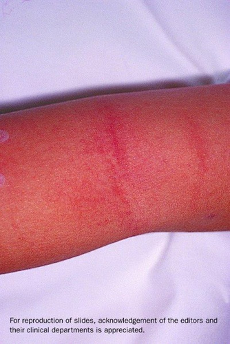
Scarlet fever: 103 fever for a week, generalized red skin rash, pastia lines, strawberry tongue, and abdominal pain, headaches and nausea
Rheumatic fever: antibodies are made to the cell wall of the bacteria and the antibodies react with different tissues causing inflam involving heart, joints, and CNS, permanent damage to heart valves
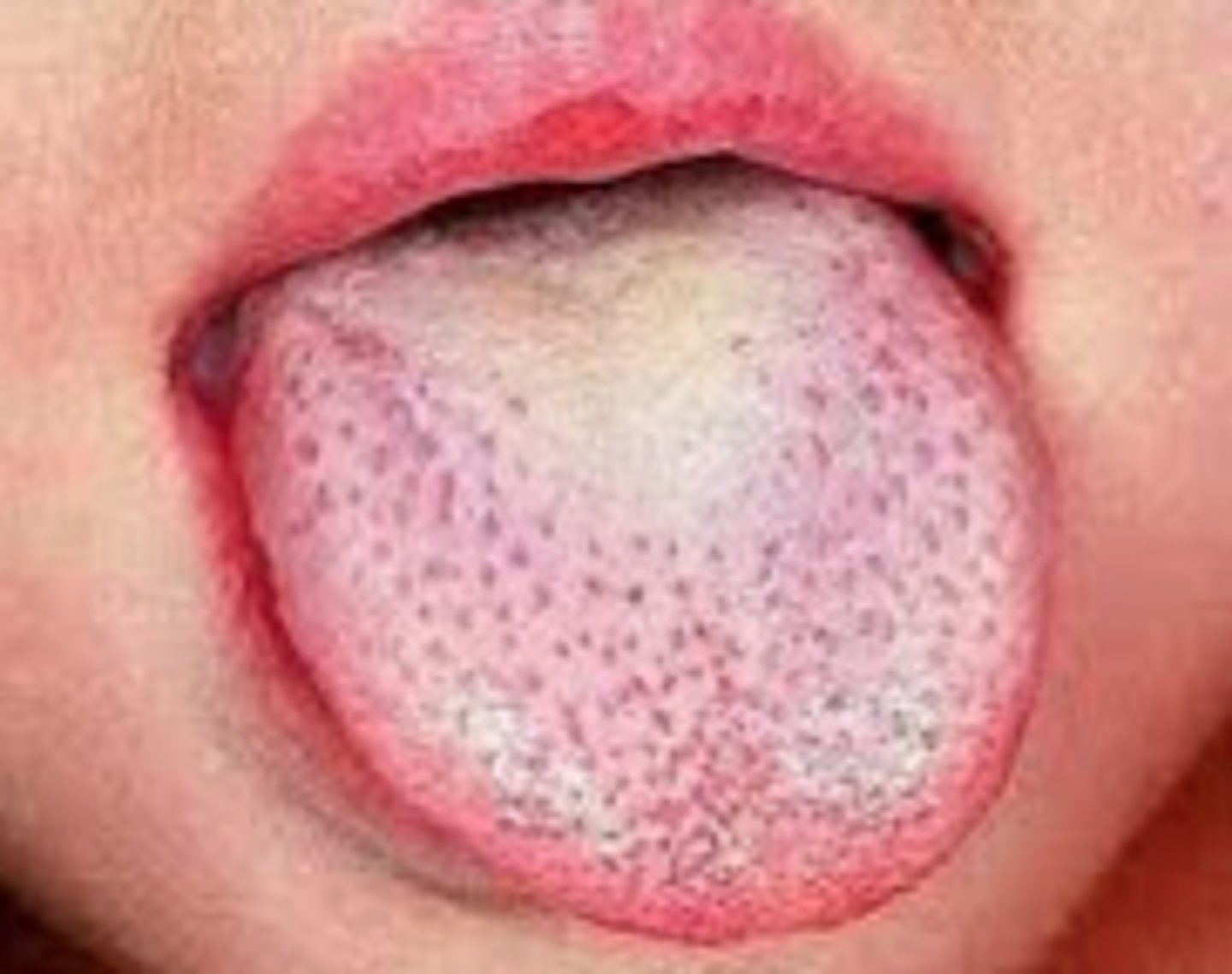
LO6: What disease are pastia lines are strawberry tongue associated with?
scarlet fever
LO6: What is strawberry tongue?
fungiform papillae are red and prominent
dorsal surface initially has a white coating and then sheds and becomes red
LO6: What are pastia lines?
transverse red streaks seen in areas of skin folds
caused by capillary fragility
What type of infection is impetigo?
bacterial infection
What causes impetigo?
Staphylococcus aureus or Streptococcus pyogenes
What part of the body does impetigo affect?
skin - face and extremities
areas of trauma
What are the signs of impetigo?
red blisters and sores
fluid filled vesicles or crusted lesions
may look like finger nail scratches
itchy, swollen lymph nodes, fever
How do we treat impetigo?
topical or systemic antibodies
What kind of infection is tonsillitis and pharyngitis?
bacterial vs viral infection
What bacteria causes tonsillitis and pharyngitis?
streptococcus pyogenes
What are the signs of tonsillitis and pharyngitis?
enlarged tonsils
erythema mucosa
onset of fever, sore throat, dysphagia
How do we treat tonsillitis and pharyngitis?
systemic antibody if bacterial --> penicillin V or amoxicillin
azithromycin or clindamycin is allergic to the above
What is necrotizing ulcerative gingivitis?
chronic, painful erythematous gingivitis with punched out papilla
bad breath, spontaneous bleeding, rapid infection, decreased resistance to infection, fever, swollen lymph nodes
What causes necrotizing ulcerative gingivitis?
spirochetes and fusiform bacilli
How do we treat necrotizing ulcerative gingivitis?
debridement and antibiotic rinse and medications
What is pericoronitis?
an inflammation of the gingiva around a partially erupted tooth
What causes pericoronitis?
result of infection by bacteria that proliferate in the pocket between the soft tissue and crown
having a suppressed immune system increase the risk
What is the most common area for pericoronitis and what happens here?
md third molars
an operculum --> soft tissue flap covering the distal portion of the tooth, becomes inflamed due to opposing molar and impaction of food under the soft tissue flap
How do we treat pericoronitis?
debridement and irrigation of the pocket, antibiotics, extraction of tooth
What is acute osteomyelitis?
acute inflammation of the bone and bone marrow
What are the signs of acute osteomyelitis?
pain, swollen lymph nodes
What causes acute osteomyelitis in the jaw?
periapical abscess as a result of fracture, surgery, or bacterium
How do we treat acute osteomyelitis?
draining exudate and antibiotics
What is chronic osteomyelitis?
long standing inflammation of bone
may be related to pts taking biphosphonates
What are the signs of chronic osteomyelitis?
bone is painful and swollen
x rays show a diffuse and irregular radiolucency that can become radiopaque due to bone growth
How do we treat chronic osteomyelitis?
debridement and systemic antibiotics
What type of infection is candidiasis?
fungal infection
most common oral lesion associated with immunodeficiency
What causes candidiasis?
overgrowth of candida albicans
What can cause the overgrowth of candida albicans?
antibiotics, corticosteroids, diabetes, cell mediated immune system deficiency
What are the types of oral candidiasis?
Pseudomembranous candidiasis
Erythematous candidiasis
Denture stomatitis
Chronic hyperplastic candidiasis
Angular cheilitis
Chronic mucocutaneous candidiasis
Describe pseudomembranous candidiasis
white, curd like material that can be wiped off the mucosa
underlying mucosa is red and may bleed
burning and metallic taste may occur
Describe erythematous candidiasis
red, painful mucosa
irregular, patchy, depapillation of tongue
Describe denture stomatitis
also called chronic atrophic candidiasis
limited to the area covered by denture, typically palate or mx alveolar ridge
red mucosa, petechias (small colored lesion), or more generalized and granular
asymptomatic
Describe chronic hyperplastic candidiasis
(also called candidal leukoplakia and hypertrophic candidiasis)
white lesion that does not rub off mucosa
if caused by candidiasis, it will respond to antifungal medication
Describe angular cheilitis
swollen, red patches at the corners of the mouth
Describe chronic mucocutaneous candidiasis
severe form of candidiasis occurring in immunocompromised pts
skin lesions, nails, skin folds, chronic oral and genital mucosal candidiasis
oral lesions appear psuedomebranous, erythematous, hyperplastic candidiasis, or angular cheilitis
Are median rhomboid glossitis and candidiasis related?
in some cases, median rhomboid glossitis is treated with antifungal medications, but other times it doesn't work
so not associated and the cause median rhomboid glossitis is not clear
How can we treat candidiasis?
antifungal medication
What are deep fungal infections?
Histoplasmosis
Coccidioidomycosis
Blastomycosis
Cryptococcosis
usually involves the lungs --> oral lesions are caused by implantation of organism carried by sputum from lungs to oral mucosa
systemic antifungal medications to treat
What are the rare fungal infections?
Mucormycosis: m/os in soil and affect diabetic or debilitated patients, affects nasal cavity / hard palate
Aspergillosis: mold, affects immunocompromised pts in lungs and sinuses and appears as a mycetoma - mass of aspergillosis
What kind of infection is HPV?
viral, 130 diff types
creates neoplasia or benign lesions
How does HPV occur?
direct contact affecting skin and oral mucosa
must infect the basal cells of epithelium, requiring a break in surface of tissue
matures in spinous layer where virus is then released onto surface of the tissue
What are HPV-infected cells called?
Koilocytes
irregular nucleus surrounded by clear cytoplasm
What is verruca vulgaris?
common wart, papillary oral lesion or skin lesion caused mainly by HPV 2
What is a common site for verruca vulgaris?
lips
What does verruca vulgaris appear as?
white, papillary, exophytic lesion
How do we treat verruca vulgaris?
excision
What is condyloma acuminatum? Causes?
benign papillary lesion/ warty growth on genital skin
due to HPV type 6 or 11
sexual contact
What does condyloma acuminatum look like?
pink, papillary, bulbous masses anywhere in oral mucosa
How do we treat condyloma acuminatum?
excision
What is multifocal epithelial hyperplasia? What does it look like?
associated with HPV types 13 and 32
multiple white/pink nodules in oral mucosa
resolve on their own
What is the difference between HSV type 1 and 2?
Type 1 causes oral lesions, initial and secondary forms
Type 2 causes genital lesions
What is primary herpetic gingivostomatitis? (acute herpes simplex infection)
initial oral infection with the herpes simplex type 1 virus
What does primary herpetic gingivostomatitis look like?
painful, red, swollen gingiva and multiple tiny vesicles on perioral skin, vermillion border of lips, and oral mucosa
become ulcers
systemically appears as fever, malaise, and swollen lymph nodes
Define subclinical infections
infection that is present without symptoms
herpes simplex virus can be this
What is recurrent herpes simplex infection?
after primary infection, herpes simplex virus persists in a latent state in the trigeminal ganglion and causes localized recurrent infections
What are signs of recurrent herpes simplex infection?
appear on vermilion border of the lips called herpes labials or cold sore/fever blister
reappear due to sunlight, menstruation, fever, etc
intraorally, ulcers attached to keratinized tissue and bone and irregular borders
can affect eyes and fingers
With herpes simplex virus, the amount of virus present is highest in what stage?
vesicle stage
How do you treat herpes simplex virus?
antiviral drugs --> acyclovir
What is varicella zoster virus?
human herpescirus 3, causes CHICKEN POX or SHINGLES
What is chickenpox?
highly contagious disease appearing as vascular and pustular eruptions on skin and mucosa membranes
systemic signs are headache, fever, and malaise
mostly in children
What is shingles? (also called herpes zoster)
highly contagious disease appearing as unilateral, painful eruptions of vesicles along the sensory nerve
mostly in adults, often occurs in association with immunodeficiency or malignancies
What are the phases of shingle?
INITIAL: severe neuralgia and acute burning and paine
MIDDLE: clusters of vesicles along affected sensory nerve
FINAL: postherapeutic neuralgia
What branches of the trigeminal neuralgia are responsible for oral lesions when affected by shingles?
mx and md
What branches of the trigeminal neuralgia are responsible for skin lesions when affected by shingles?
opthalmic
How do you treat varicella zoster viruses?
antiviral or corticosteroids
preventative vaccinations
What is Epstein-Barr virus (EBV)?
human herpesvirus 4, disease in the oral cavity
infectious mononucleosis, hairy leukoplakia, mucocutaneous ulcer
What is infectious mononucleosis?
kissing disease, associated with atypical activated T lymphocytes
sore throat, fever, swollen lymph nodes, splenomegaly, malaise, fatigue
palatal petechiae, skin rashes
What is hairy leukoplakia?
Irregular, corrugated (ridges, grooves) white lesion most commonly occur on the lateral borders of the tongue
EBV found in epithelial cells
associated with HIV or immunocompromised pts
What is mucocutaneous ulcer?
when associated with EBC, it is a persistent, nonhealing ulcer
seen in immunocompromised pts that have received transplants, are HIV positive, or in older adults
What are the types of coxsackievirus infections? How are they spread?
herpangina, hand/foot/mouth disease, and acute lymphonodular pharyngitis
spread thru fecal/oral contamination, saliva, and respiratory droplets
usually resolved on their own
What is herpangina?
vesicles on soft palate, fever, malaise, sore throat, difficulty swallowing, erythematous pharyngitis
What is hand/foot/mouth disease?
children under 5 yrs old
oral lesions anywhere in the mouth, painful ulcers and vesicles
multiple macules and papules on skin
What is acute lymphonodular pharyngitis?
Hyperplastic lymphoid tissue of the soft palate or tonsillar pillars appears as yellowish or dark pink nodules
fever, sore throat, headaches
Can measles and mumps cause oral manifestations?
yes
Measles --> koplik spots or small red macules with with necrotic centers
Mumps --> infection of salivary glands and swelling of parotid glands and epithelial tissues
How is HIV transmitted?
blood, semen, vaginal secretions, breast milk
How are AIDS and HIV related?
HIV causes AIDS
What immune system cell does HIV target?
helper T cell, CD4 carrier