Examples of Joint II
1/24
There's no tags or description
Looks like no tags are added yet.
Name | Mastery | Learn | Test | Matching | Spaced | Call with Kai |
|---|
No analytics yet
Send a link to your students to track their progress
25 Terms

Hip Joint
Components: Head of femur ↔ acetabulum of hip bone
Classification: Synovial (ball-and-socket)
Movement: Flexion, extension, abduction, adduction, medial rotation, lateral rotation, circumduction

Hip Joint: Articular Capsule
Strong capsule surrounding the ball-and-socket joint of the hip, reinforced by several ligaments
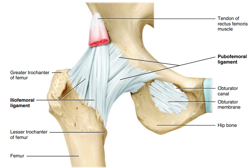
Hip Joint: Iliofemoral Ligament
Strongest, thickened ligament in the body to prevent hyperextension of the hip.

Hip Joint: Pubofemoral Ligament
Thickened portion of the articular capsule that prevents excessive abduction of the thigh and strengthens the joint.

Hip Joint: Ischiofemoral Ligament
Thickened portion of the articular capsule that strengthens the hip joint and limits excessive movement.
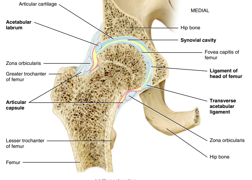
Hip Joint: Ligament of the Head of the Femur
Flat, triangular band that connects the femur to the acetabulum and carries a small blood vessel to the femoral head.

Hip Joint: Acetabular Labrum
Fibrocartilage rim of acetabulum that deepens the acetabulum and increases hip stability.

Hip Joint: Transverse Acetabular Ligament
Strong ligament that bridges the acetabular notch and supports the acetabular labrum.

Knee Joint
Components: Femur ↔ tibia and patella
Classification: Synovial (modified hinge)
Movement: Flexion, extension, and medial and lateral rotation
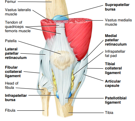
Knee Joint: Articular Capsule
A mostly incomplete capsule of the knee joint formed primarily by surrounding muscle tendons and their expansions, with only some true capsular fibers directly connecting the articulating bones.
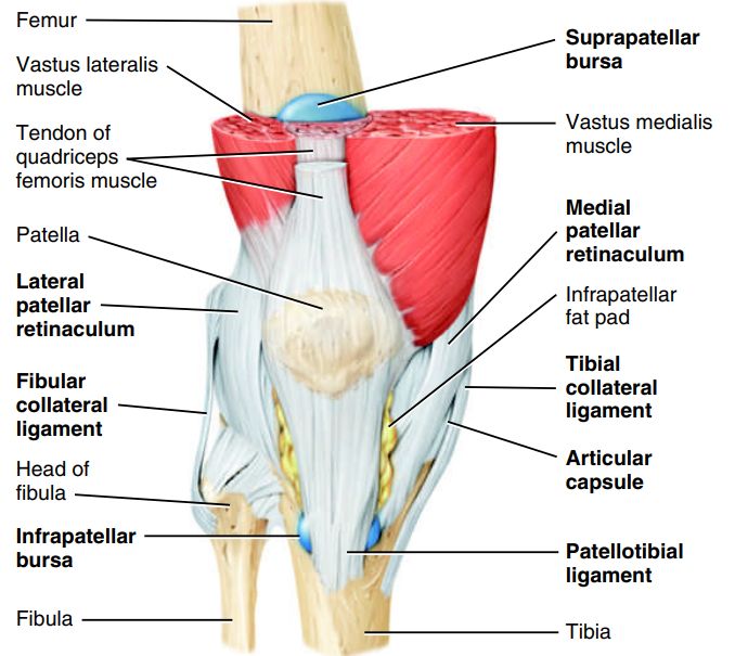
Knee Joint: Medial and Lateral Patellar Retinacula
Fused tendons from the quadriceps femoris and fascia lata (thigh fascia) that help stabilize and strengthen the front (anterior) of the knee joint.
Knee Joint: Patellotibial Ligament
Continuation of the quadriceps tendon from patella to tibial tuberosity that strengthens the front of the knee and is separated from the joint by an infrapatellar fat pad.
Knee Joint: Oblique Popliteal Ligament
Broad, flat ligament extending from the femur (intercondylar fossa and lateral condyle) to the tibia (head and medial condyle); strengthens the posterior knee joint.
Knee Joint: Arcuate Popliteal Ligament
Ligament extending from the lateral femoral condyle to the styloid process of the fibular head; strengthens the lower lateral posterior knee joint.
Knee Joint: Tibial Collateral Ligament
Broad, flat, medial knee ligament from femur to tibia; stabilizes inner knee and attaches to the medial meniscus.
Knee Joint: Fibular Collateral Ligament
Strong, rounded ligament on the lateral knee ligament from lateral condyle femur to fibular head; stabilizes outer knee.
Knee Joint: Intracapsular Ligaments
Two strong, crossed ligaments inside the knee joint capsule (ACL and PCL) that connect the tibia and femur and cross each other to help stabilize the knee.
Knee Joint: Anterior Cruciate Ligament (ACL)
Ligament inside the knee that connects the intercondylar tibia to the lateral condyle femur and prevents the tibia from sliding too far forward.
Knee Joint: Posterior Cruciate Ligament (PCL)
Ligament inside the knee that connects the intercondylar tibia to the medial condyle femur and prevents the tibia from sliding too far backward, especially when walking downhill or downstairs.
Knee Joint: Articular Discs (Menisci)
Two fibrous cartilage discs between the tibial and femoral condyles to absorb shock, circulate synovial fluid, and distribute weight
Knee Joint: Medial Meniscus
Knee Joint: Lateral Meniscus
Knee Joint: Prepatellar Bursa
Knee Joint: Infrapatellar Bursa
Knee Joint: Suprapatellar Bursa