Nervous System PP’s
1/116
There's no tags or description
Looks like no tags are added yet.
Name | Mastery | Learn | Test | Matching | Spaced | Call with Kai |
|---|
No analytics yet
Send a link to your students to track their progress
117 Terms
Nervous System Divisions
Central Nervous System (CNS)
• Brain
• Spinal Cord
Peripheral Nervous System (PNS)
• All neural tissue outside of the CNS (Commonly referred to as your nerves)
Central Nervous System CNS
Integrates, processes and coordinates sensory data and motor commands
Peripheral Nervous System PNS
Carries sensory info from periphery to CNS
Carries responsive Info from CNS to peripheral tissues + systems
PNS Subdivisions
Sensory
• Brings sensory information to CNS
Motor
• Brings motor commands from CNS
Afferent (SENSORY)
toward CNS from receptors
Somatic sensory receptors
Position, touch, pressure, pain and temperature sensations.
Special sensory receptors
Smell, taste, vision, balance and hearing
Part of somatic sensory division
Visceral sensory receptors
Monitors internal organs
Efferent (MOTOR)
away from CNS to effectors
Somatic NS (SNS)
Provides voluntary control over skeletal muscles
Autonomic NS (ANS)
Provides involuntary regulation of smooth muscles, cardiac muscle, glands and adipocytes
Nervous System Cells
NEURONS
Receive stimuli and transmit action potentials
Can’t divide
GLIAL
Support, nourish, and protect neurons
Can divide
Neuron Anatomy
Cell body (Soma)
Dendrites
input portion, receives
Axons
output portion, sends
Axoplasmic Transport
Movement of materials through axon along neurotubules
Bidirectional
Retrograde Flow
From synaptic terminal to cell body
Synapse
Site of communication between neuron and another cell
Neurons: Structural Classes
Anaxonic neuron
Bipolar neuron
Unipolar neuron
Multipolar neuron
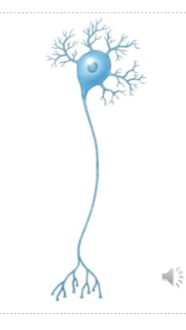
Multipolar Neuron
2/+ dendrites
Single axon
Most common CNS neuron
PNS motor neurons
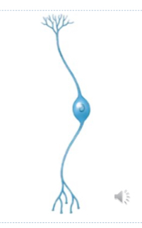
Bipolar Neuron
One dendritic process
W/ extensive distal branch
One axon
W/ several telodendria and synaptic terminals
Rare; special senses
Eyes, Nose, Ears
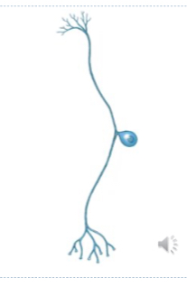
Unipolar Neuron
Dendrite Continuous with axon
Soma off to one side
Initial segment located where dendrites fuse
PNS sensory neurons
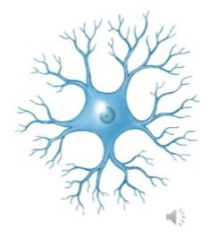
Anaxonic Neuron
Cannot differentiate between axons and dendrites (star like)
Brain, Special sense neurons
Function not yet understood
local, graded potentials
Which nervous system division would be responsible for receiving touch and pressure information from your fingers?
(Afferent) Somatic Nervous System
Subdivision of PNS
What type of neurons would you be likely to find within your nose?
Bipolar Neurons
(Olfactory Receptor Neurons)
If one's neurons within said nose were to be damaged or lost, can they be replaced? Why or why not?
Yes, Olfactory Sensory Neurons within nose can be replaced
Neurons: Functional Classes
Sensory Neurons
Interneurons
Motor Neurons
Sensory (Afferent) Neurons
PNS Afferent division
Carry info from sensory receptors to CNS
Unipolar
Cell bodies in sensory ganglia
Afferent fibers from sensory receptor to CNS
Afferent Neurons
Somatic Sensory Neurons
Monitor external conditions
Monitor body/limb position
Visceral Sensory Neurons
Monitor internal conditions
Monitor organ systems
Sensory Receptors
Process of specialized sensory neurons OR cells monitored by sensory neurons
Interoceptors
Proprioceptors
Exteroceptors
Interoceptors
Monitor digest, resp, cardiovasc, urinary, & reproduct systems
Provide Sensations of distention, deep pressure & pain
Exteroceptors
Carry info about external environment
Touch, Temp, Press, Sight, Smell, Taste, Hear, Balance
Proprioceptors
Monitor position and movement of skeletal muscles and joints
Motor (Efferent) Neurons
PNS efferent division
Carry instruct from CNS to peripheral effectors
Somatic motor neurons
Visceral motor neurons
Somatic Motor Neurons
Somatic subdivision of efferent PNS
Multipolar Neurons
Innervate skeletal muscles
Under voluntary control
Somatic Motor Neurons
Soma
In CNS
Axon
Extends to periphery to innervate skeletal muscles
Visceral Motor Neurons
Autonomic subdivision of efferent PNS
Innervate smooth muscle, cardiac muscle, glands, and adipose tissue
Under involuntary control
Visceral Motor Neurons (VMN)
Soma in CNS
Axon Innervate second set of VMN at autonomic ganglion
2nd VMN
Solar in autonomic ganglion
Axon Innervates peripheral effectors
Interneurons
Brain, Spinal cord, Autonomic ganglia
Interconnect other neurons
Receive/distribute sensory info
Coordinate motor commands
Higher brain functions
The more complex response, more Interneurons involved
Neuroglia of CNS
Ependymal cells
Astrocytes
Microglia
Oligodendrocytes
Ependymal Cells
Line brain ventricles and central canal of spinal cord
Produces CSF
Some may be ciliated
Astrocytes
Largest and most numerous
Create structural framework
Preform repairs
Guide neuron development
Control interstitial environment
Maintain homeostasis:
Secrete chem that make capillaries of CNS impermeable
Lipid soluble substances cross freely
Water-soluble substances must be actively transported
Microglia
Smallest and rarest
Mobile cells:
Migrate to areas of injured nerve tissue
Engulf invading microbes and dead cell debris
Oligodendrocytes
Have cytoplasmic extensions that wrap around axons
Form the myelin sheaths of neurons
Neuroglia of PNS
Satellite cells
Schwann Cells
Satellite Cells
Amphicytes
Surround neuron cell bodies in ganglia
Regulate interstitial environment
Schwann Cells
Neurilemmocytes
Surround axons
Form a sheath around the axon
Myelin Sheath (CNS & PNS)
Internodes
Areas of axons covered by myelin sheath
Nodes of Ranvier
Gaps between internodes
White Matter Vs Gray
White: Myelinated axons
Gray: Unmyelinated axons
Cell bodies and dendrites
Why do you think it can be important for nerves to have a sheath around them?- what happens to those whose myelin sheathes are damaged/destroyed?
crucial for Insulating nerves, increasing speed of signal, and protecting nerve fibers
-Slow/Block signals, MS Disease
Transmembrane Potential
Difference in electrical charge between inside and outside of cell membrane (voltage)
Outside +
Inside -
Banana in Ocean
Element Charges
K +
Na +
Ca +
Cl - Only negative
ECF VS ICF
OUT
High Na, Cl-, Ca
IN
High K, proteins-
Ion Channels
Protein tunnels for specific ions
3 Types
Leak, Gated, Active Transport proteins
Leak (PASSIVE) channels
Always open
More K leak than Na leak
50-100x more permeable to K than Na
More K leaks out than Na in
Gated Channels
Open and close in response to stimuli
Usually closed at resting potential
Types of Gated Channels
3: Chemically, Voltage, Mechanically
Chemically (LIGAND) Gated Channels
Open in response to specific channels
Locations: cell body and dendrites
Voltage Gated Channels
Open in response to changes to transmembrane potential
have an activation gate and inactivation gate
Found in excitable membranes
Axons (as well as skeletal muscles cells and cardiac muscle cells)
Mechanically Gated Channels
Open response to membrane distortion
Locations: sensory receptors
Touch, press, vibration
Gated (ACTIVE) Channels Summary
Chem Gated (Dendrites, Cell Bodies)
Mech Gated (Sensory receptors)
Voltage Gated (Axons, Axon Terminals)
Active Transport Proteins
Use ATP to push molecules against conc. gradient
ex: Na+/K+ pump
Transmembrane Potential
Passive forces acting across plasma membrane
Chemical Gradients
Diff conc. of specific ions (Na, K, Ca, Cl)
Chem gradient tries to equalize concentrations via diffusion
Electrical Gradients
Difference in positive and negative charges and outside the cell
Result in potential difference / resting potential
Neurons -70mv
Active forces acting across plasma membrane
Na/K ATPase/exchange pump
Establishing Transmembrane Potential
Established and maintained at -70mv by
Sodium potassium ATPase pump
Ion channels
Na+ / K+ Exchange Pump
Moves 3 Na ions out for every 2 K in
Inside gets less positive or more negative
Na/K pump
Na wants to move into cell
K wants to move out of cell
Must move through ion channels
TMP & electrochemical gradients
K ions
Chemical gradient stronger than electrical gradient
Wants to move out
Na ions
Chemical gradient combined with electrical gradient
Wants to move in
Equilibrium Potential
Neurons transmembrane potential at which there is no net movement of ions across a membrane
Resting Membrane Potential
Transmembrane potential of a resting (Unstimulated) neuron
Usually about -70 mV
Changes in Transmembrane Potential
Rises or falls in response to temporary changes in membrane permeability to Na and K
Open/Close Na and K gated Channels
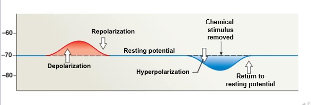
Membrane Potential Changes
Depolarization
Repolarization
Hyperpolarization
Depolarization
Gated Na Channels open
Na enters cell
Membrane potential becomes more positive (less negative)
Repolarization
Gated Na channels close
Na/K pumps move Na back out of cell
Membrane potential returns to resting level -70 mV
Hyperpolarization
Gated K channels open
K exits cell
Membrane potential becomes more negative (-80 mV)
Return to resting potential
Gated K channels close
Na/K pumps move K back into cell
Membrane potential returns to resting level (-70 mV)
Electrical Signals
Graded potentials
Affect only small portion of cell membrane
Allows communication over short distances
Action potentials
Affect the entire surface of cell membrane
Allows communication over long distances
Electrical Signals Sections
Graded Potentials
Dendrites, Cell bodies, Synaptic terminals
Action Potentials
Axons
Graded Potential Phases
4:
Resting,
Stimulation,
Depolarization
Repolarization
OR
Hyperpolarization
Return to Resting
GP: Resting Cell
Transmembrane potential -70mV
Chem regulated Na Channels closed
GP: Stimulation
Membrane exposed to chemical
Chem regulated Na Channels open
Na ions begin to enter cell
GP: Depolarization
Movement of Na into cell depolarizes membrane (At stimulation site)
Potential moves from -70mV to towards 0 (more positive, less negative)
Movement of Na into cell also produces local current that depolarizes adjacent membrane segments
GP: Repolarization
Chemical (stimulus) removed
Chem regulated Na Channels close
Na/K pump removes Na from cytosol
Transmembrane potential -70mV
GP Stimulation Strength & Distance
The stronger stimulation:
more chem regulated Na channels open
more Na ions enter cell
greater degree of depolarization
larger area is depolarized
The greater distance from stimulation:
smaller effect on adjacent membrane segments
Only produce local effects
Electrical Signals
Graded Potentials
Affect only small portion of cell membrane
Allows communication over short distances
Action Potentials
Affect entire surface of cell membrane
Allows communication over long distances
Threshold
Transmembrane potential at which action potential begins
Happens around -55mV
Degree of depolarization that must occur by GP to start an action potential
Once threshold reached, action potential triggered
All or Nothing
Action Potentials
6 Phases:
Resting
Threshold
Depolarization/ Activate
Repolarization/ Inactivate
Hyperpolarization
Return to Rest
Resting Phase
Transmembrane potential around -70mV
Voltage gated Na channels are closed
Activation gate is closed
Inactivation gate is open (still no movement of Na ions)
Threshold Phase
Graded Potential (in AXON HILLOCK) depolarizes initial segment of axon to threshold (-55mV)
Depolarization/ Activation
Voltage gated Na channels open
Activation gate opens
Na ions rush through membrane, causing depolarization within axon
Repolarization/ Inactivation
When membrane potential reaches about +30mV
Voltage gated Na ion channels inactivate
(Inactivation gate closes)
Membrane potential reaching +30mV,
Voltage gated K ion channels open
K rushes out of cell, repolarizing membrane of axon
Repolarization Phase Membrane
As membrane potential reaches around -40mV,
Voltage gated Na channels reset:
Activation Gate closes
Inactivation Gate opens
Hyperpolarization
As membrane potential reaches around -90mV,
All voltage gated K ion channels close
membrane is still hyperpolarized
Return to Resting Phase Membrane
Na/K pumps returns transmembrane potential back to resting point (-70mV)
Pumps Na back out, pumps K back in

Refractory Period
Period of time during which another action potential cannot be generated
Begins with Depolarization
Ends when Repolarization is near completion
Absolute Refractory Period
Membrane cannot respond to another stimulus
Why? - Na Channels are already open / inactivated
Relative Refractory Period
Membrane potential almost normal
Na channels in ready state
K channels are not yet closed
Larger than normal stimulus can initiate action potential
Membrane is still hyperpolarized (-90mV)
Action Potential Propagation
Movement of action potential
From axon hillock, along axon, ends at synaptic terminals
Continuous or Saltatory
Action Potential Propagation Cont VS Salt
Continuous
Unmyelinated axons
Slower
Every Portion of Axon membrane must depolarize
Saltatory
Myelinated axons
Faster
Only Axon membrane at nodes of Ranvier must depolarize
Continuous Propagation EXP
Domino effect
Propagation can only occur in one direction. Why do you think thats the case? (Keep in mind steps of involved in action potential)
Due to refractory period of voltage gated Na
Saltatory Propagation EXP
‘Jumping’ to each Node of Ranvier only with sections covered in Myelin
Myelin prevents flow of ions across membrane
local current skips over internode and depolarizes node of Ranvier
Saltatory = Saltar- Leap
Multiple Sclerosis
Causes damage to myelins and supporting cells
Affects normal neural signaling and possible degradation of axon itself
Common clinical symptoms
Vision probs, Sensory disturb, Motor weak, Imbalance
Propagation Speed
The larger the axon diameter, the faster the action potential moves.
Type A (position, balance, vision, motor, commands)
Large; myelinated; over 300mph
Type B (sensory, other than vision)
Medium; myelinated; around 40mph
Type C (smooth muscle, gland secretion)
Small; Unmyelinated; about 2mph
If action potentials are all or nothing, how does sensation become more intense?
Through frequency coding and population coding
Stronger stimulus = neurons fire more frequently/S, recruits larger number of sensory neurons to fire simultaneously
Synapse
Site of communication between neuron and another cell
Synapse Sections
Presynaptic neuron
Synaptic cleft
Postsynaptic neuron
Types of Synapses
Electrical
Direct physical contact between cells
NS (rare) - eye, areas of brain, some ganglia
Chemical
Signal transmitted across a gap (synaptic cleft) by chemical neurotransmitters
Electrical Synapse
Action potential always produced in postsynaptic cell
Chemical Synapse
Most common - In all synapses between neurons and other cells
Cells not in direct contact → synaptic cleft