Anterior Compartment
1/28
There's no tags or description
Looks like no tags are added yet.
Name | Mastery | Learn | Test | Matching | Spaced | Call with Kai |
|---|
No analytics yet
Send a link to your students to track their progress
29 Terms
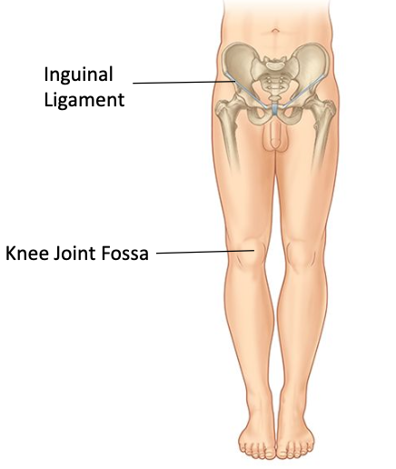
Where does it start and end?
Inguinal ligament to knee joint
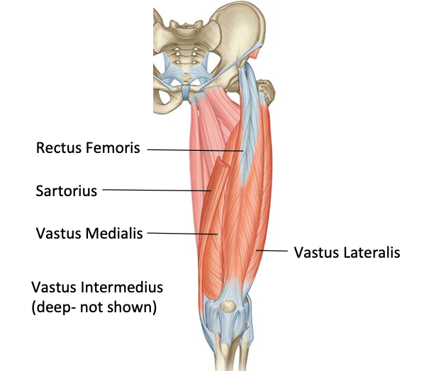
Anterior thigh muscles
Sartorius, quadriceps femoris (rectus femoris, vastus lateralis, vastus medialis, vastus interdmedius). Psoas major and iliacus
Anterior thigh - muscle action
Hip flexion and knee extension
Anterior thigh - nerves
Femoral nerve (supplies all muscles except the psoas major)
Anterior thigh - blood supply
Profunda femoris artery
Psoas major
O: transverse process, vertebral bodies, discs of T12 - L5
I: lesser trochanter
Nerve supply: Lumbar plexus
Nerve roots: anterior rami of L1, L2, L3

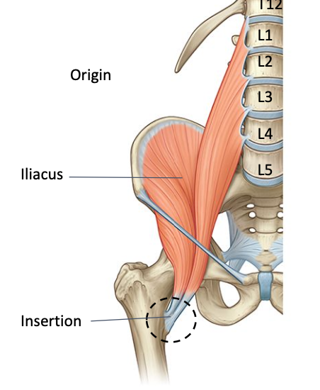
Iliacus
O: iliac foss
I: lesser trochanter
Nerve supply: femoral nerve
Nerve roots: L2, L3
Iliopsoas - action
hip flexion (from standing). Sit ups - where hip flexes on a fixed femur
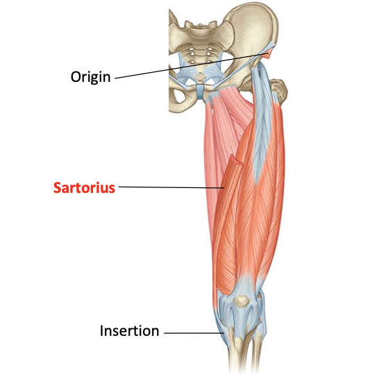
Sartorius
O: anterior superior iliac spine
I: anteromedial surface of proximal tibia
Nerve supply: femoral nerve
Nerve roots: L2, L3
Quadriceps
Recturs femoris, vastus lateralis, vastus medialis, vastus intermedius
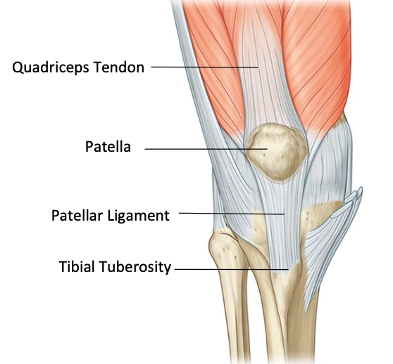
Quadriceps insertion - extensor mechanism
All 4 insert onto tibial tuberosity via extensor mechanism: quadriceps tendon, patella, patellar ligament, tibial tuberosity
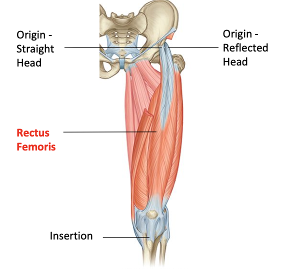
Rectus femoris
O: Straight head (anterior inferior iliac spine) reflected head (upper margin of acetabelar rim)
I: tibial tuberosity via quadriceps mechanism
Nerve supply: femoral
Nerve roots: L2, L3, L4
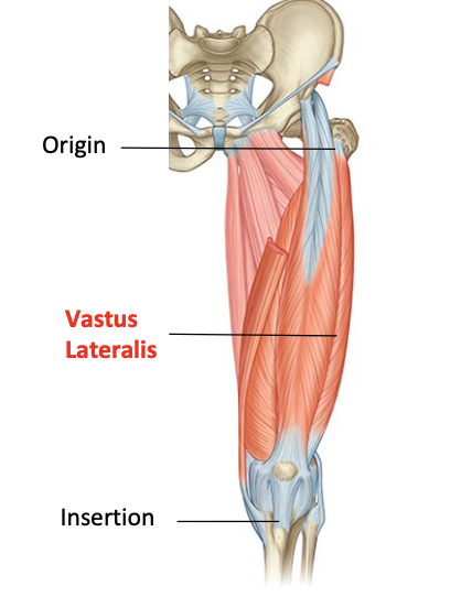
Vastus lateralis
O: gluteal tuberosity, lateral intertrochanteric line, ½ of linea aspera
I: tibial tuberosity via quadriceps mechanism
Nerve supply: femoral
Nerve roots: L2, L3, L4
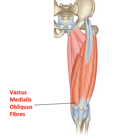
Vastus medialis
O: inferomedial intertrochanteric line, medial lip of linea aspea, 1/3 of medial supracondylar line
I: tibial tuberosity via quadriceps mechanism
Nerve supply: femoral
Nerve roots: L2, L3, L4
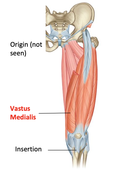
vastus medialis - obliquus fibres
Fibres run horizontally
Some consider this vastus medialis obliquus muscle
Helps track the patella medially
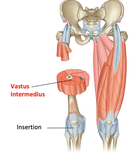
Vastus intermedius
O: upper 2/3 of anterior and lateral shaft of femur
I: tibial tuberosity via quadriceps mechanism
Nerve supply: femoral
Nerve roots: L2, L3, L4
Action - quadriceps muscle
All 4 does knee extension
Action - rectus femoris
Hip flexion/knee extension
Functions - quadriceps muscle
Walking, stairs, in and out of chair
The femoral nerve - motor supply
Anterior compartment of thigh: rectus femoris, vastus intermedius, vastus medialis, vastus lateralis, pectineus, sartorius
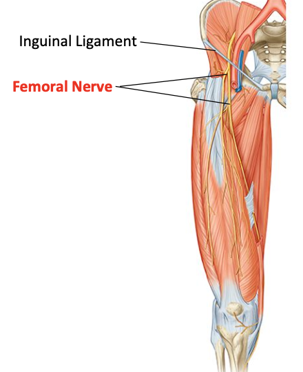
Femoral nerve - cutaneous supply
Skin on front of anterior thigh
Femoral and profunda femoris artery
Femoral and profunda main in anterior thigh compartment
Profunda: anterior, medial and posterior compartments
Femoral artery
Continuation of external iliac artery, going to anterior thigh femoral triangle, descends to anteromedial thigh
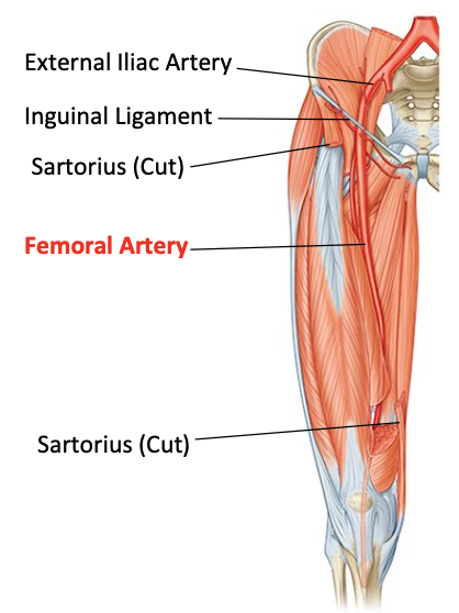
What is the profunda artery also called?
deep artery of the thigh
Profunda femoris artery - perofrating branches
Descends deep into thigh in 3-4 branches
Travels to medial compartment of thigh
pierce openings in adductor magnus to enter posterior compartment of thigh
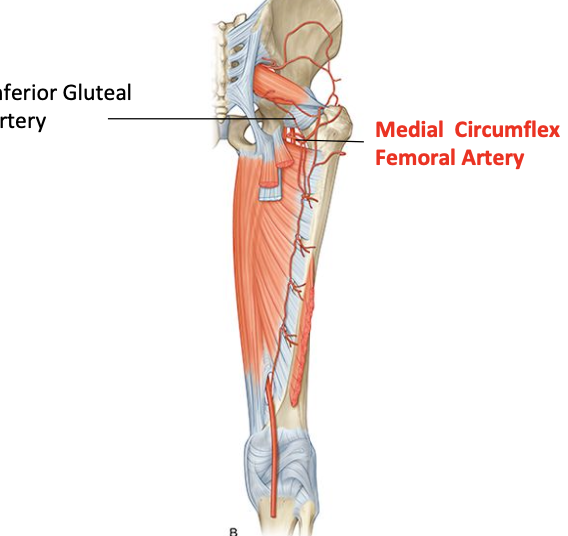
Medial circumflex femoral artery
Travels into back of thigh
Joins with inferior gluteal artery
Provides important branches to back of femur
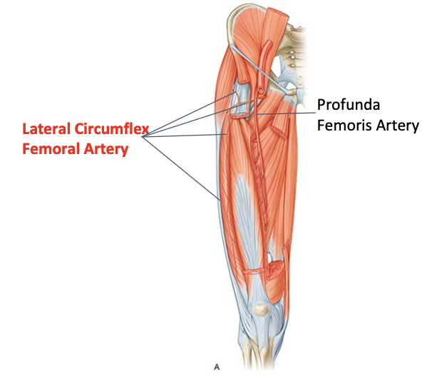
Lateral circumflex femoral artery
Tramsverse, ascending and descending branches
Supplies adjacent quadriceps, head and neck of femur, anastomes around the knee
What muscles crosses the hip joint?
Iliacus and psoas major
What muscles crosses the knee?
3 vasti (only extend the knee joint)
Rectus femoris crosses both hip and knee joint