animal health - diagnostics
1/62
There's no tags or description
Looks like no tags are added yet.
Name | Mastery | Learn | Test | Matching | Spaced | Call with Kai |
|---|
No analytics yet
Send a link to your students to track their progress
63 Terms
what is diagnostic testing
the process by which we can identify the presence of a disease or condition
what is diagnostic testing used for
detecting diseases or pathogens
identify a specific type or strain
monitor the progression of a disease
assess the severity of the disease
guide treatment plans and biosecurity
monitor success of treatment
types of diagnostic testing
clinical exam
blood tests - counts, smears, biochem
urine + faecal tests
cytology + histopathology
more types of diagnostic testing
imaging techniques
microbiological tests - bact + viral culture, fungal and parasite ID, PCR
molecular diagnostics - PCR, sequencing
immunological tests - serology, ELISA
clinical exam
initial process - vet will evaluate animal health - sings, symptoms + physical findings
history
observation
palpation
auscultation
percussion
how does diagnostic testing support clinical exam
confirm clinical suspicions
differentiate between sim conds
identify sub clinical or hidden conds
informs approp treatment plans
diagnostic imaging
visualise the internal structs of animals to aid in diagnostics + treatment
detect conds which not visible
non invasive
X-ray - assess?
bones
chest cavity
abdomen
inc: fractures, infections, lung diseases + tumours
pro of xrays
widely available
cost effective
quick results
con of x-rays
2D imaging - makes examining soft tissue difficult
ultrasound
sound waves produce real time images
abdominal + cardiac imaging
ideal for soft tissues + liver, kidneys, heart + rep
pro of ultrasound
no radiation exposure
non invasive
con of ultrasound
limited view of bone structs
CT imaging
cross sectional 3D images
detailed imaging of the brain, bones, soft tissues
good for detecting tumours, fractures + neurological disorders
MRI imaging
uses magnetic fields + radio waves
create highly detailed images of soft tissues
used for imaging the brain, spinal cord + joints
Light microscopes
only objects larger than wavelength of light are visible - e.g. nucleus
sides stained to enable colour to be viewed
process of staining + fixing kills cells
transmission electron microscopes
smaller objects visible - mitochondria, ribosomes
usually monochrome
not possible to view living specimens
expensive
scanning electron microscope
coats the sample w molecular layer of heavy metal e.g. gold
monochrome
not poss to view living specimens
3D
histology
microscopic exam of tissues from the body
histology - biopsy sample
collect from live animal - e.g. in surgical procedure
samples are abnormal areas of growth
histology - necropsy sample
samples from dead animal - from post mortem
can inc wide array of tissue w suspected pathologies/lesions
tissue collection
should be from fresh specimens
should use sharp blade to cut specimen out
tissue should be approp thickness w lesion inc
should fix immed after collection
aims of tissue fixation
preserves a sample as close to its nat state
prevent postmortem degeneration
stop bact growth
harden the tissues - fix causes coagulation of proteins
facilitate subsequent tissue staining
a good fixative
preserve cell quick w/ shrinkage or swelling
penetrate tissue rapidly
inhibit bact decay + autolysis
harden tissue + render it insensitive to subsequent treatment
allow tissue to be stored for long time
simple to prep + economical to use
tissue cassette
cut small sections of fixed tissue w sharp knife
put in a special tissue cassette
wash 3h in water
tissue processing
dehydration in acl series - 8h
clearing in xylene - 3h
wax infiltration - 1h
dehydration
water must be removed before it is infiltrated w wax
serues of acl solutions used
sequence ensures water is replaced w alc progressively to avoid tissue distortion
embedding
molten wax
helps orientate tissue for sectioning
avoid air bubbles in the section
sectioning
wax block is fixed on to the microtome
expose tissue by trimming wax out
section 5-7nm thick sections
lay on to a water bath at 40C
lift onto glass slides
dry on drying rack + then in oven at 60C
staining
haematoxylin - basic dye - stains acidic structures a purplish blue - nucleus
eosin - acidic dye - stains basic structs red or pink e.g. cytoplasm
special staining
used to explore tissues/specimens in detail - e.g. presence of certain molecules/microorgs, distribution + intensity
microorgs special staining
gram
giemsa
acid fast - mycobacteria
connective tissue special staining
toluidine blue
trichrome stain
carbohydrates special staining
alician blue
acid shiff
histopathology
microscopic exam of diseased tissue allowing for accurate diagnosis of certain diseases
histopathology fixation + staining
tissues must initially be fixed onto the slide so that the cells dont decompose after cell death
trad use formalin
brain tissue, adrenal gland + eye tissue staining
bouins
karnovsky
diseases could be picked up thru histopathology
arthritis/osetoarthritis
leukaemia
immunohistochemistry
labelled antibodies used to detect antigens win tissues
can be used to check for signs of disease often following a biopsy
serology
exam of blood - cells, antibodies, antigens
what is serology used for
to detect whether an animal has been infected w a pthogen
determine immunity
test for prev exposure
e.g. Feline Leukemia Virus to detect proteins from the virus
types of serology tests 1
enzyme linked immunosorbent assay (ELISA)
western blot
agar gel immunodiffusion AGID
types of serology tests 2
Indirect fluorescent antibody test IFAT
complement fixation test CFT
rapid diagnostic tests
serology - ELISA
used to detect antigens or antibodies in the serum
highly sensitive + widely used for disease diagnosis
examples of when ELISA used
test for parvovirus
leptospirosis
heartworm
serology - western blot
used to detect specific proteins from a pathogen
gel electrophoresis will sep proteins for visual identification
microbiological testing
bacterial + viral culture - selective media
parasite identification
PCR
PCR - polymerase chain reaction
rapid technique of amplifying a specific DNA sequences
DNA heated to high temp
sep into single strands but reforms helix when cooled
uses in vitro enzyme catalysed DNA synthesis to create mills of identical copies of DNA
PCR Method 1
extracted DNA is added to a solution containing:
Primers
nucleotide bases
DNA polymerase
primers
short DNA fragments complimentary to the target region
nucleotide bases
A
C
G
T
DNA polymerase
e.g Taq polymerase isolated from Thermus Aquatics found in hot water springs
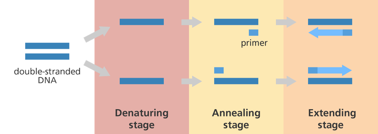
PCR Method step 1
Denaturing
heating to 94C causing DNA to separate into single strands
PCR method step 2
Annealing
cooling to 45C allowing primers to bind to the ends of ssDNA
PCR method step 3
Extension
heating to 72C causing DNA polymerase to synthesise new complementary strands to all ssDNA
PCR cycles
the 3 steps are cycled over + over to reach the required amplification
the DNA is doubled at each cycle
32 cycles = 1 bill times amplification
1 cycle = 17 secs
identification
gel electrophoresis
sequencing/ DNA profiling
DNA profiling
the use of molecular genetic methods to determine the exact genotype of a DNA sample to distinguish one org from another
how might DNA profiling be helpful
species determination for diagnosis
crime scene investigations
genetic testing
steps in DNA profiling
sample collection
DNA extraction
DNA amplification - PCR
identification + analysis
considerations
importance of contamination
use of controls - pos and neg control
also good to inc a DNA extraction control
why choose one over another
type of disease or infection
stage of disease
reliability of tests - sensitivity + specificity
cost
availability
speed
should we use more than one?
when clinical signs are ambiguous we may use more than one tests
make test results more reliable
to confirm the prev diagnosis
monitor disease progression
surveillance purposes