Exchange surfaces
1/17
There's no tags or description
Looks like no tags are added yet.
Name | Mastery | Learn | Test | Matching | Spaced | Call with Kai |
|---|
No analytics yet
Send a link to your students to track their progress
18 Terms
The need for specialized exchange surfaces
The need for specialized exchange surfaces
SA:V ratio = Surface Area ÷ Volume
As organisms get larger, SA:V decreases.
Single-celled organisms: high SA:V, diffusion is sufficient.
Multicellular organisms: low SA:V and higher metabolic activity, so diffusion alone is insufficient.
Multicellular organisms need specialised exchange surfaces to efficiently absorb substances and remove wastes.
Features of efficient exchange system
Large surface area → increases rate of exchange (e.g. root hair cells).
Thin surface → short diffusion distance (e.g. alveoli - one cell thick).
Good blood supply and/or ventilation → maintains a steep concentration gradient (e.g. alveoli, gills).
Mammalian gaseous exchange system
Nose/Mouth → Trachea → Bronchi → Bronchioles → Alveoli
Cartilage: C-shaped rings in the trachea (plates in bronchi); keep airways open and prevent collapse.
Smooth muscle: Surrounds airways; contracts to narrow them, relaxes to widen them (constriction and dialation).
Ciliated epithelium: Cilia beat mucus up towards the throat to be swallowed or removed.
Goblet cells: Produce mucus to trap dust and pathogens.
Elastic fibres: Stretch during inhalation and recoil during exhalation.
Trachea
C-shaped cartilage rings keep airway open and prevent collapse.
Goblet cells secrete mucus to trap dust and pathogens.
Ciliated epithelium moves mucus towards the throat.
Smooth muscle allows flexibility.
Elastic fibres stretch and recoil during breathing.
Bronchi
Cartilage plates support airways and prevent collapse.
Goblet cells produce mucus.
Ciliated epithelium wafts mucus to the throat.
Smooth muscle regulates airflow.
Elastic fibres aid recoil.
Bronchioles
No cartilage as support is unnecessary in small airways.
Little/no goblet cells to prevent mucus blockage.
Some ciliated epithelium present.
Thick smooth muscle layer controls airway diameter (bronchodilation/bronchoconstriction).
Elastic fibres help air movement.
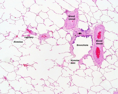
Alveoli
One-cell-thick squamous epithelium gives a short diffusion distance.
Very large surface area from millions of alveoli.
Dense capillary network maintains steep concentration gradients.
Elastic fibres allow expansion and recoil.
No cartilage, cilia, goblet cells, or smooth muscle as they would hinder gas exchange.
Inhalation mechanisms in mammals
External intercostal muscles contract; internal intercostals relax.
Rib cage moves up and out.
Diaphragm contracts and flattens.
Thoracic volume increases → lung pressure decreases below atmospheric pressure.
Air moves into the lungs.
Exhalation mechanisms in mammals
External intercostal muscles relax; internal intercostals contract (forced exhalation).
Rib cage moves down and in.
Diaphragm relaxes and domes upwards.
Thoracic volume decreases → lung pressure increases above atmospheric pressure.
Air moves out of the lungs.
Functions of respitory structure
Rib cage: Protects lungs and changes thoracic volume.
External intercostal muscles: Raise ribs during inhalation.
Internal intercostal muscles: Lower ribs during forced exhalation.
Diaphragm: Flattens to increase thoracic volume and domes to decrease it.
Vital Capacity, Tidal Volume, Breathing Rate & Oxygen Uptake
Tidal volume (TV): Volume of air inhaled or exhaled in one normal breath.
Breathing rate (BR): Number of breaths per minute.
Vital capacity (VC): Maximum volume of air that can be exhaled after a maximum inhalation.
Oxygen uptake: Amount of oxygen absorbed by the body per unit time.
Minute ventilation = Tidal volume × Breathing rate
Exercise → ↑ oxygen demand → ↑ oxygen uptake → ↑ breathing rate + ↑ tidal volume.
Spirometer
A device that measures lung volumes and capacities by recording the volume of air inhaled and exhaled (exhaled breath CO2 often absorbed by soda lime to reduce toxicity)
Produces a spirogram (graph of lung volume against time)
Height of waves = tidal volume.
Number of waves per minute = breathing rate.
Maximum breath volume = vital capacity.
During exercise: tidal volume and breathing rate increase.
Data Logger
An electronic device that records physiological data continuously using sensors (e.g. oxygen uptake, breathing rate, heart rate).
Higher readings = greater value of the variable being measured.
Steeper increase = faster change.
During exercise:
Oxygen uptake increases.
Breathing rate increases.
Often tidal volume increases.
Look for trends, peaks, and comparisons between rest and exercise
Bony fish structures
Buccal cavity: Chamber in the mouth that pumps water over the gills.
Operculum: Bony flap covering the gills; helps create pressure changes to move water.
Gill filaments: Long structures providing a large surface area.
Gill lamellae (gill plates): Thin folds one cell thick on filaments; site of gas exchange with a rich blood supply; many lamellae on many gill filaments providing large SA:V.
Ventilation mechanism bony fish
Water enters
Mouth opens, operculum closes.
Floor of buccal cavity lowers.
Buccal cavity volume increases.
Pressure decreases.
Water flows into the mouth.
Water forced over gills
Mouth closes, operculum opens.
Floor of buccal cavity rises.
Buccal cavity volume decreases.
Pressure increases.
Water is forced over gill lamellae and exits through the operculum.
Bony fish gas exchange
Water passing over lamellae contains a higher O₂ concentration than the blood.
Oxygen diffuses from water → blood.
Carbon dioxide diffuses from blood → water.
Thin lamellae one cell thick and rich capillary network make diffusion rapid (increases concentration difference).
Water flows in one direction, blood flows in the opposite direction.
At every point along the lamella, water has a higher O₂ concentration than the blood.
This maintains a steep concentration gradient (parallel) across the entire gill.
Histology mammalian ventilation structures
Bronchus has cartilage and much larger than bronchiole