Openstax Anatomy & Physiology Chapter 8
1/109
There's no tags or description
Looks like no tags are added yet.
Name | Mastery | Learn | Test | Matching | Spaced | Call with Kai |
|---|
No analytics yet
Send a link to your students to track their progress
110 Terms
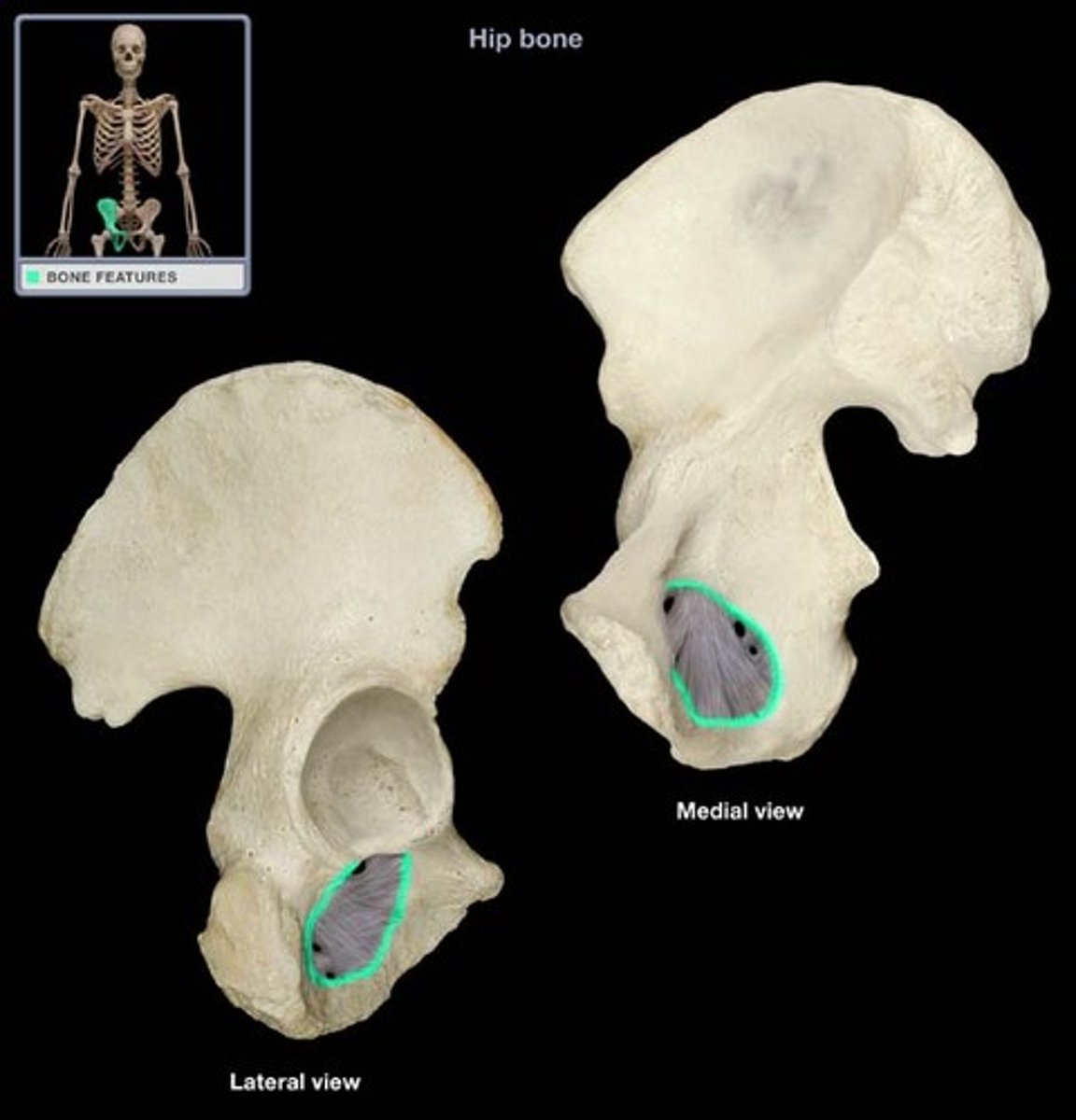
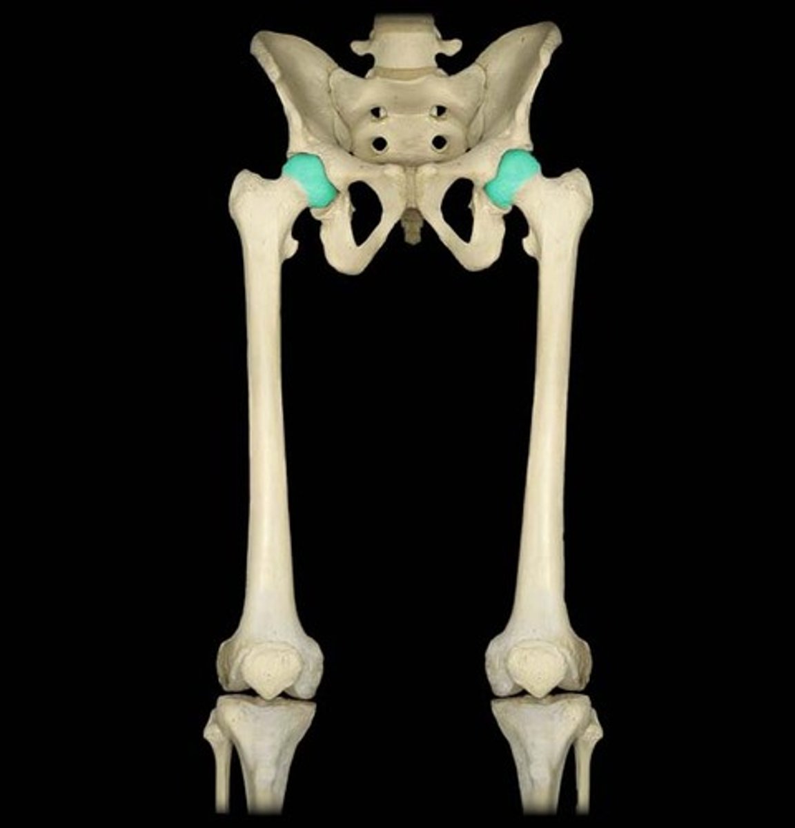
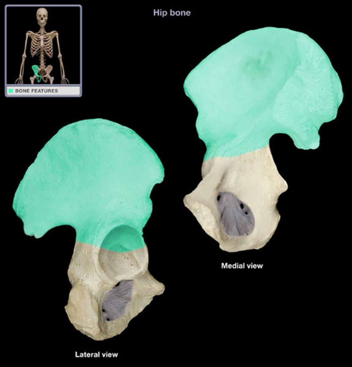
Acetabulum
large socket in the pelvic bone for the head of the femur
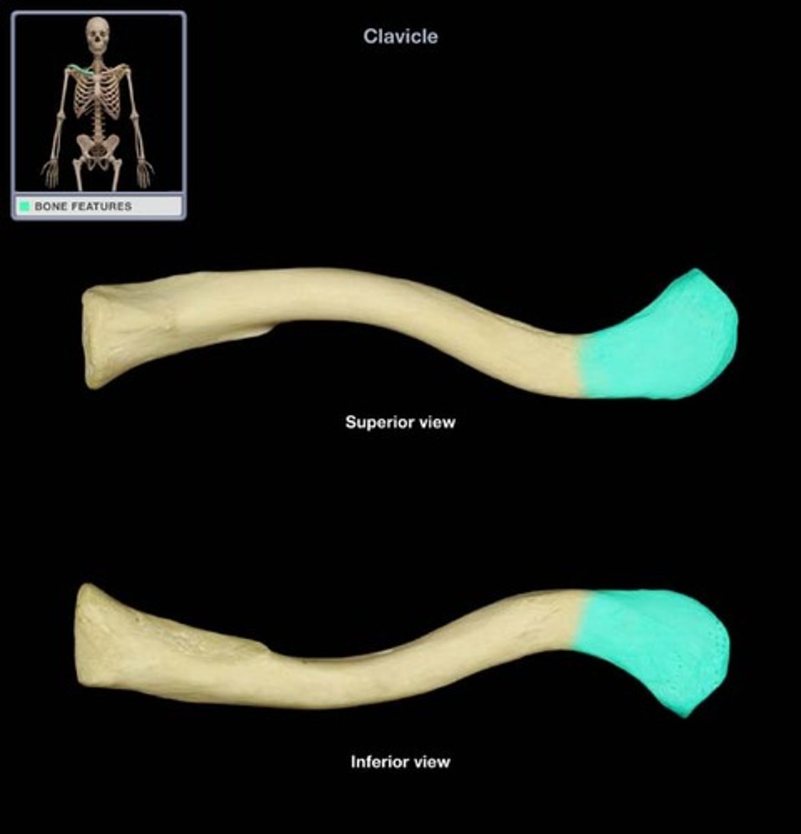
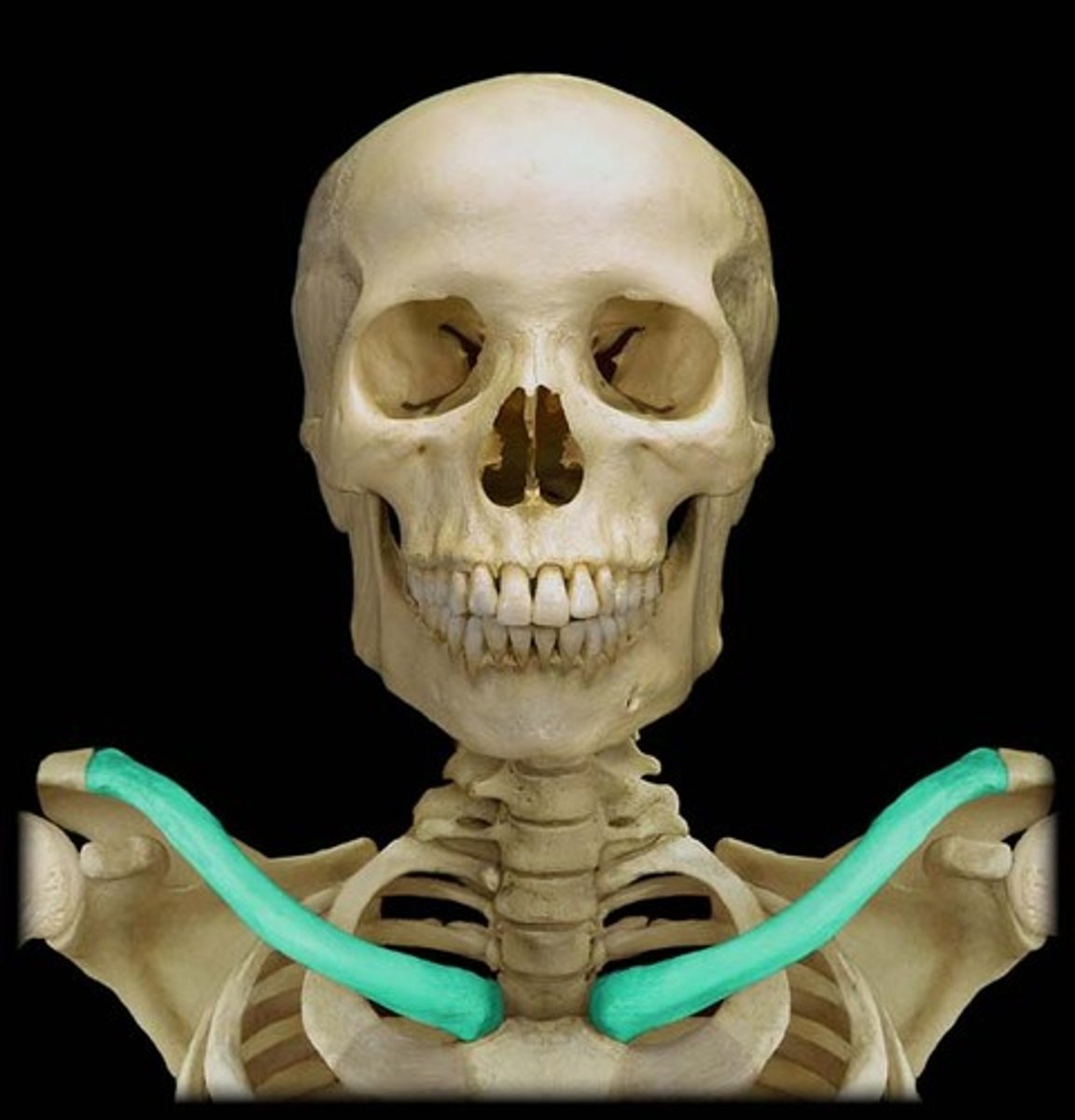
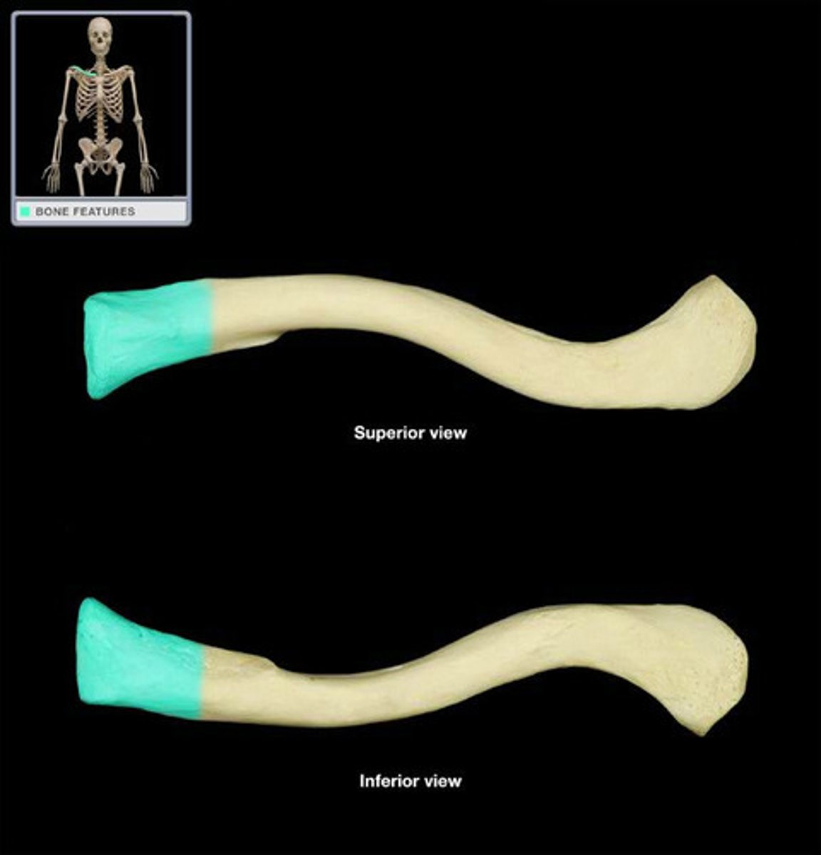
Acromial end of clavicle
articulates with scapula
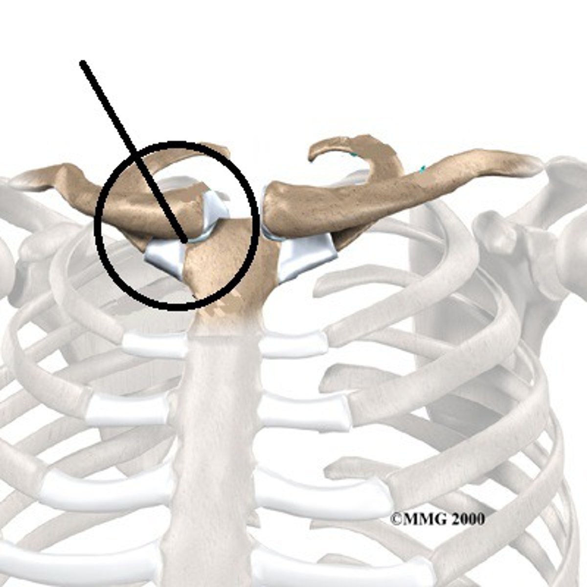
Acromioclavicular joint
acromion articulates with clavicle
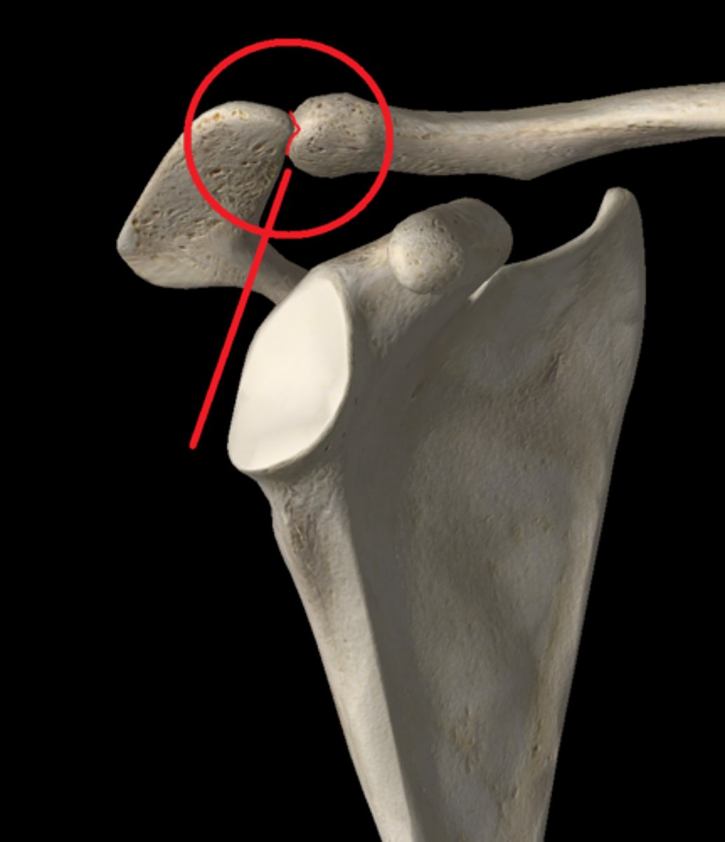
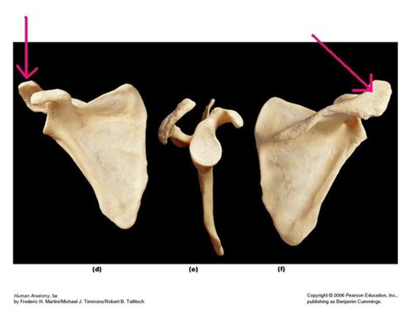
acromial process
acromion of scapula
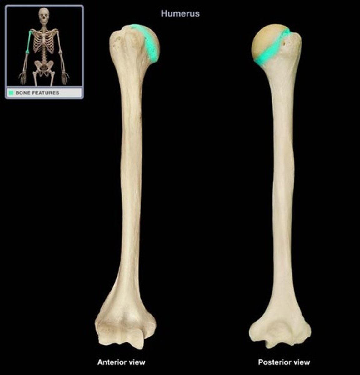
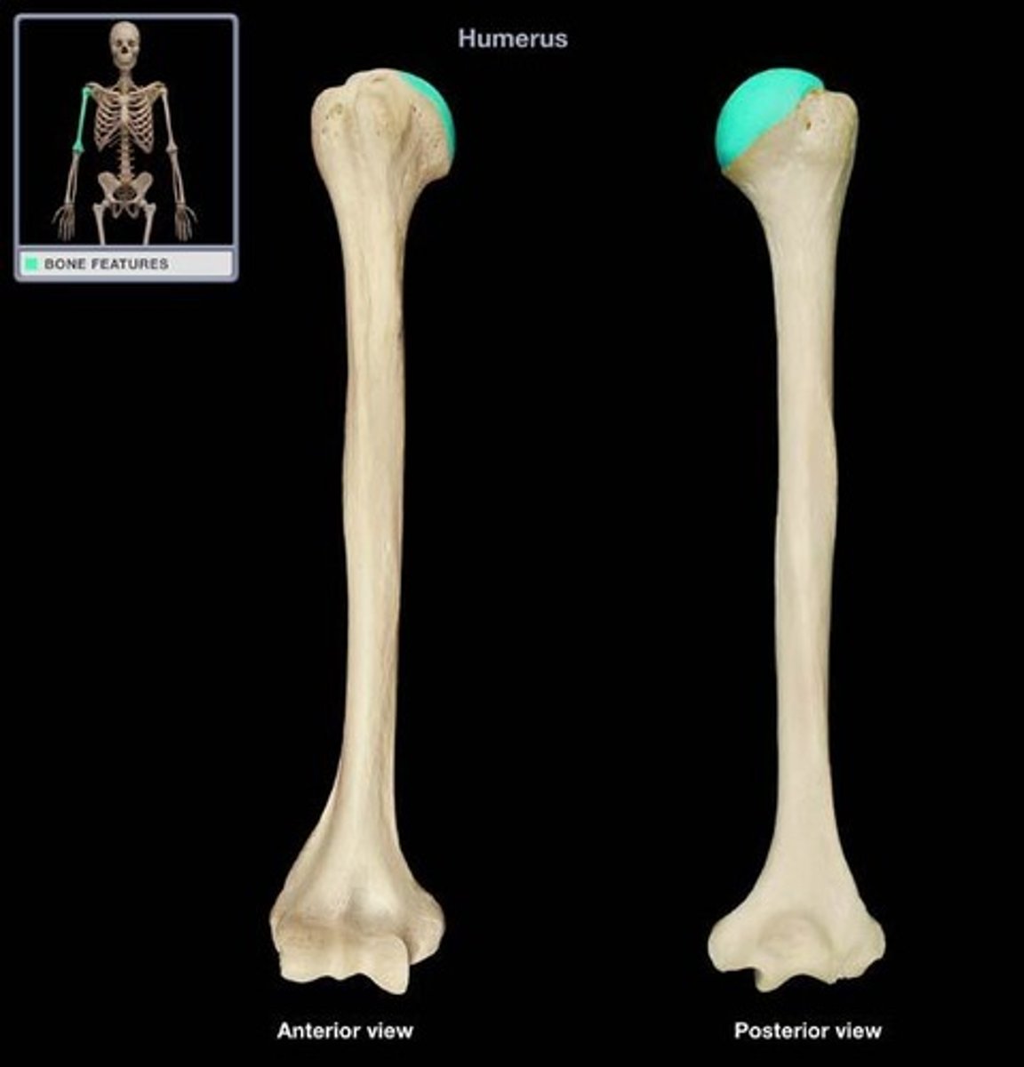
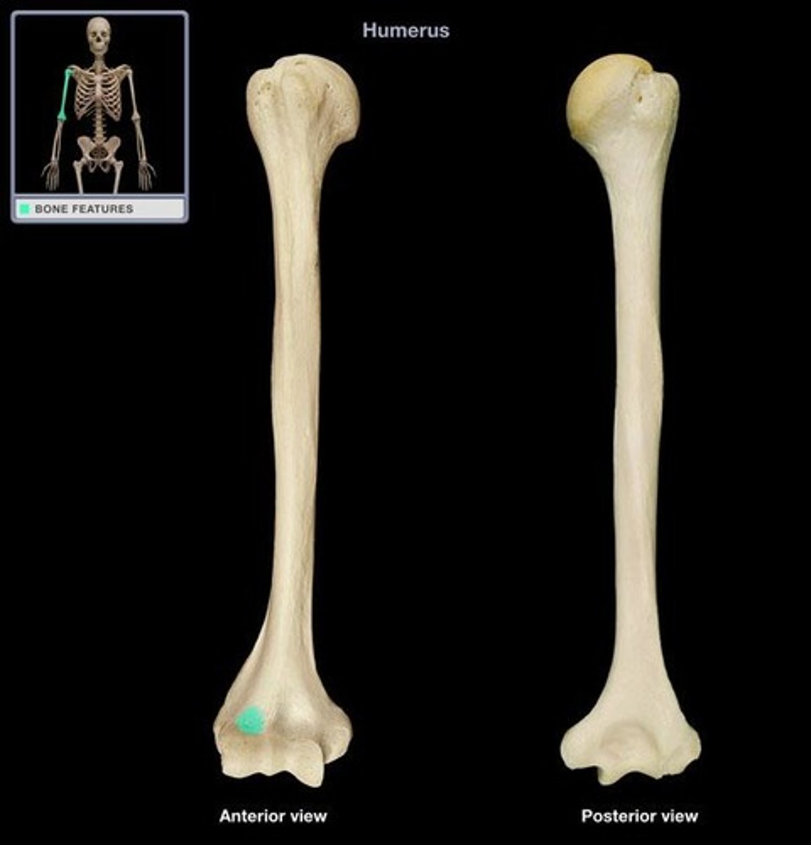
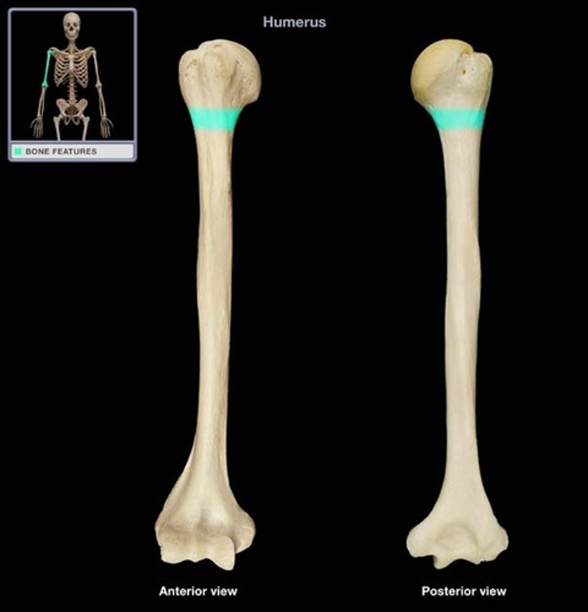
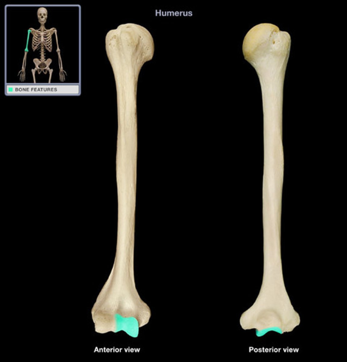
anatomical neck
line on the humerus located around the outside margin of the humeral head
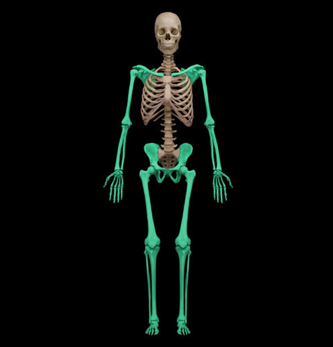
appendicular skeleton
Bones of the limbs and limb girdles that are attached to the axial skeleton
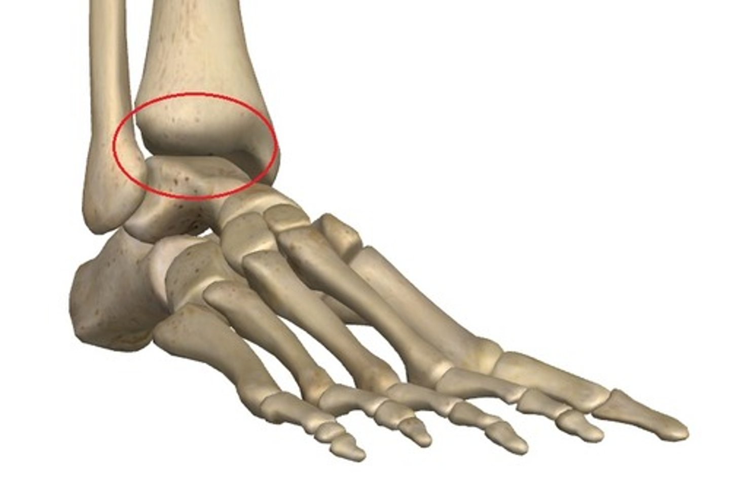
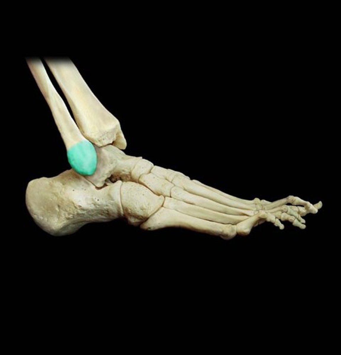
ankle joint
joint that separates the leg and foot portions of the lower limb; formed by the articulations between the talus bone of the foot inferiorly, and the distal end of the tibia, medial malleolus of the tibia, and lateral malleolus of the fibula superiorly
anterior inferior iliac spine
small, bony projection located on the anterior margin of the ilium, below the anterior superior iliac spine
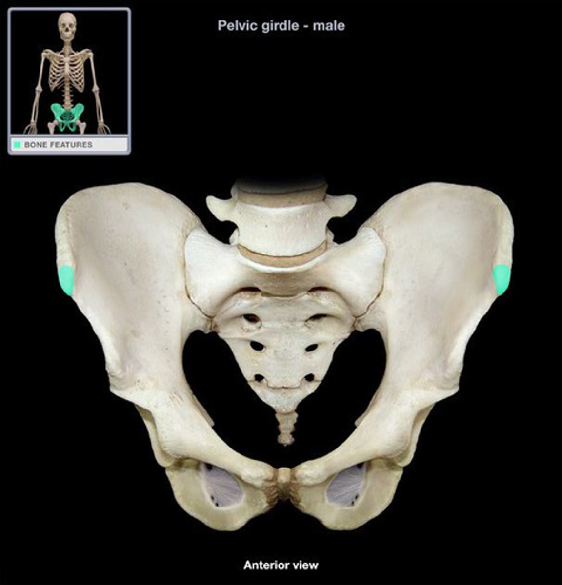
anterior superior iliac spine
rounded, anterior end of the iliac crest
Bicipital groove
intertubercular groove; narrow groove located between the greater and lesser tubercles of the humerus
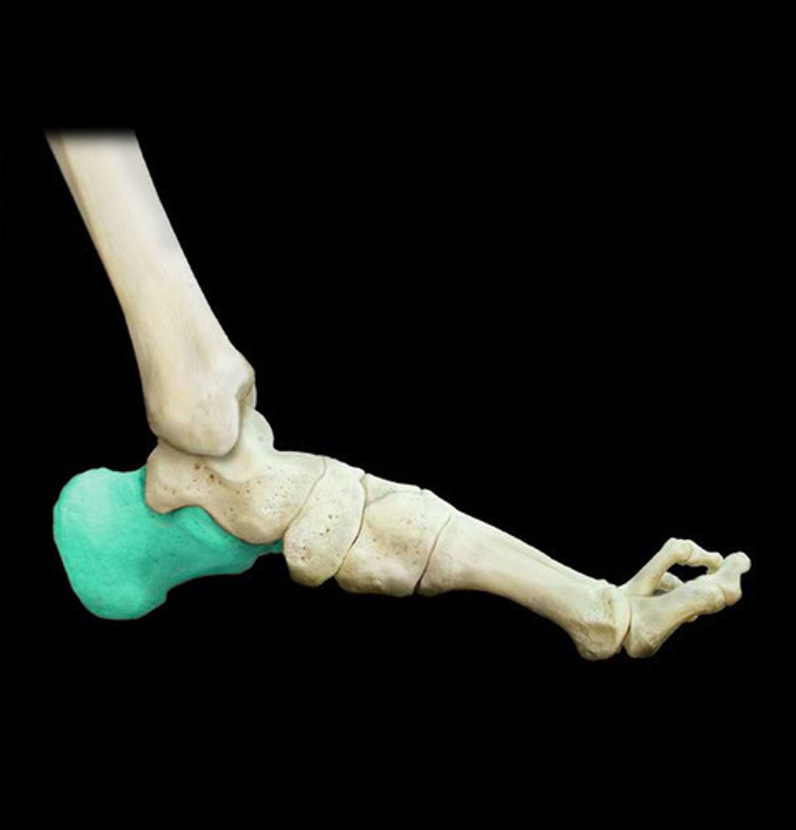
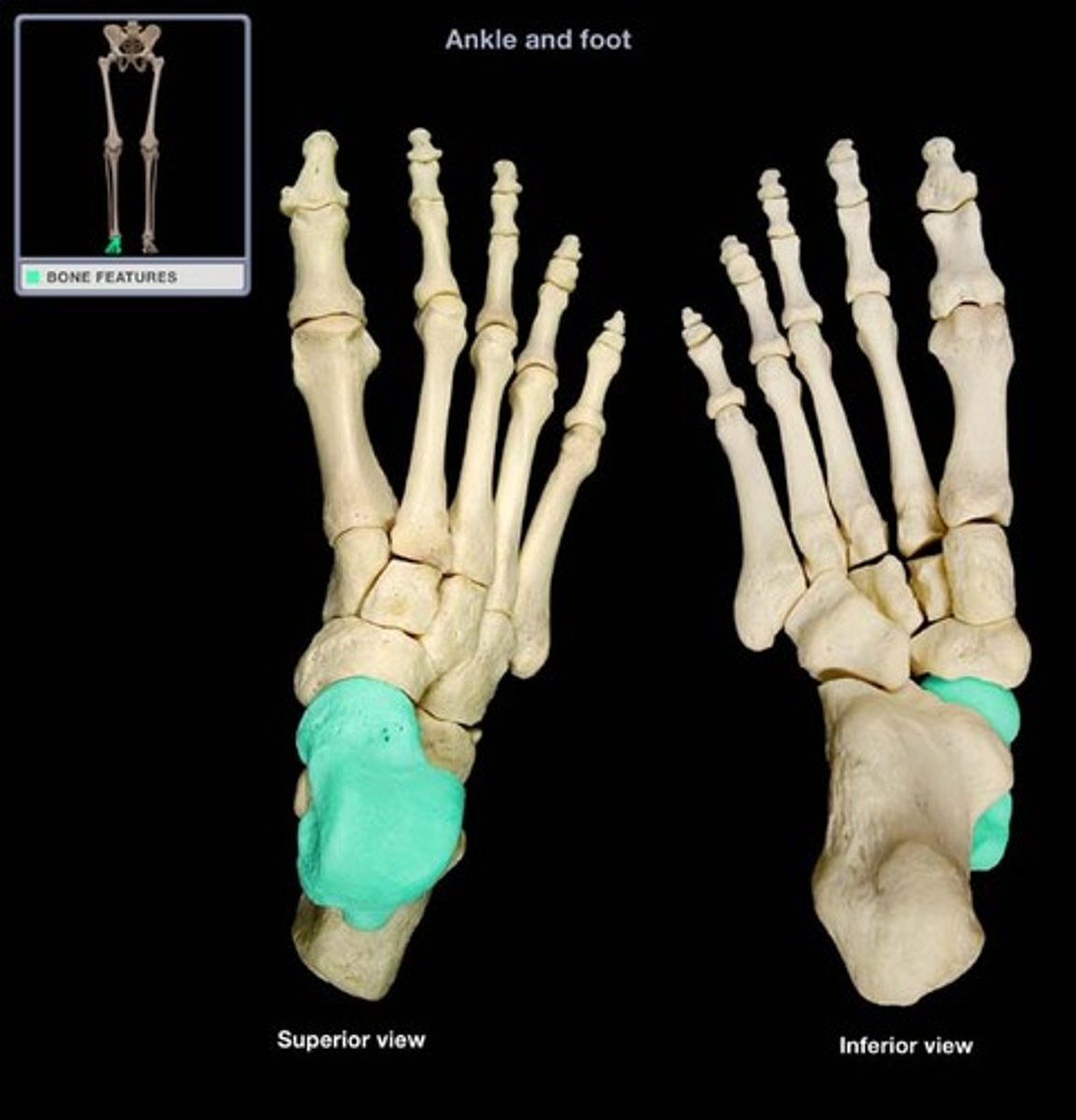
Calcaneus
heel of foot
Capitulum
knob-like bony structure located anteriorly on the lateral, distal end of the humerus

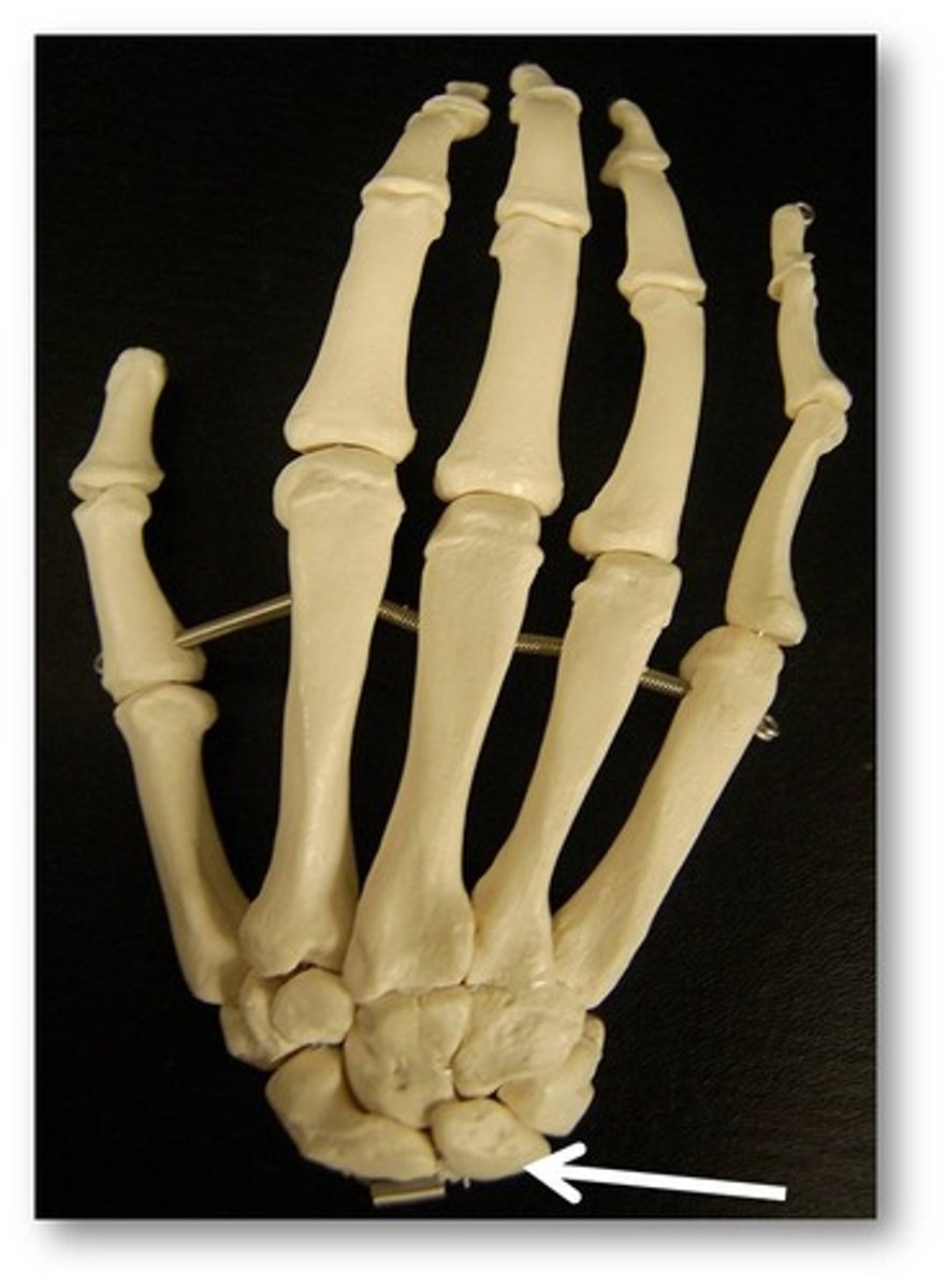
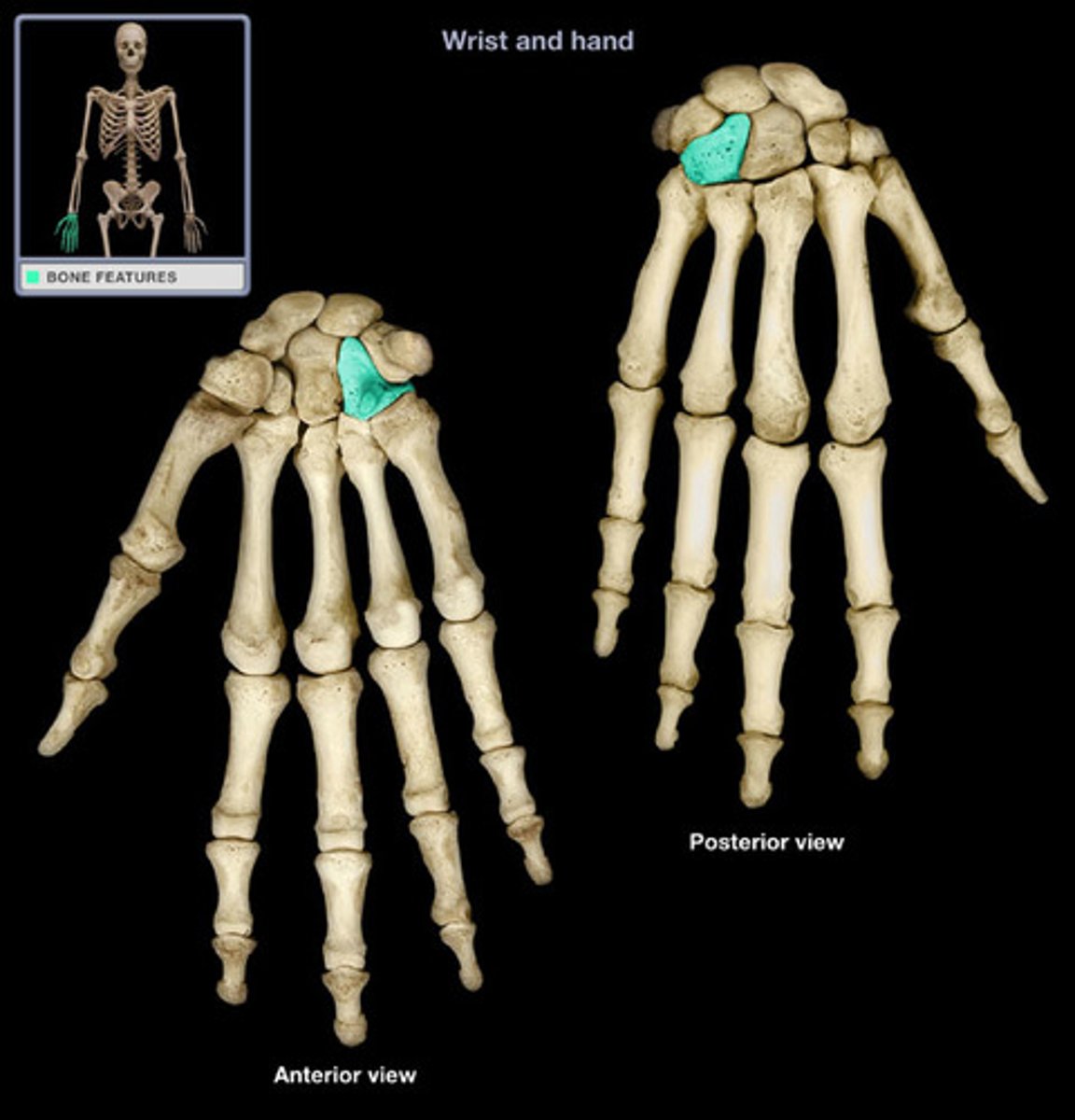
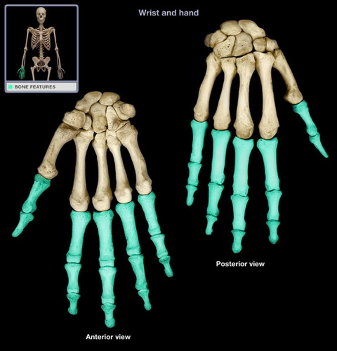
carpal bone
one of the eight small bones that form the wrist and base of the hand; these are grouped as a proximal row consisting of (from lateral to medial) the scaphoid, lunate, triquetrum, and pisiform bones, and a distal row containing (from lateral to medial) the trapezium, trapezoid, capitate, and hamate bones
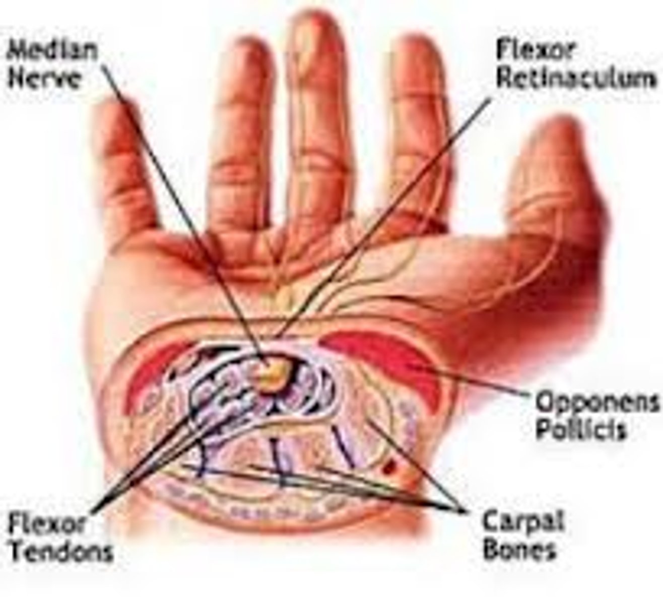
Carpal tunnel
Passageway for 9 tendons and major nerve, wall and floor are made up of carpal bones where the roof is flexor retinaculum. Overuse compresses the nerve causing pain and numbness
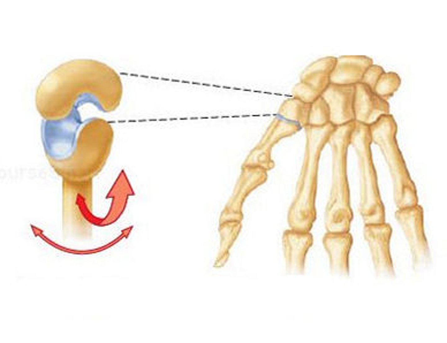
carpometacarpal joint
Connects the metacarpals to the distal carpals
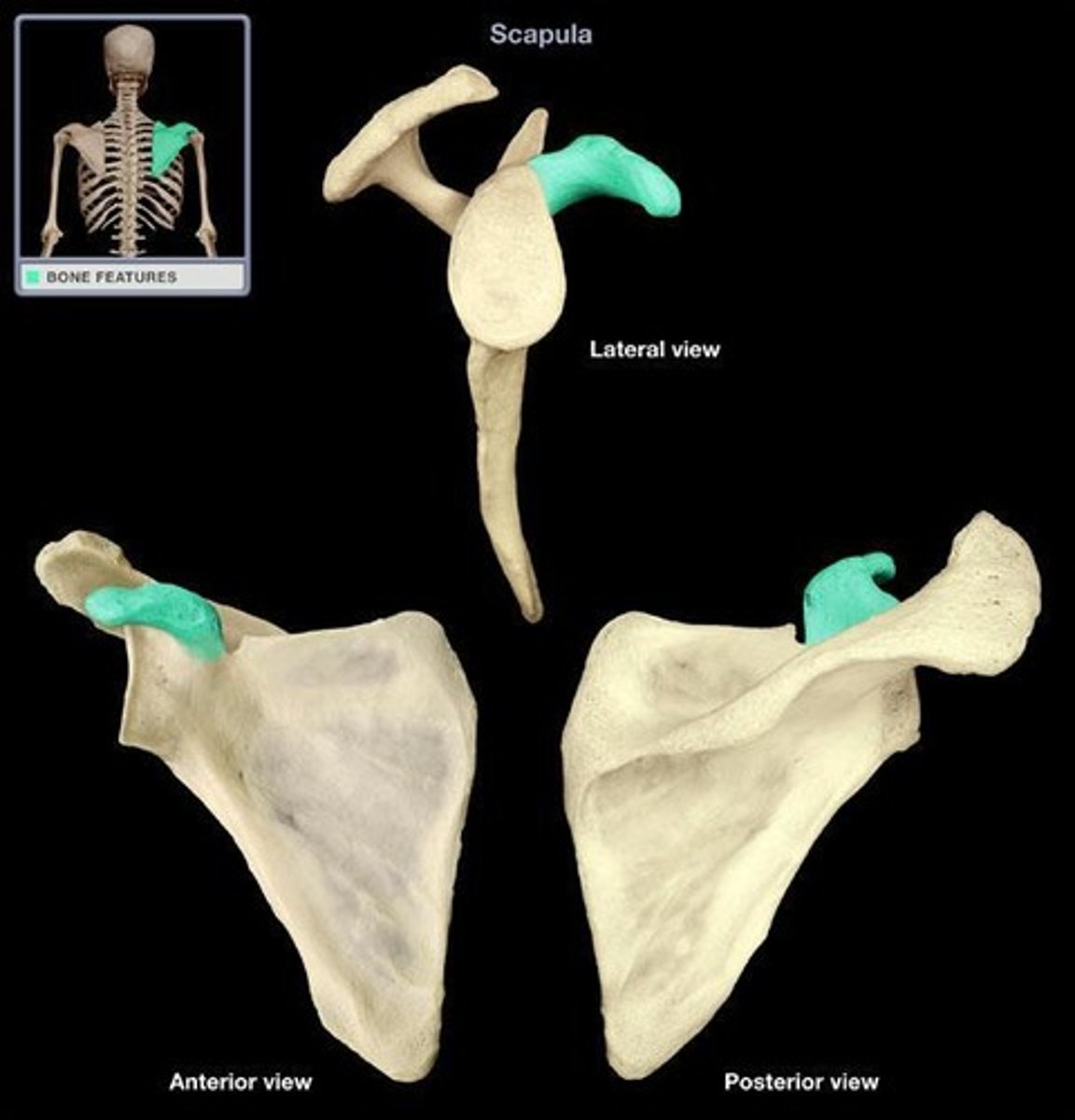
corcoid process
short, hook-like process that projects anteriorly and laterally from the superior margin of the scapula
Clavicle
articulates with acromion of the scapula and manubrium
Coronoid fossa
room for coronoid process of ulna on humerus
Coronoid process of ulna
swings into coronoid fossa at flexion
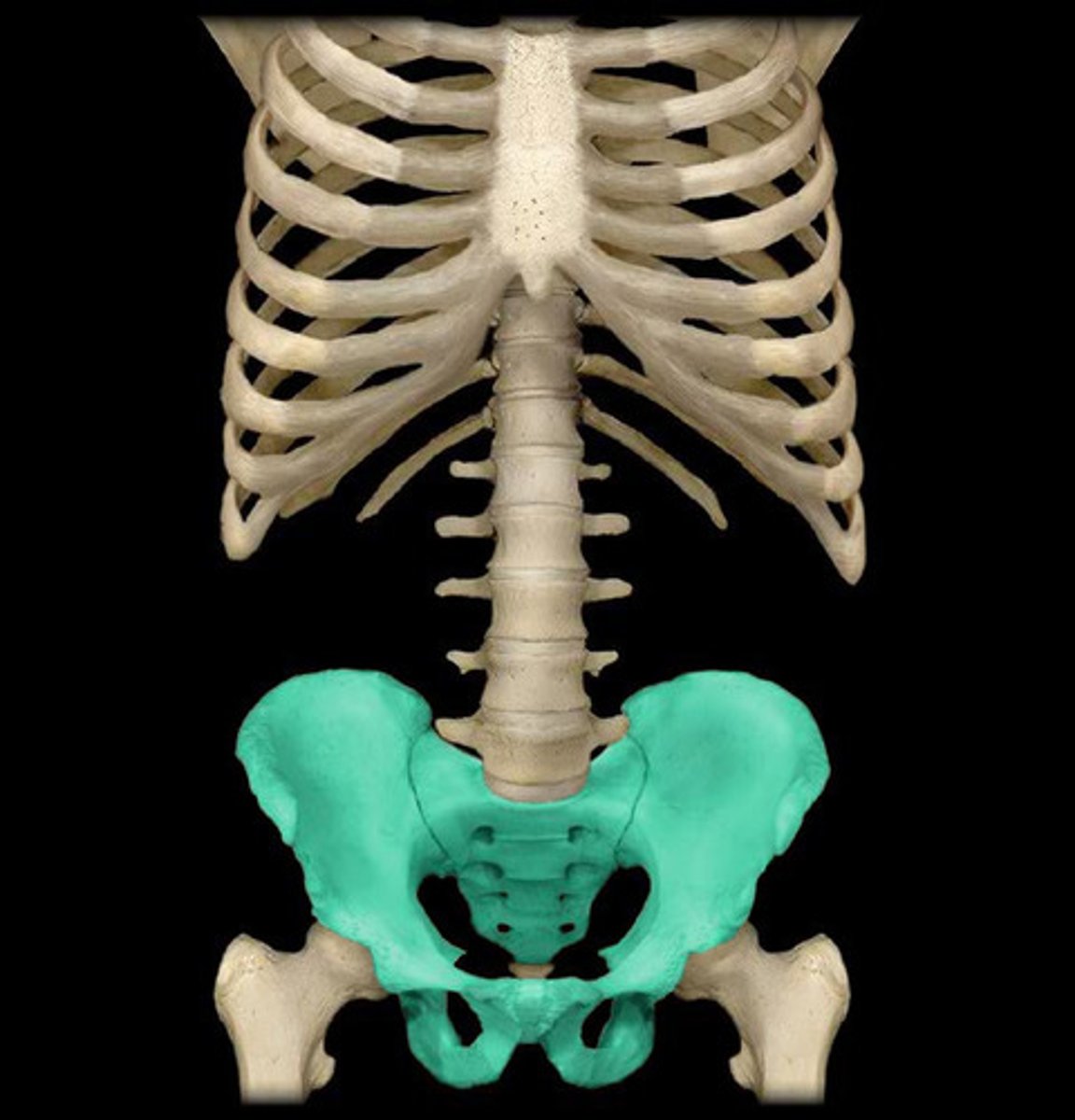
Coxal bones
bones around sacral curve
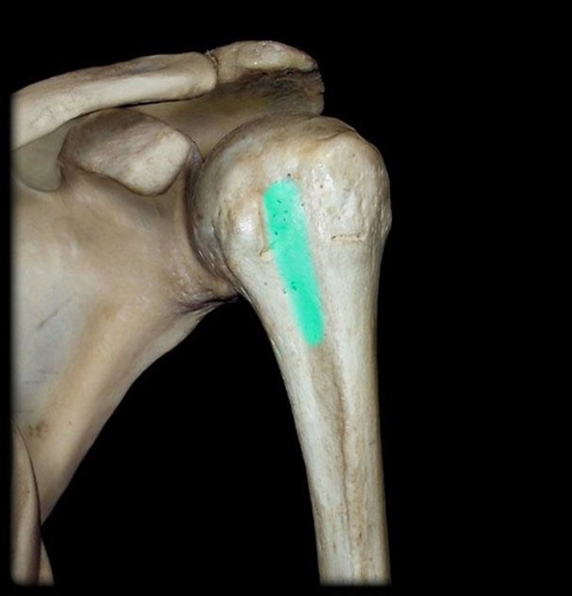
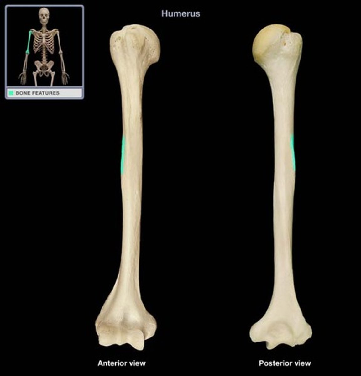
Deltoid tuberosity
connection for deltoid muscle
Distal radioulnar joint
head of ulna articulates with distal end of radius
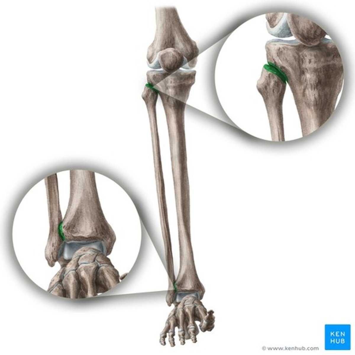
distal tibiofibular joint
articulation between the distal fibula and the fibular notch of the tibia
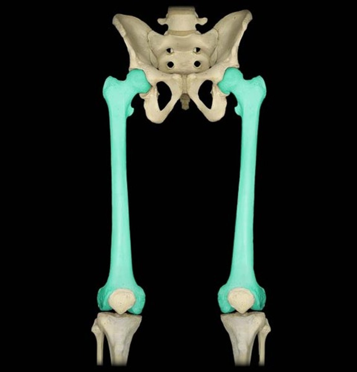
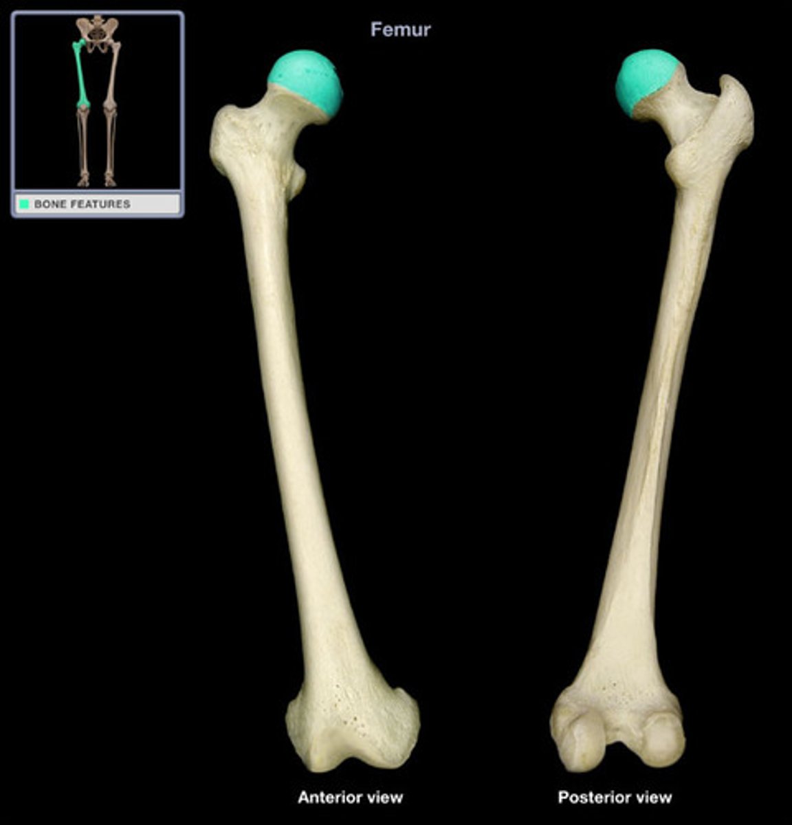
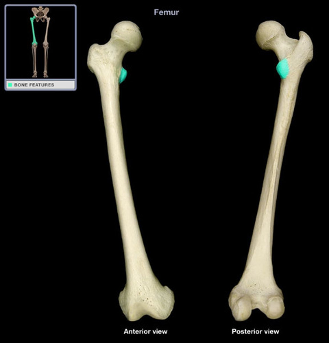
Femoral head
articulates with the pelvis at acetabulum to femur
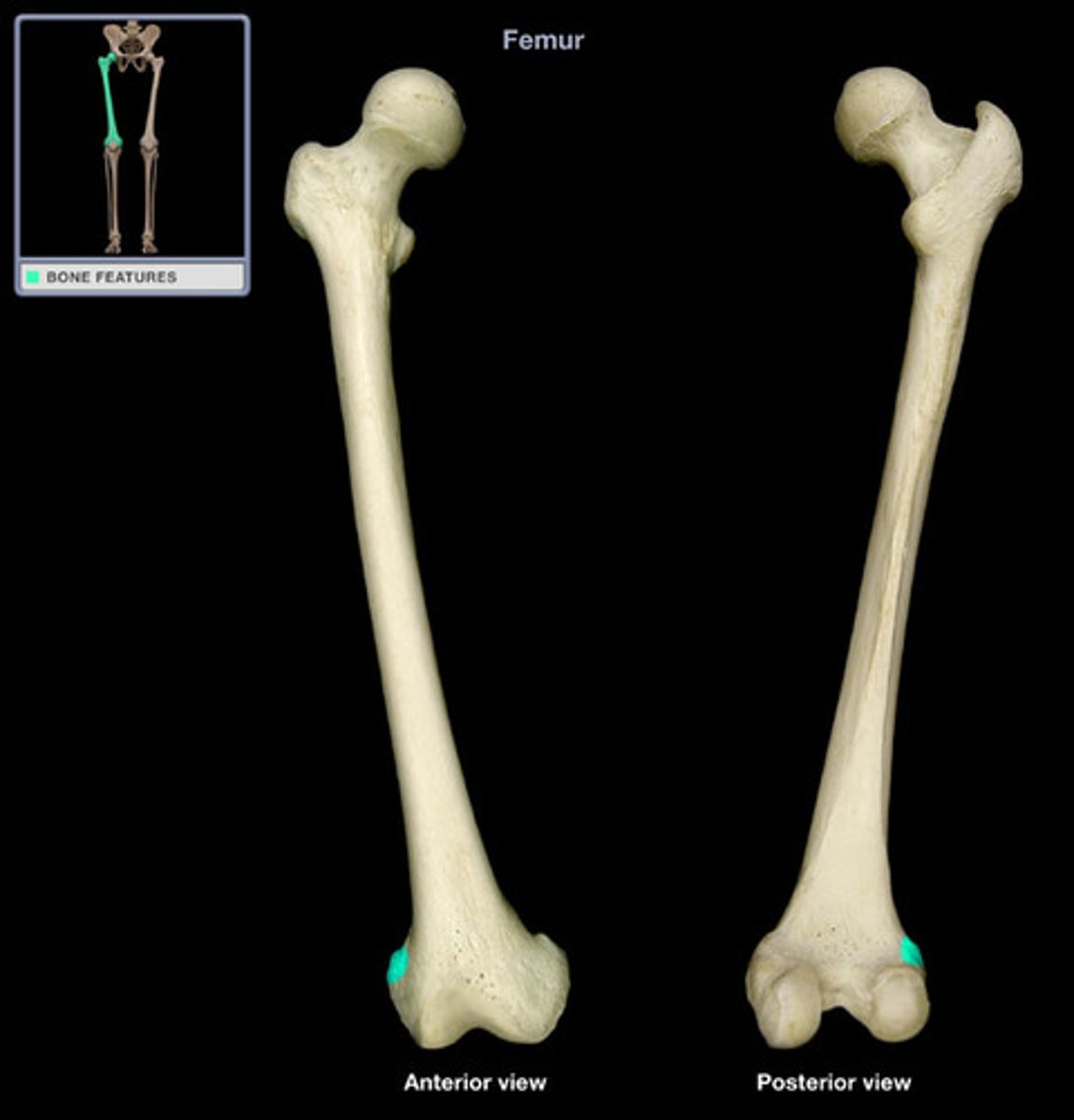
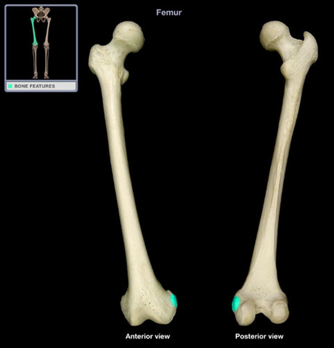
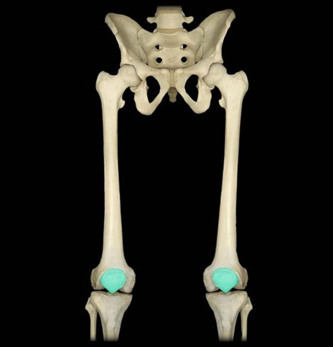
Femur
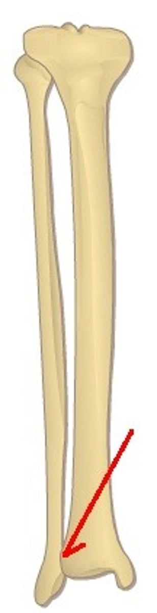
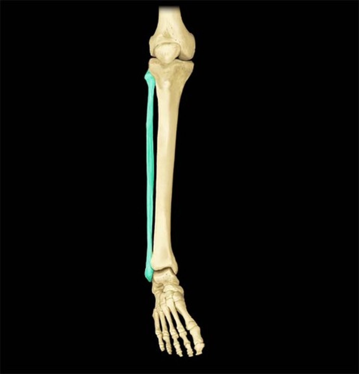
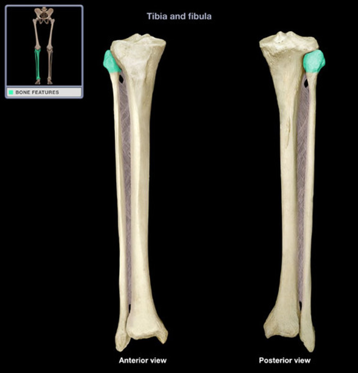
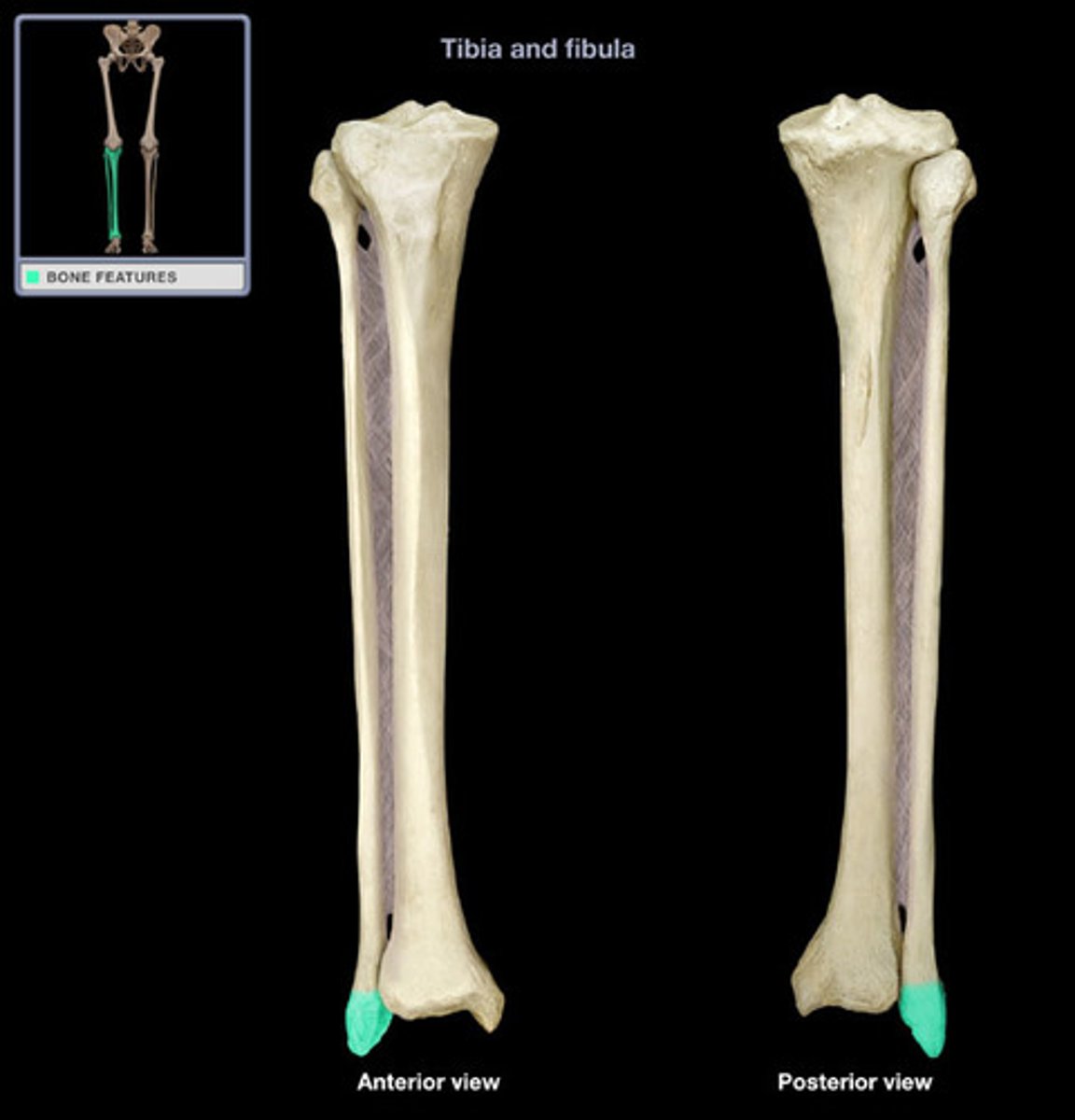
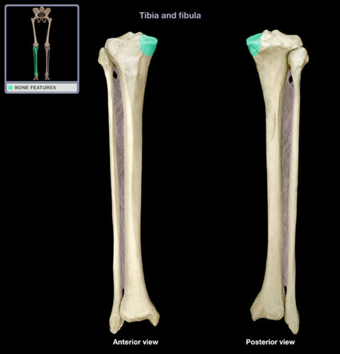
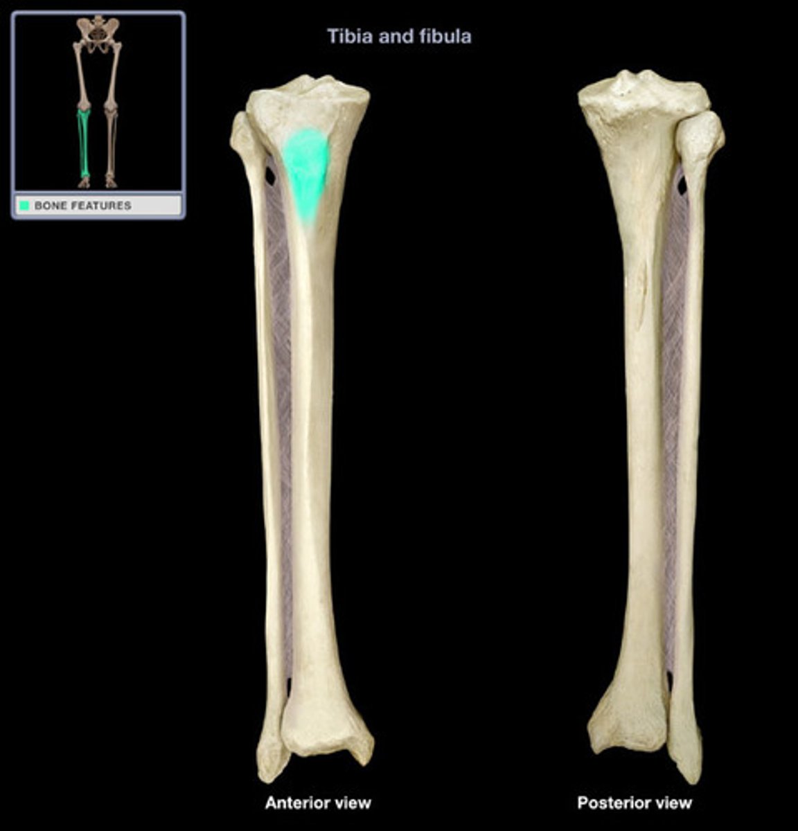
Fibula
fibular notch
wide groove on the lateral side of the distal tibia for articulation with the fibula at the distal tibiofibular joint
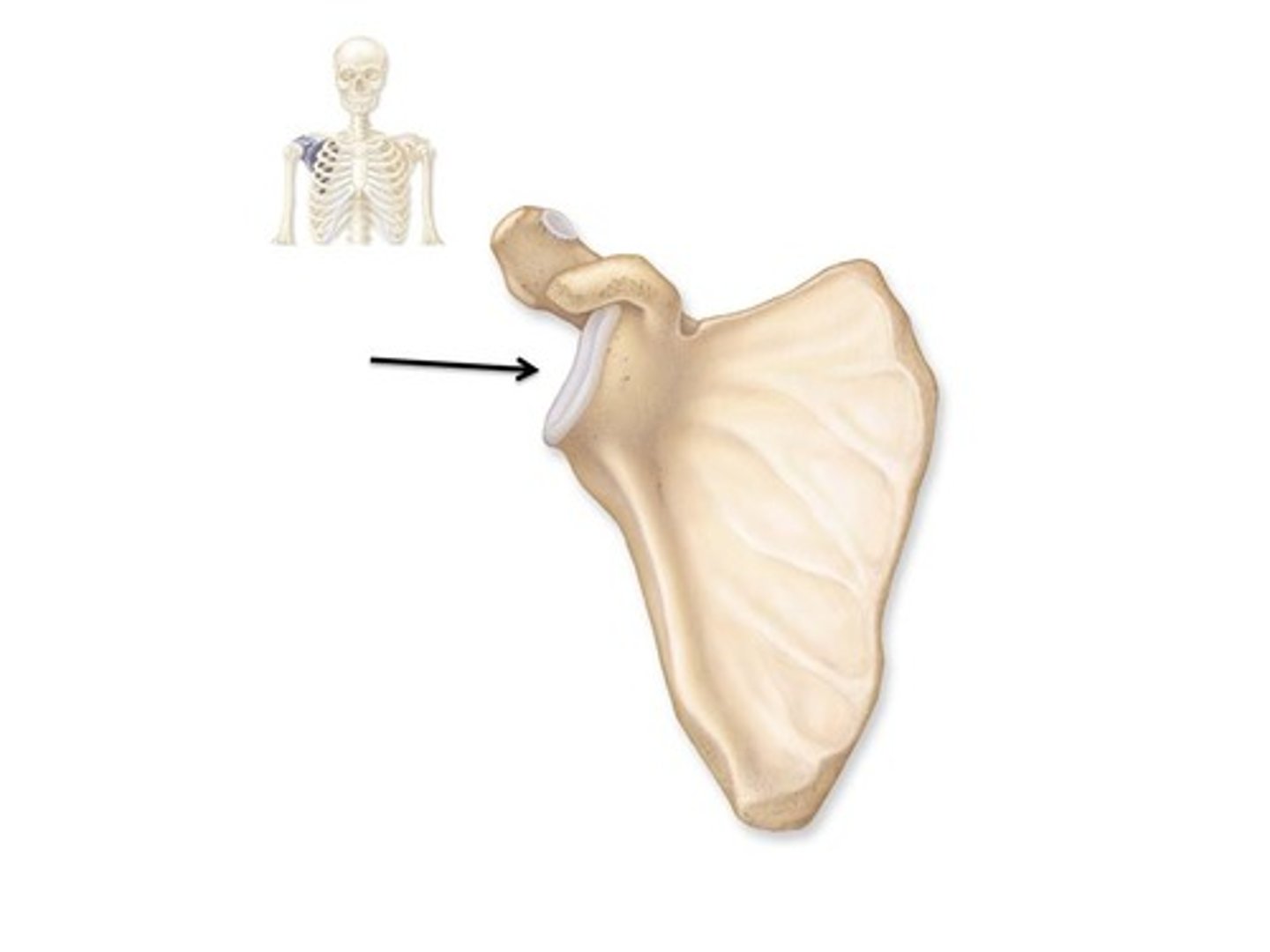
Glenhumeral joint
The synovial ball-and-socket joint of the shoulder
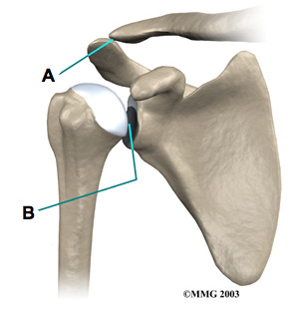
Glenoid cavity
scapula articulates with humerus
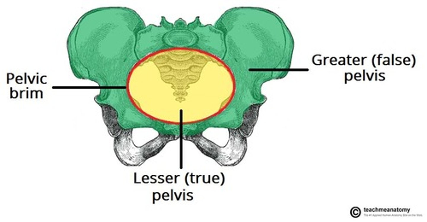
Greater pelvis
Greater sciatic/iliac notch
large, U-shaped indentation located on the posterior margin of the ilium, superior to the ischial spine

Hallux
big toe
Hamate
Head of fibula
articulates with tibia at superior tibiofibular joint
Head of Femur
rounded, proximal end of the femur that articulates with the acetabulum of the hip bone to form the hip joint
Head of humerus
articulates with scapula

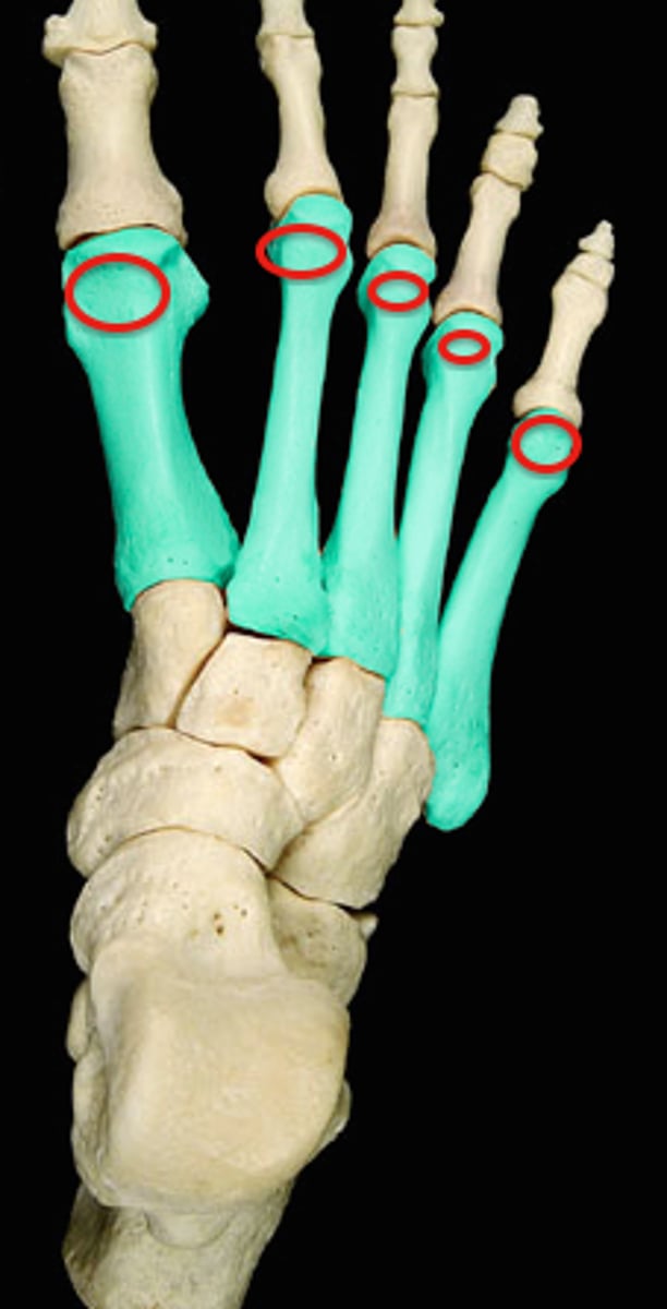
Head of metatarsals
distal end of metatarsal

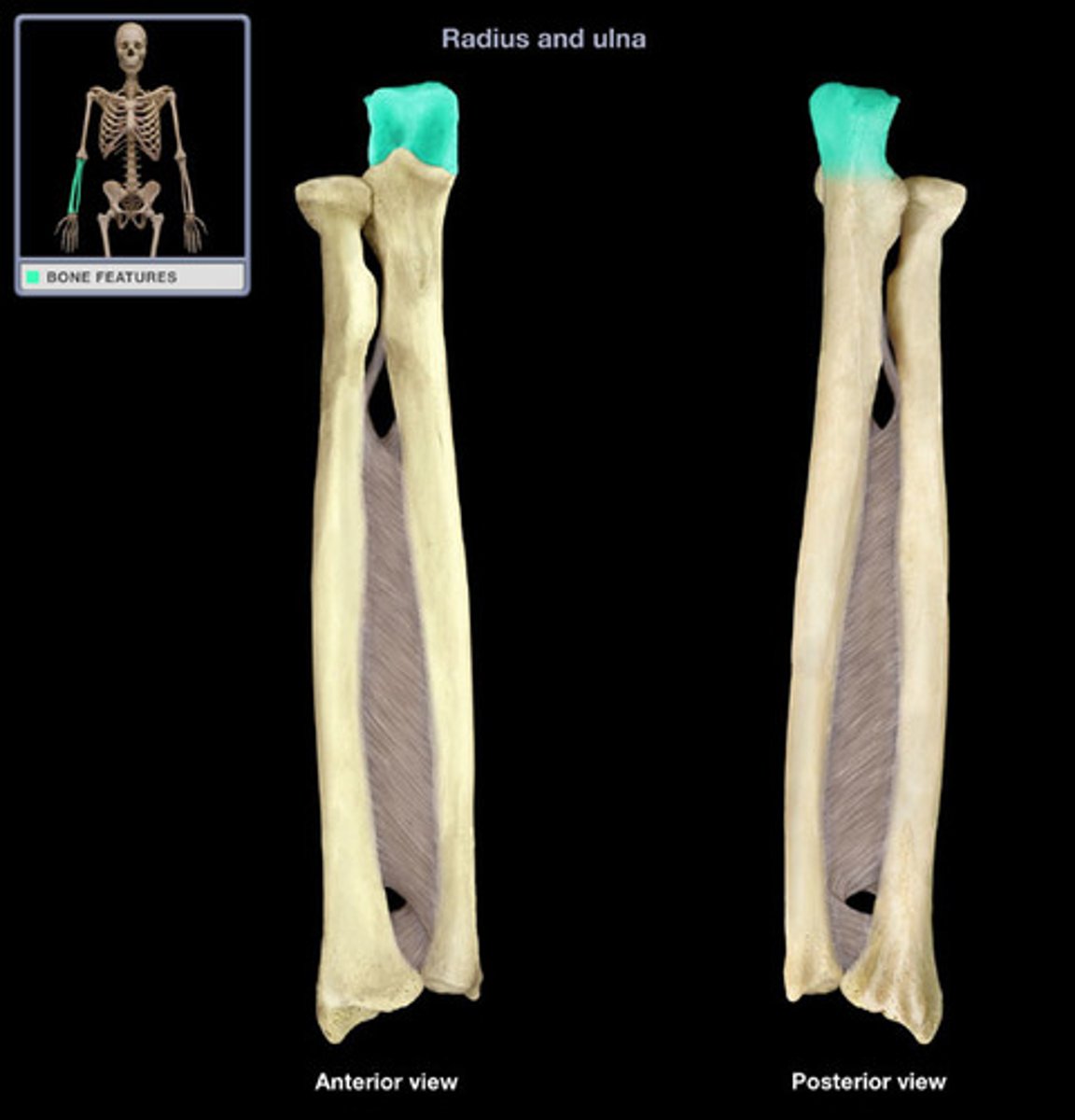
Head of ulna
small, rounded distal end of the ulna; articulates with the ulnar notch of the distal radius, forming the distal radioulnar joint
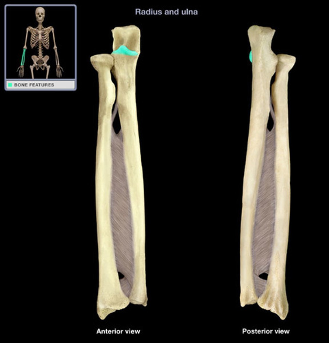
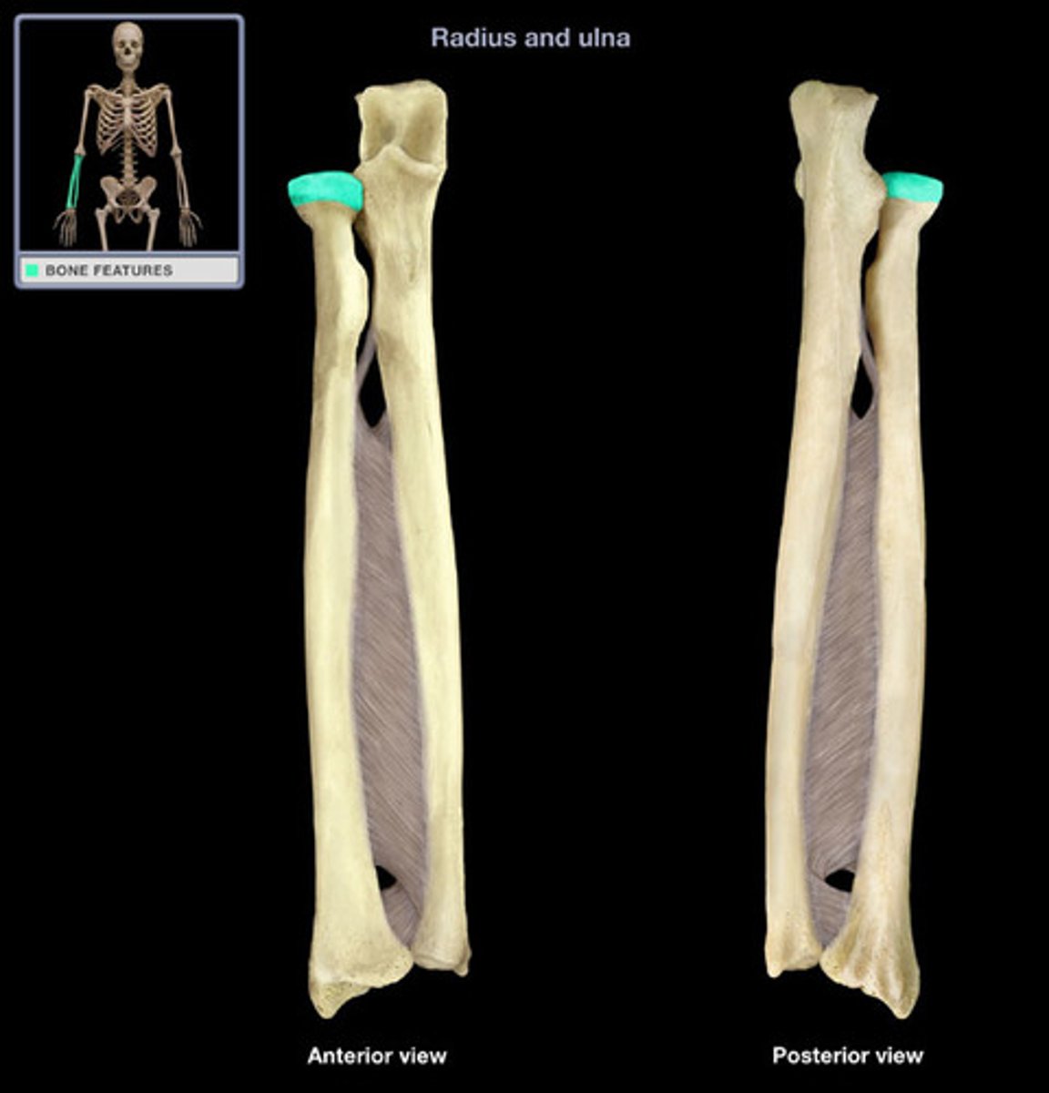
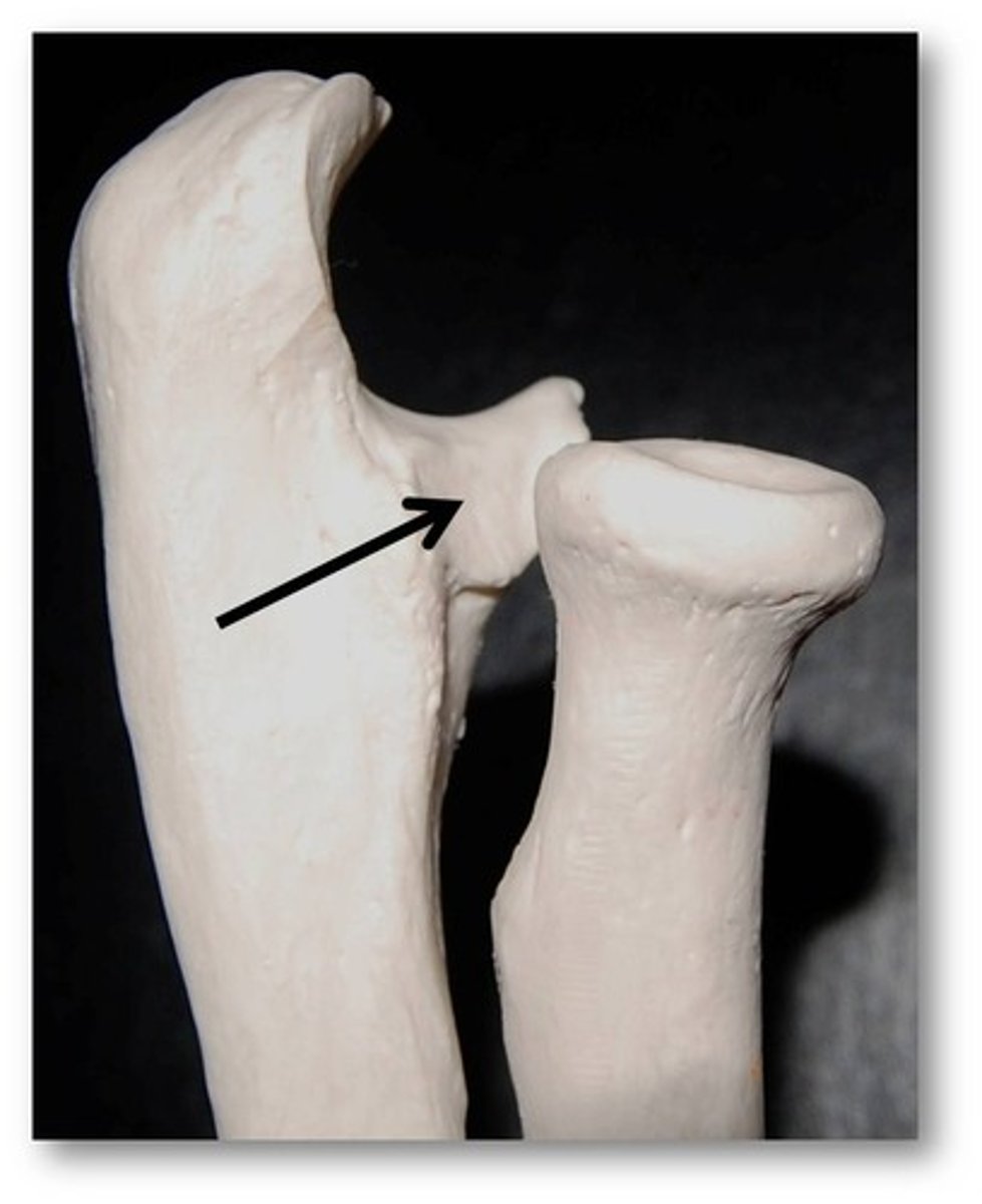
Head of radius
disc-shaped structure that forms the proximal end of the radius; articulates with the capitulum of the humerus as part of the elbow joint, and with the radial notch of the ulna as part of the proximal radioulnar joint
Head of humerus
smooth, rounded region on the medial side of the proximal humerus; articulates with the glenoid fossa of the scapula to form the glenohumeral (shoulder) joint
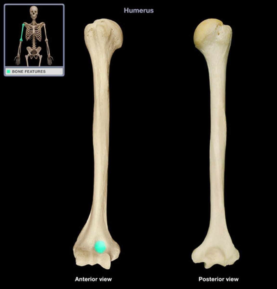
Humerus condyle
articulates with radius and ulna

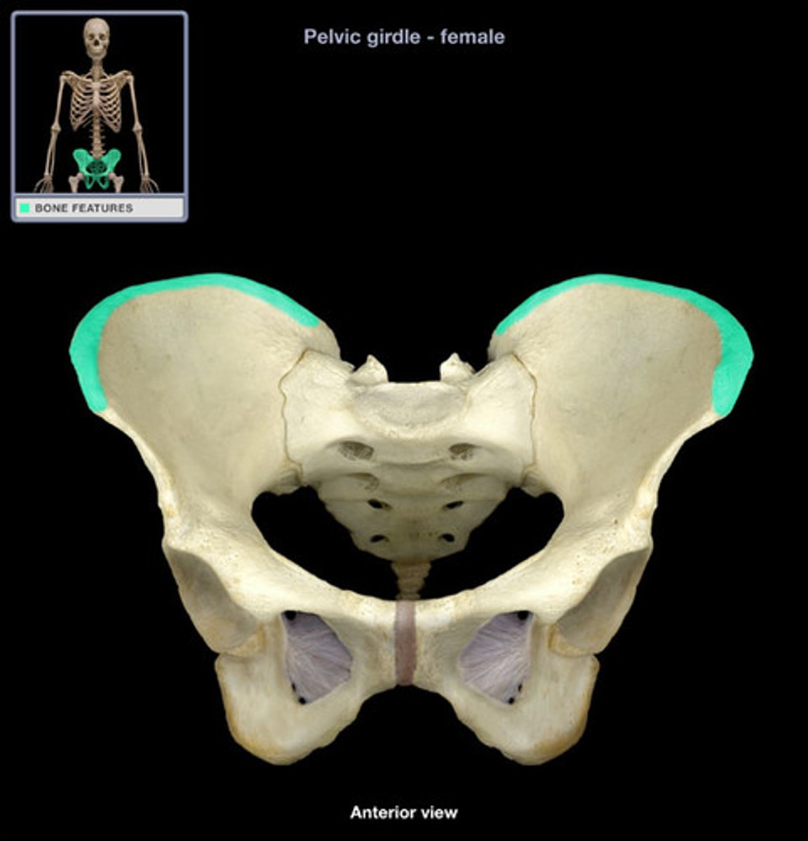
Iliac crest
upper margin of iliac bones
Iliac fossa
The broad, slightly concave inner surface of the ilium.
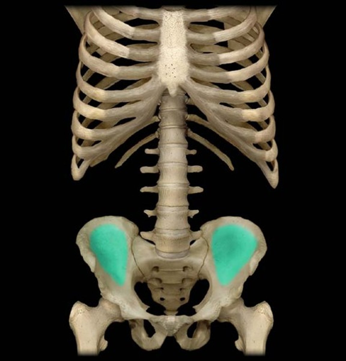
Ilium
hip bone
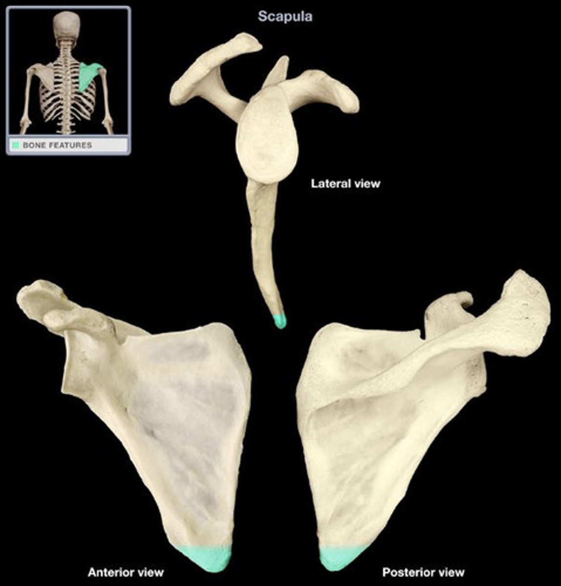
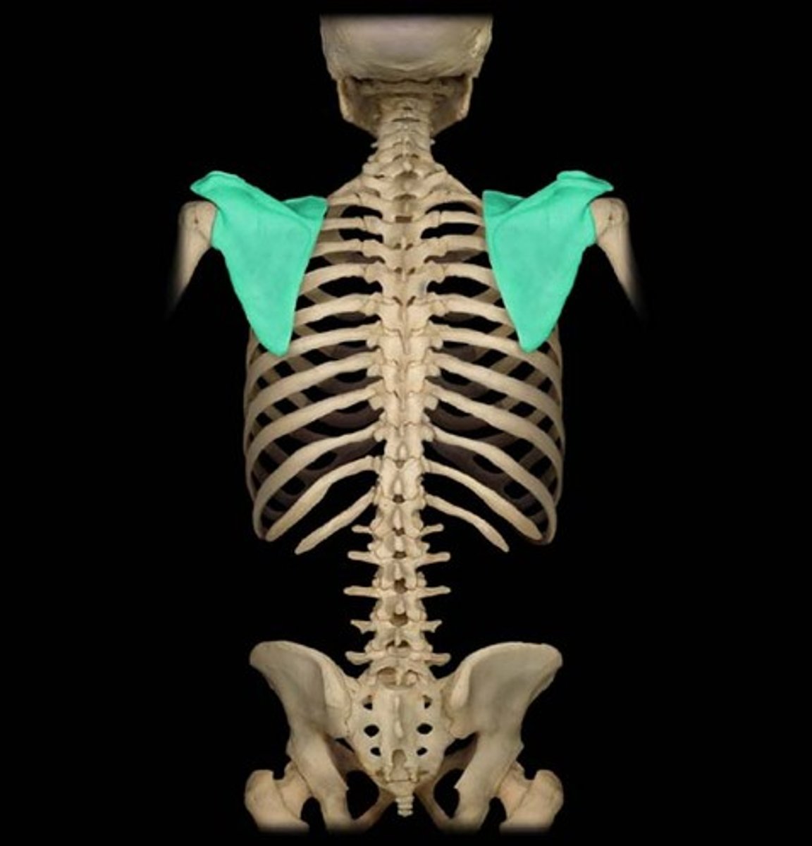
inferior angle of scapula
inferior corner of the scapula located where the medial and lateral borders meet
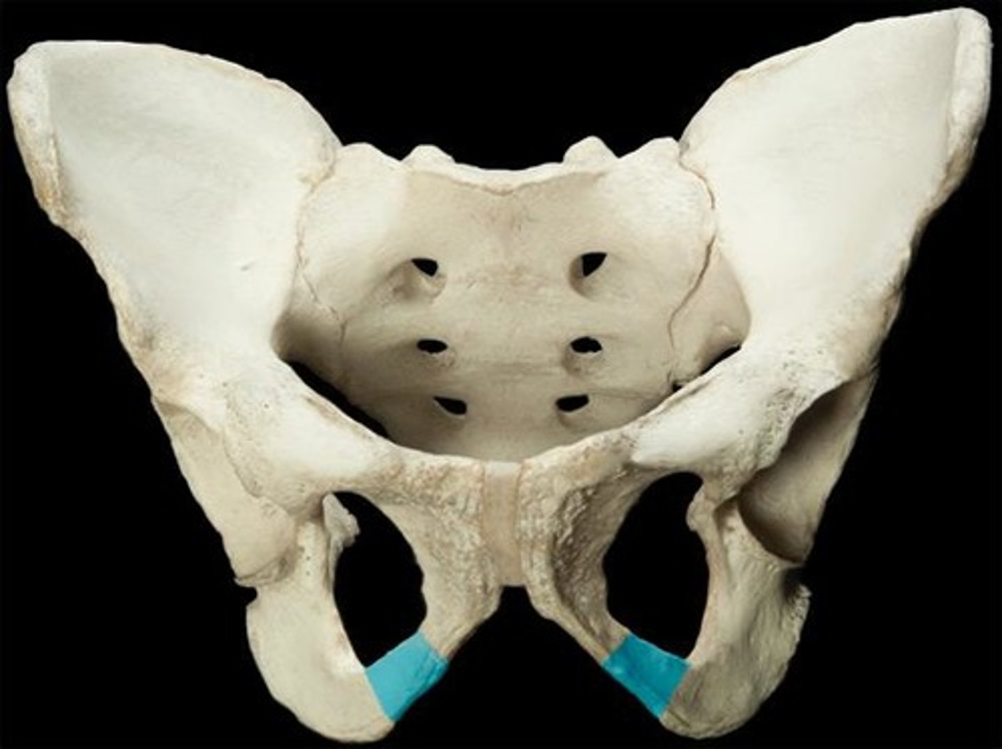
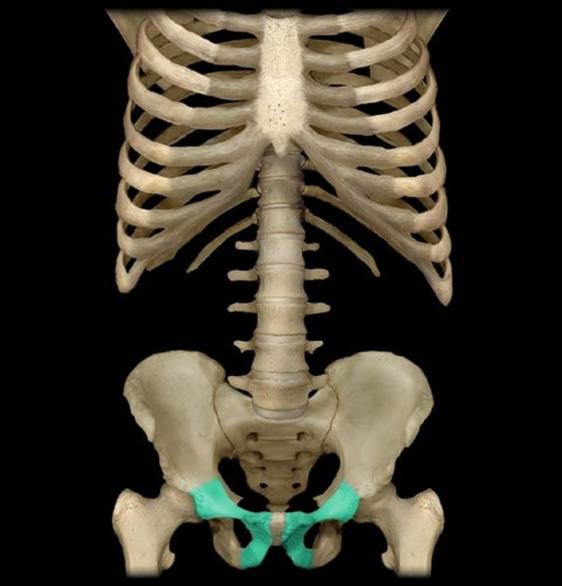
inferior pubic ramus
narrow segment of bone that passes inferiorly and laterally from the pubic body; joins with the ischial ramus to form the ischiopubic ramus
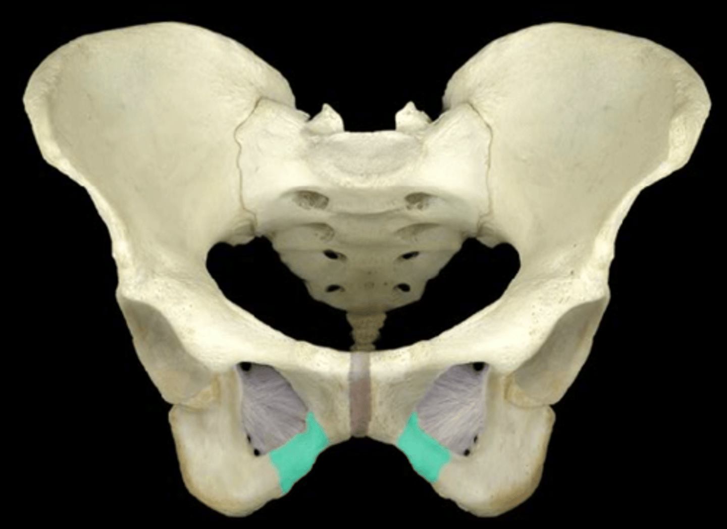
Ischial ramus
bony extension projecting anteriorly and superiorly from the ischial tuberosity; joins with the inferior pubic ramus to form the ischiopubic ramus
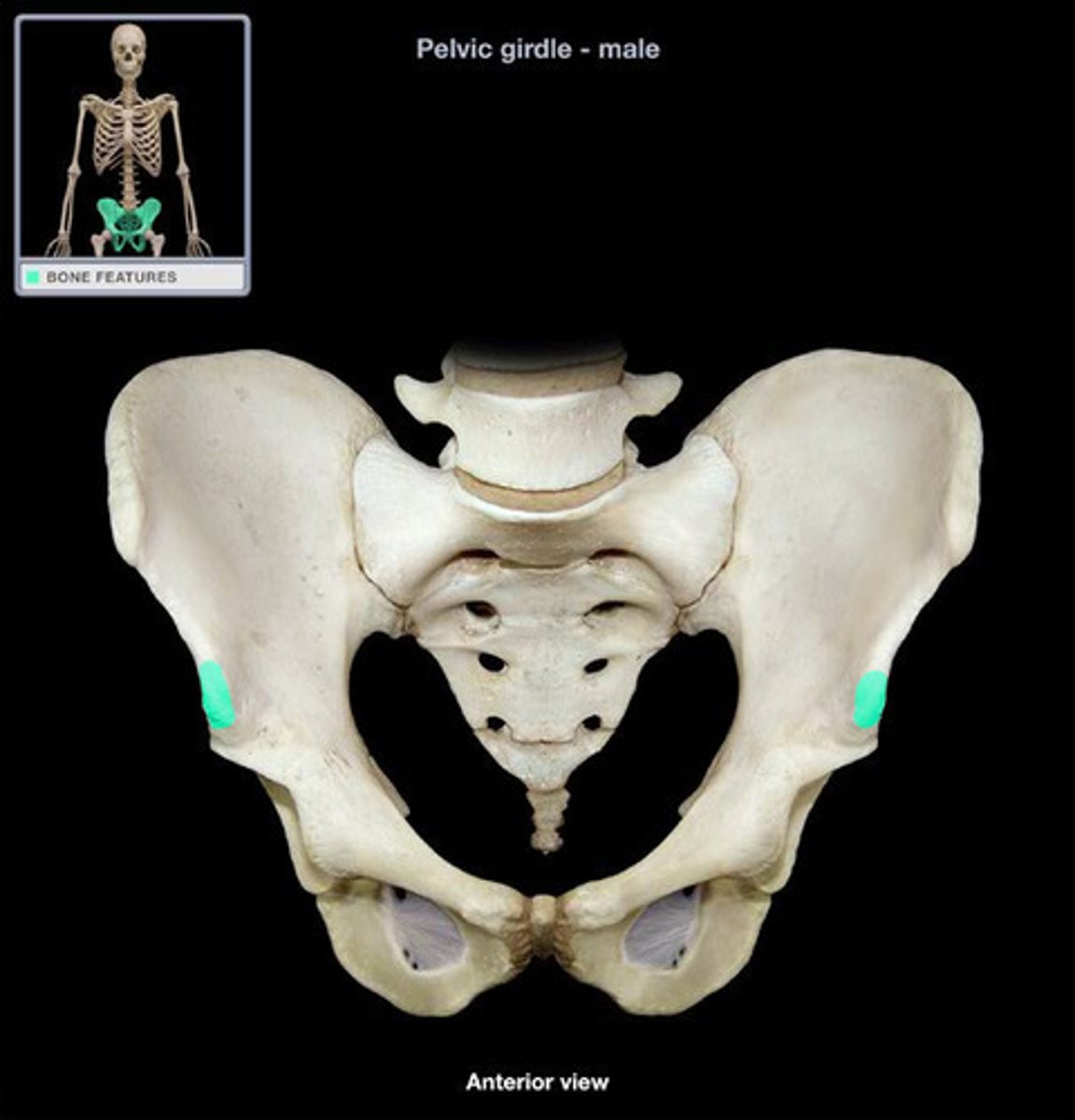
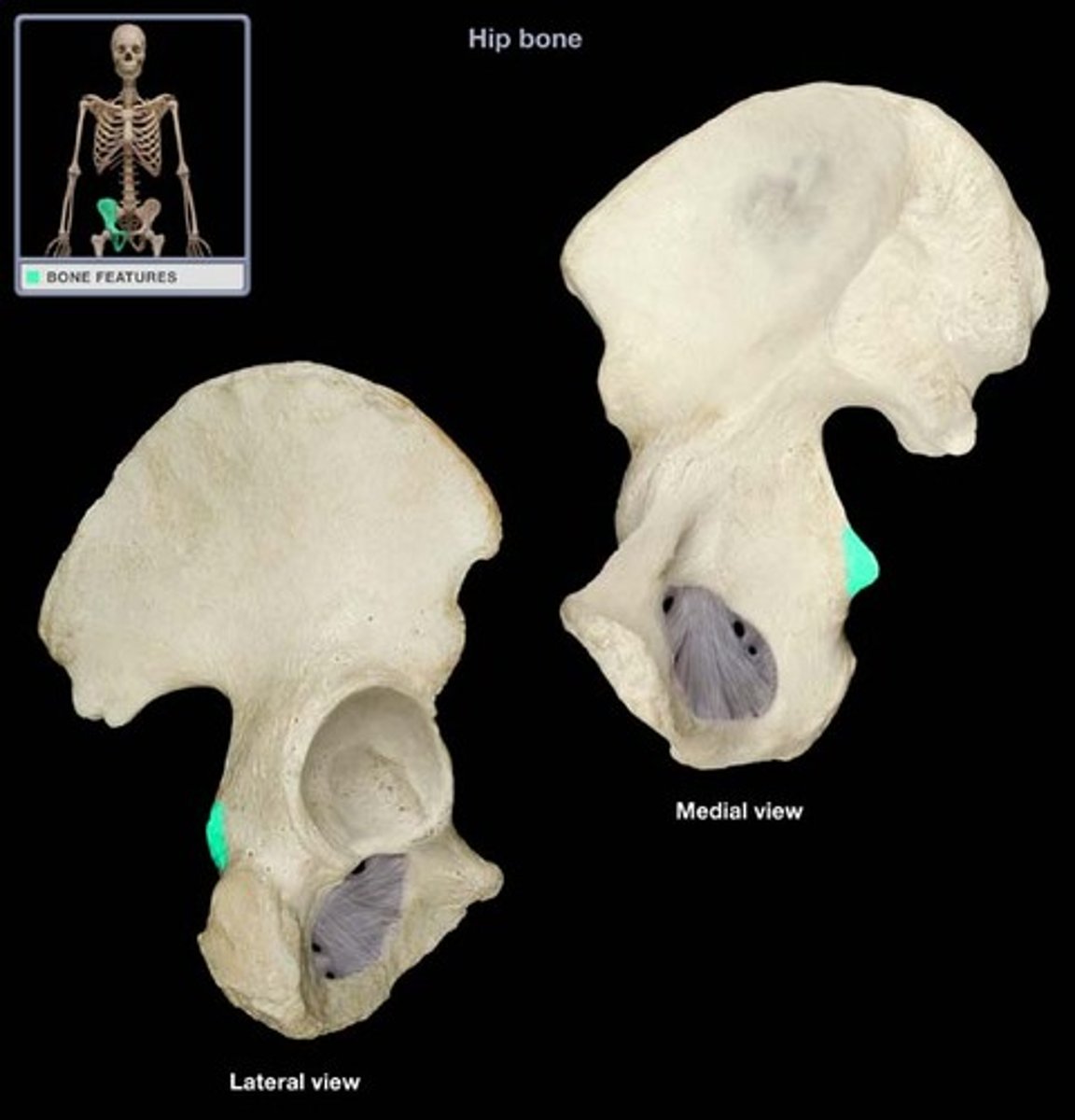
Ischial spine
pointed, bony projection from the posterior margin of the ischium that separates the greater sciatic notch and lesser sciatic notch
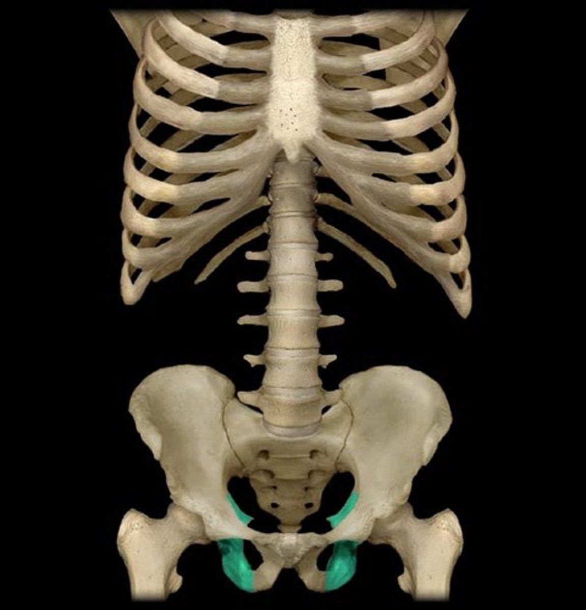
Ischial tuberosity
receives the weight of the body when sitting
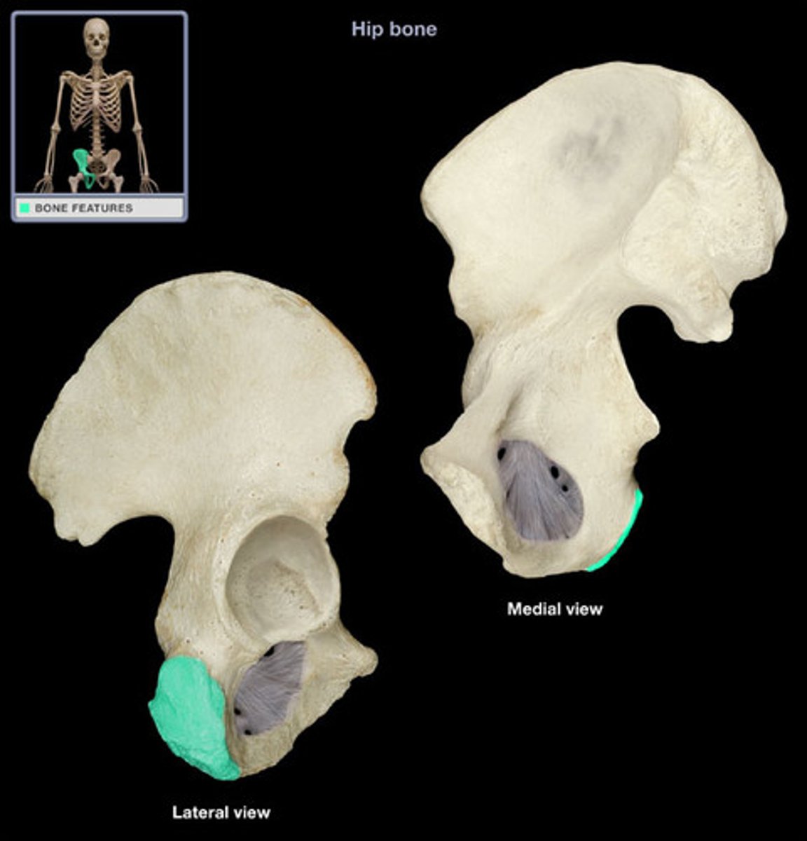
Ischium
the lower, posterior portions of the pelvis
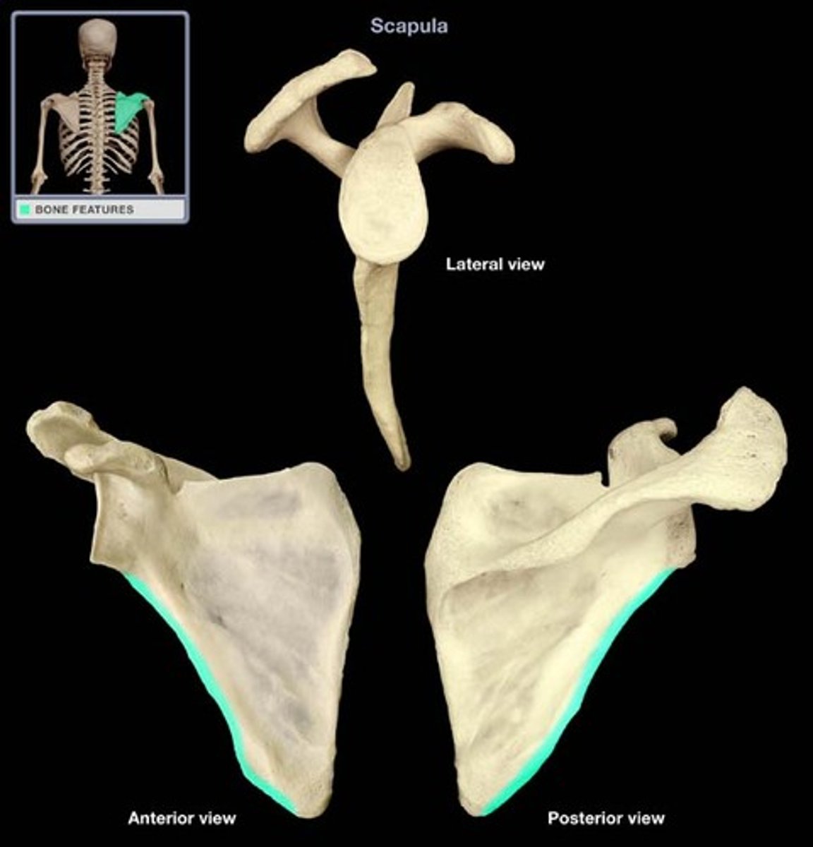
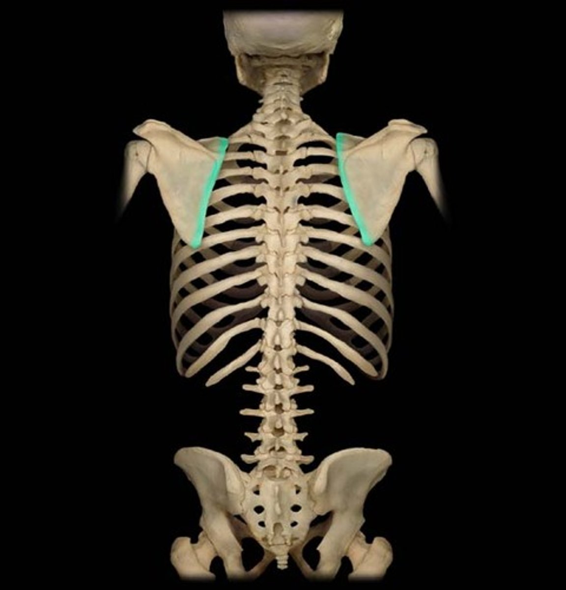
Lateral border of scapula
diagonally oriented lateral margin of the scapula
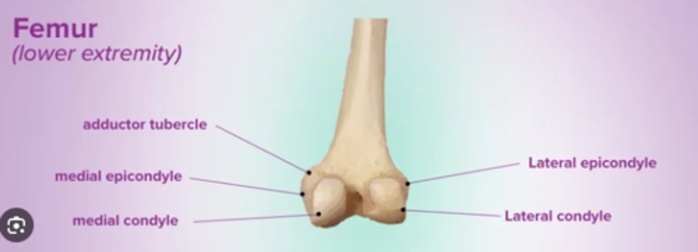
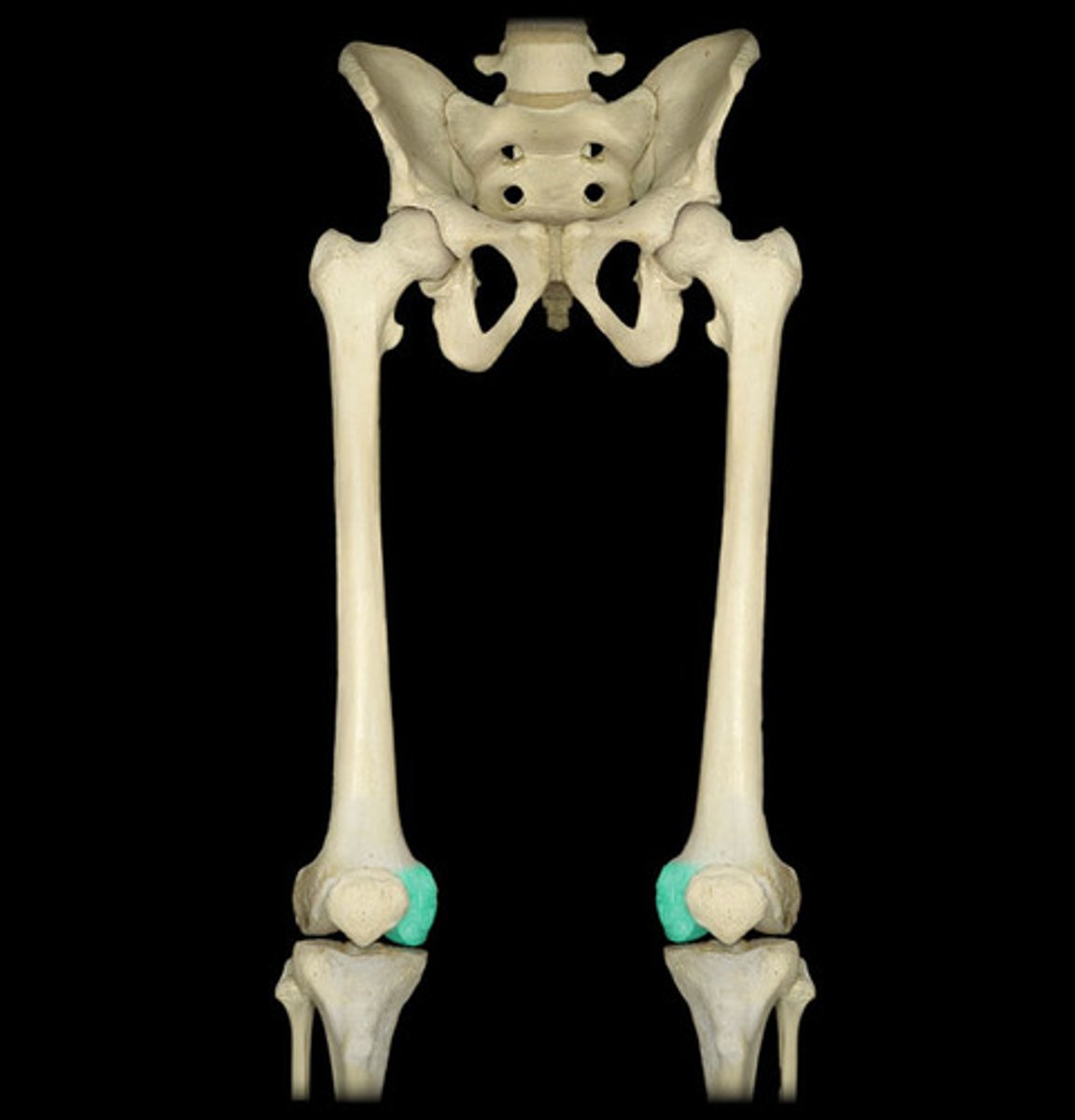
layer condyle of the femur
smooth, articulating surface that forms the distal and posterior sides of the lateral expansion of the distal femur
lateral condyle of the tibia
lateral, expanded region of the proximal tibia that includes the smooth surface that articulates with the lateral condyle of the femur as part of the knee joint
lateral epicondyle of the femur
roughened area of the femur located on the lateral side of the lateral condyle
Lateral malleolus
lateral support for ankle
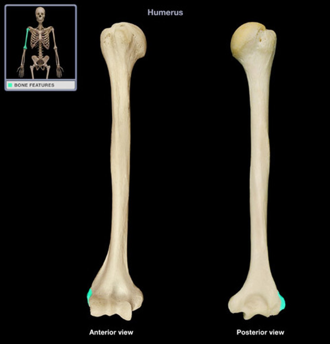
lateral epicondyle of humerus
small projection located on the lateral side of the distal humerus
lesser trochanter
small, bony projection on the medial side of the proximal femur, at the base of the femoral neck
lesser tubercle
small, bony prominence located on anterior side of the proximal humerus

Medial and lateral tibial condyles
articulates with condyles of femur
Medial border of scapula
elongated, medial margin of the scapula
medial condyle of the femur
smooth, articulating surface that forms the distal and posterior sides of the medial expansion of the distal femur
medial condyle of the tibia
medial, expanded region of the proximal tibia that includes the smooth surface that articulates with the medial condyle of the femur as part of the knee joint
medial epicondyle of the femur
roughened area of the distal femur located on the medial side of the medial condyle
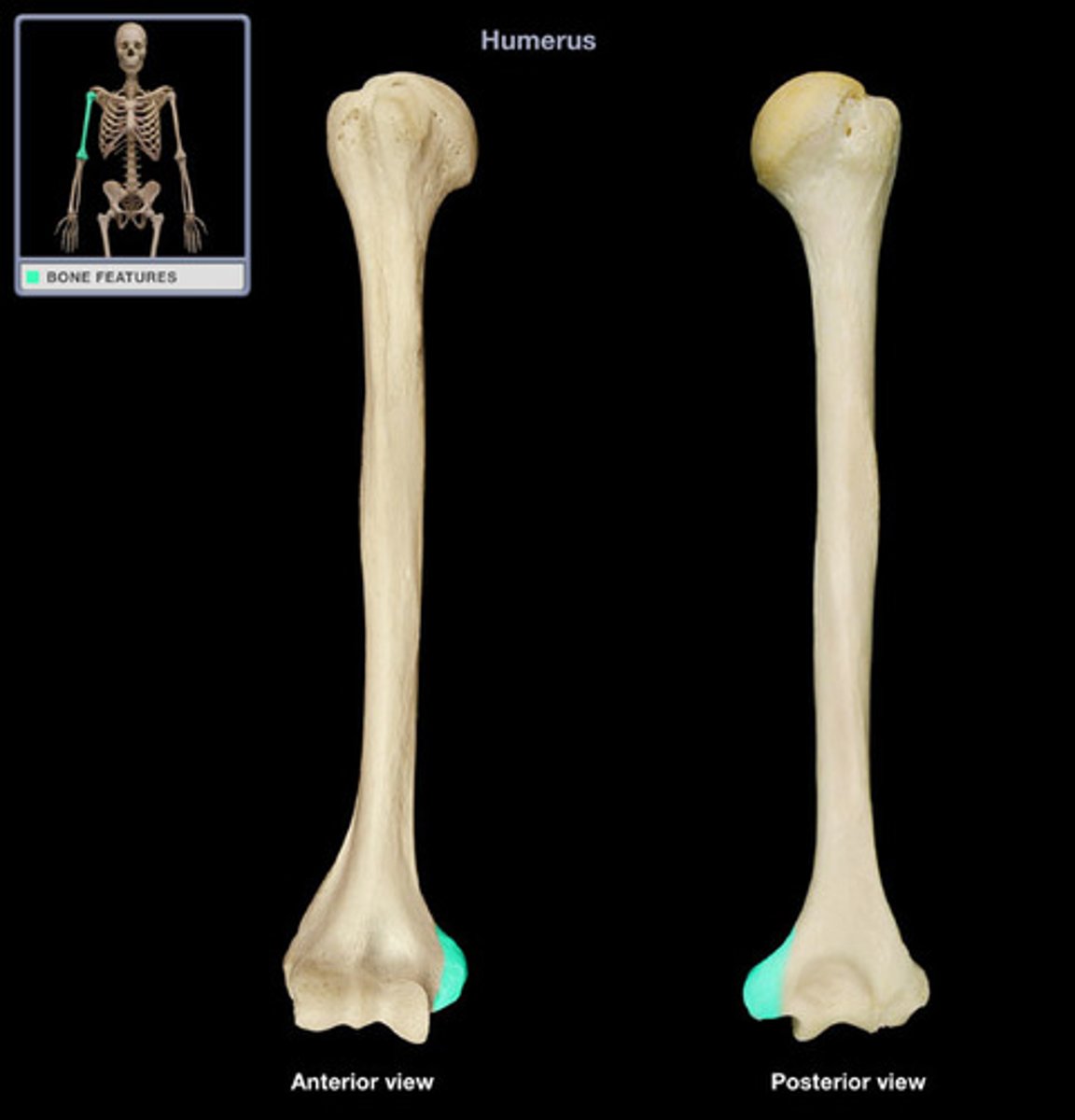
medial epicondyle of the humerus
enlarged projection located on the medial side of the distal humerus
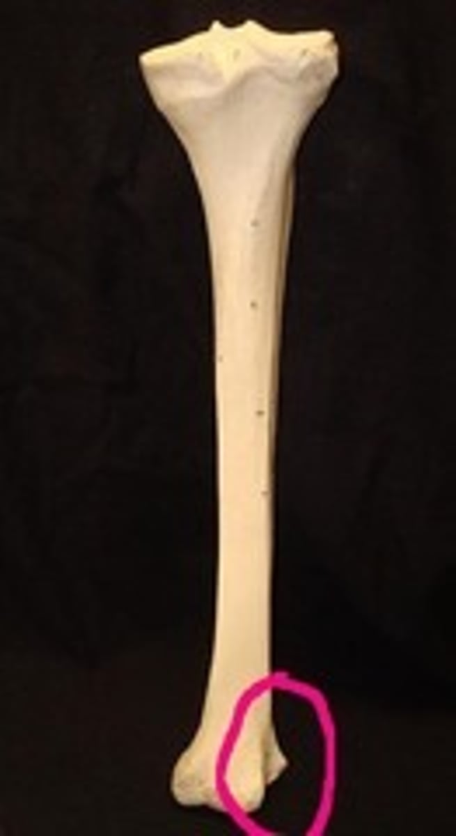
medial malleolus
bony expansion located on the medial side of the distal tibia
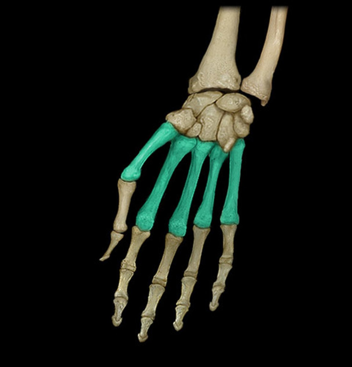
metacarpal bone
one of the five long bones that form the palm of the hand; numbered 1-5, starting on the lateral (thumb) side of the hand
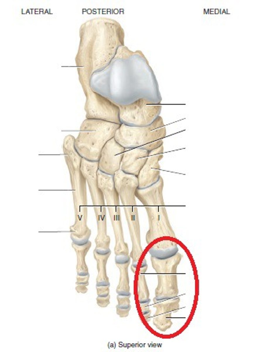
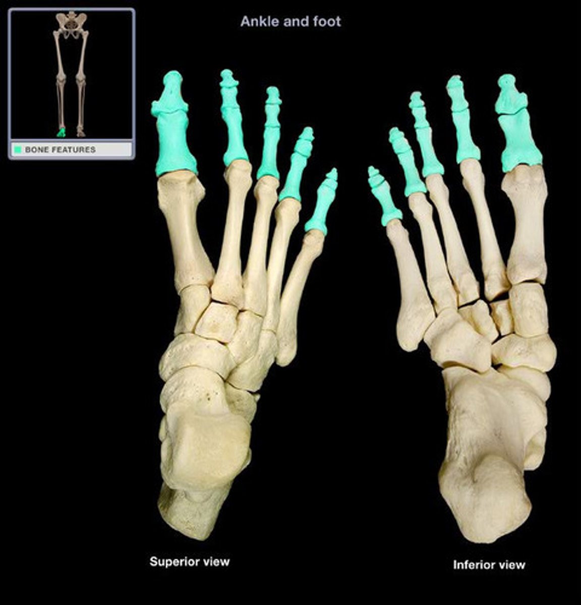
metatarsal bone
one of the five elongated bones that forms the anterior half of the foot; numbered 1-5, starting on the medial side of the foot
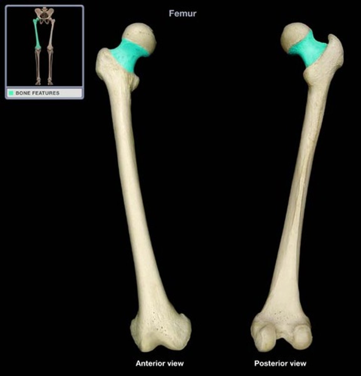
neck of femur
narrowed region located inferior to the head of the femur
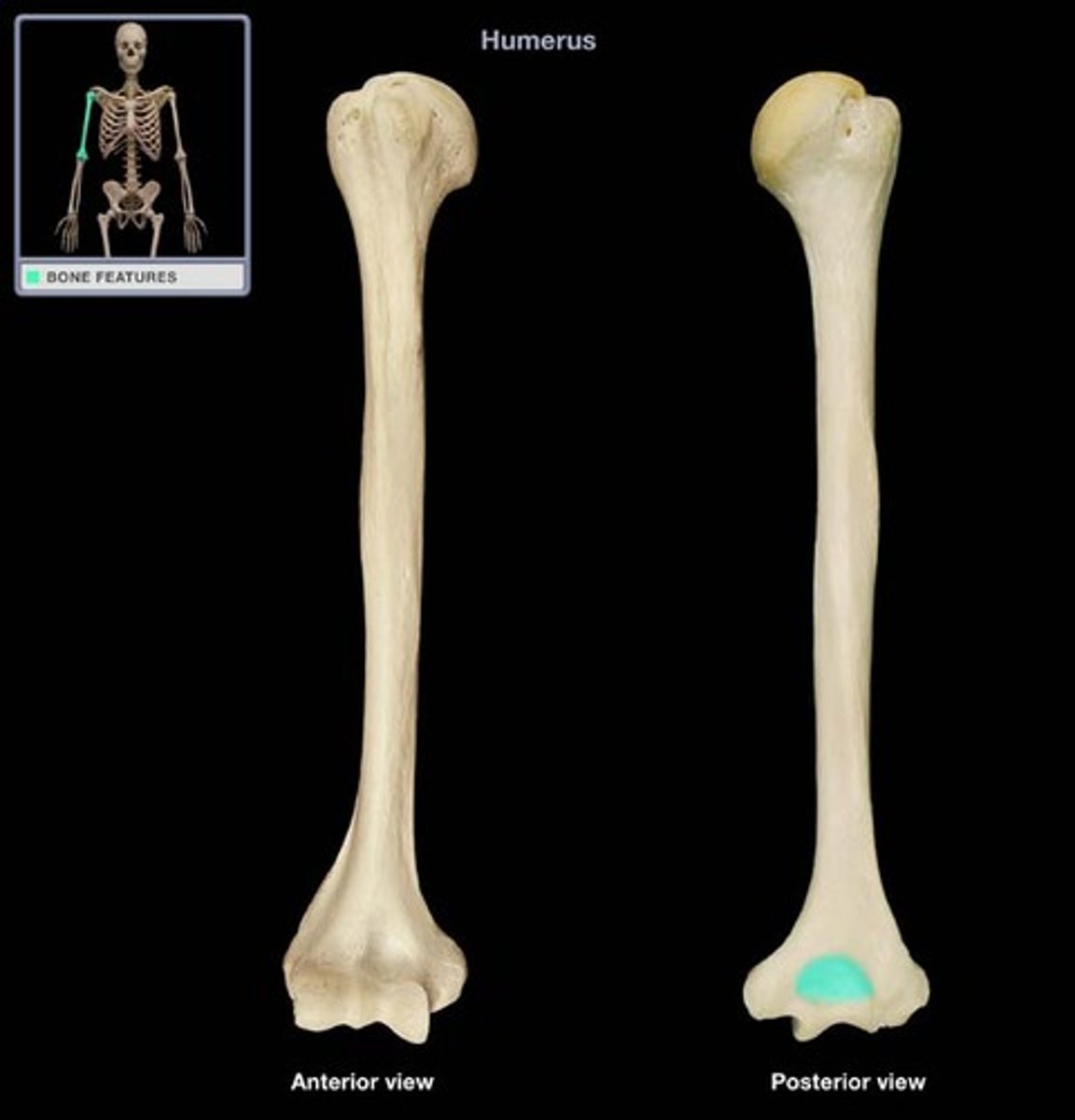
Olecranon fossa
room for olecranon of the ulna on humerus
Olecranon
swings into olecranon fossa at full extension
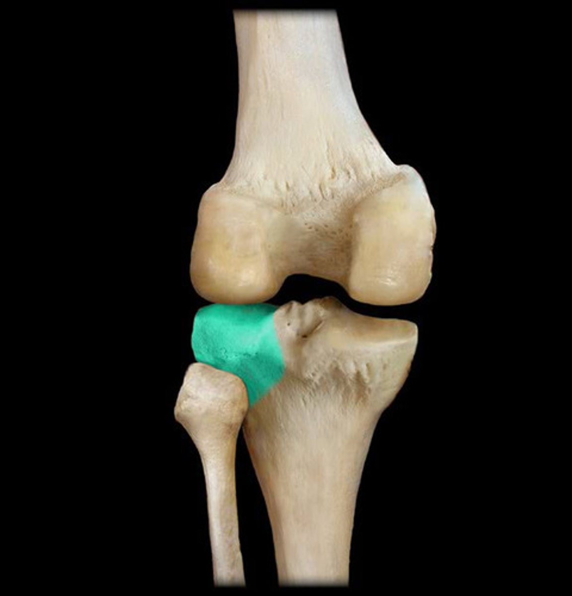
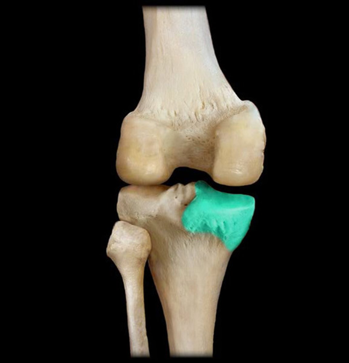
Patella
kneecap
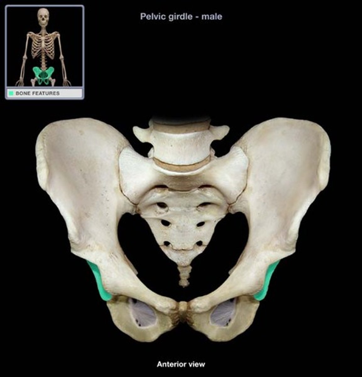
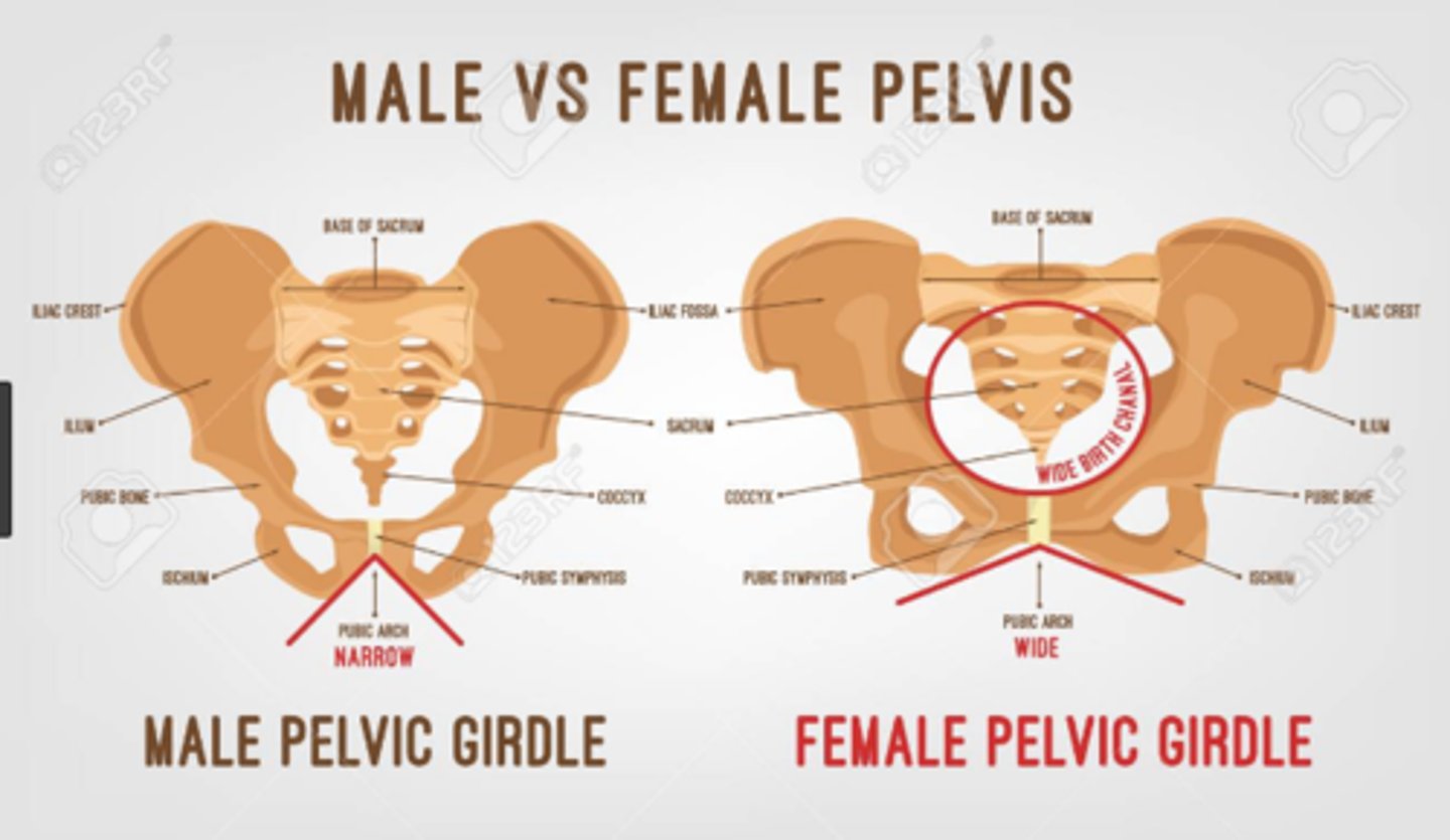
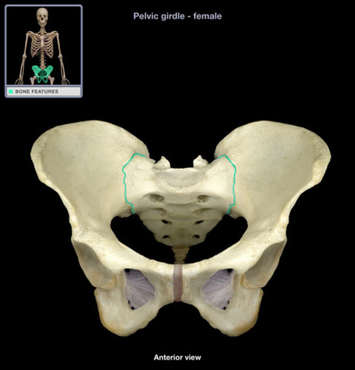
Pelvic sizes for male vs female
males have bigger pelvic bones where female have wider pelvis with greater distance between the ischial tuberosities
Phalanges of hand
14
Phalanges of the foot
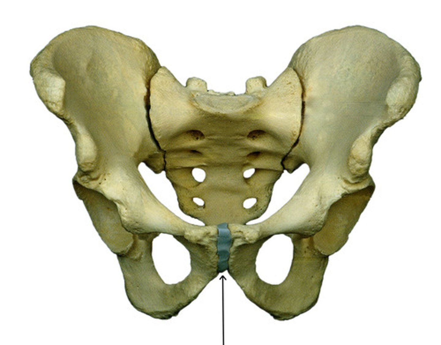
Pubic symphysis
median pad of fibrocartilage that connects the two pubic bones
Pubis
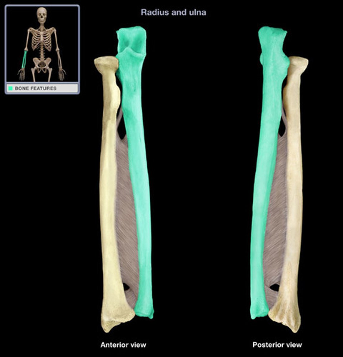
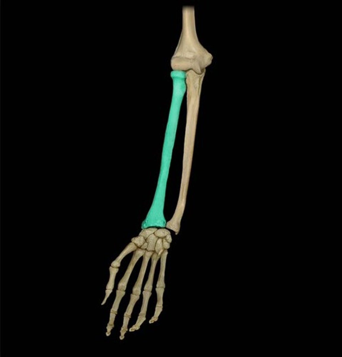
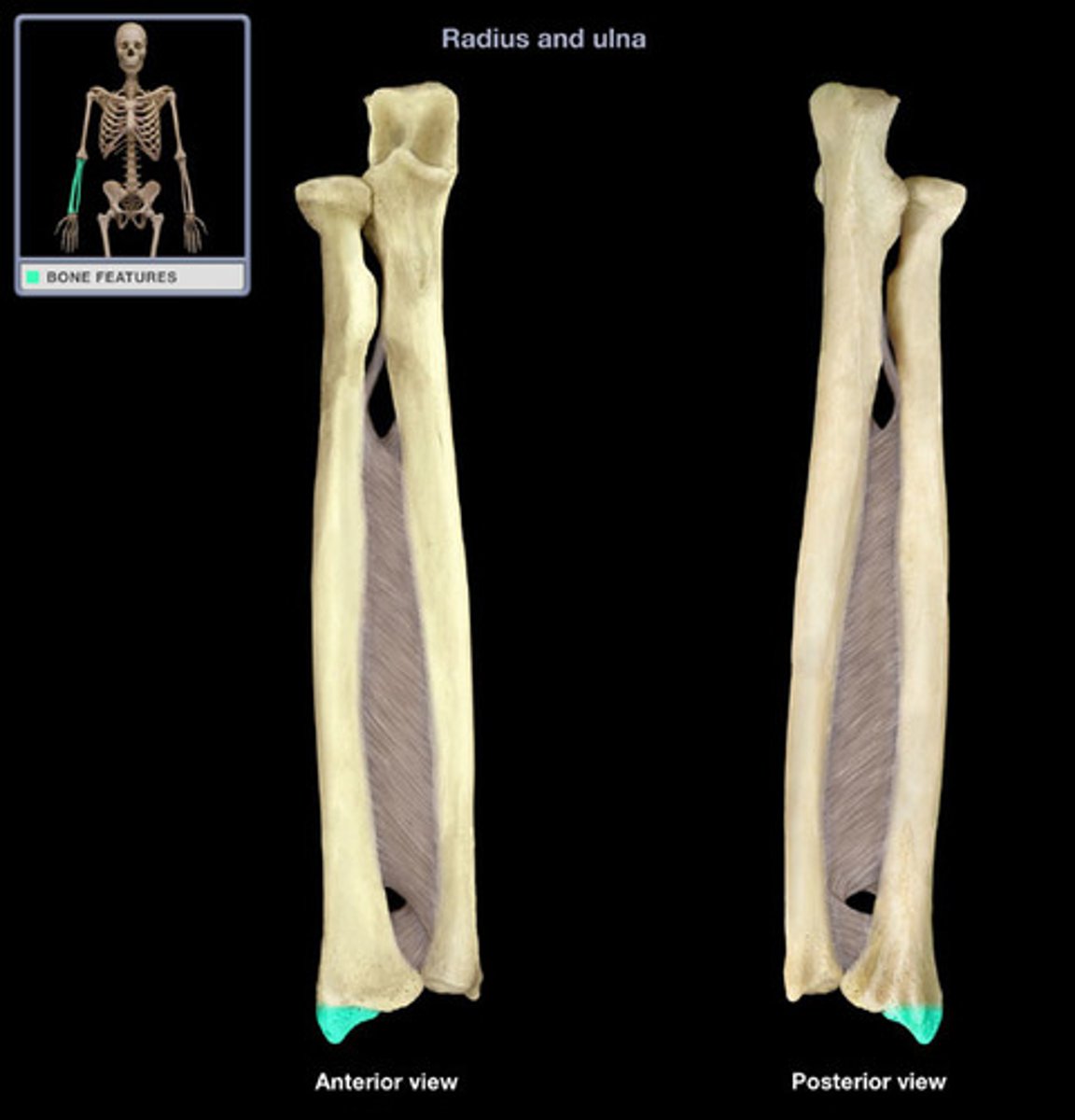
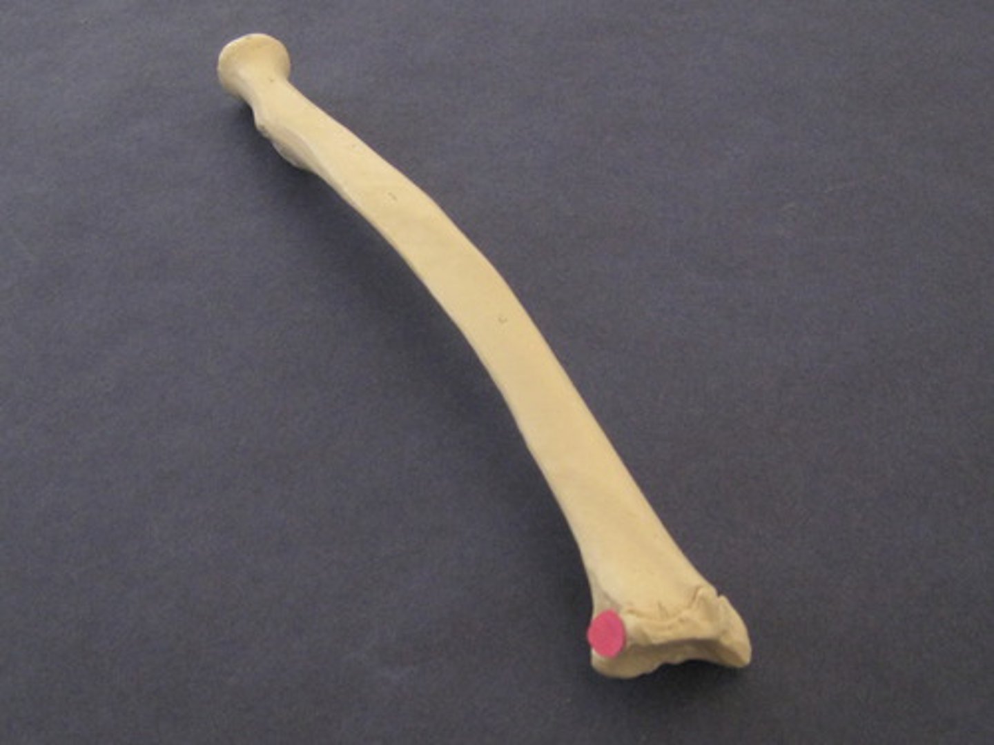
radius
bone located on the lateral side of the forearm
Radial fossa
room for radial head on humerus
Radial head
swings into radial fossa at humerus at flexion

Radial notch
room for head of radius on ulna
Sacroiliac joint
The connection point between the pelvis and the vertebral column.
Scapula
Sternal end of clavicle
articulates with sternum
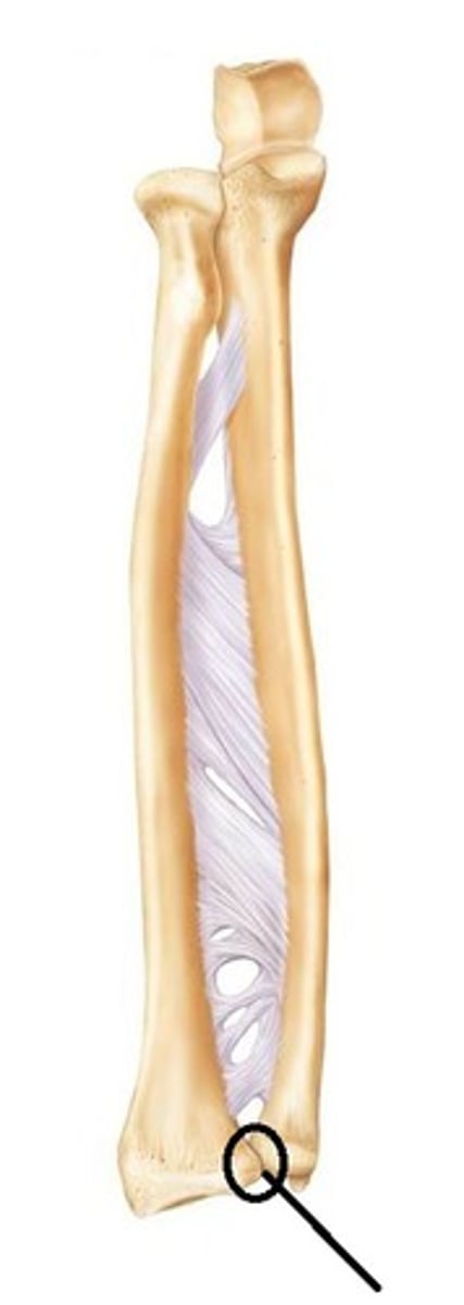
Styloid process of ulna
provides flexibility of wrist
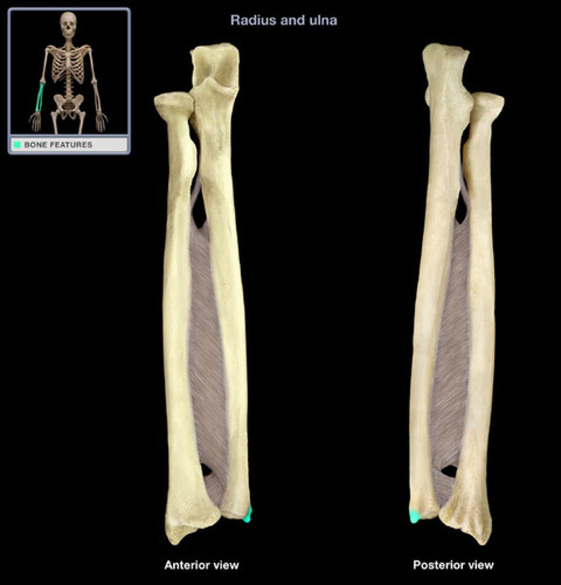
Styloid process of radius
articulates with wrist bones
Surgical neck of humerus
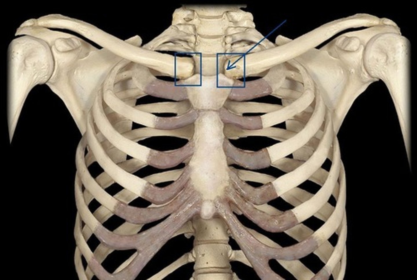
sternoclavicular joint
articulation between the manubrium of the sternum and the sternal end of the clavicle; forms the only bony attachment between the pectoral girdle of the upper limb and the axial skeleton
Talus
articulates with tibia
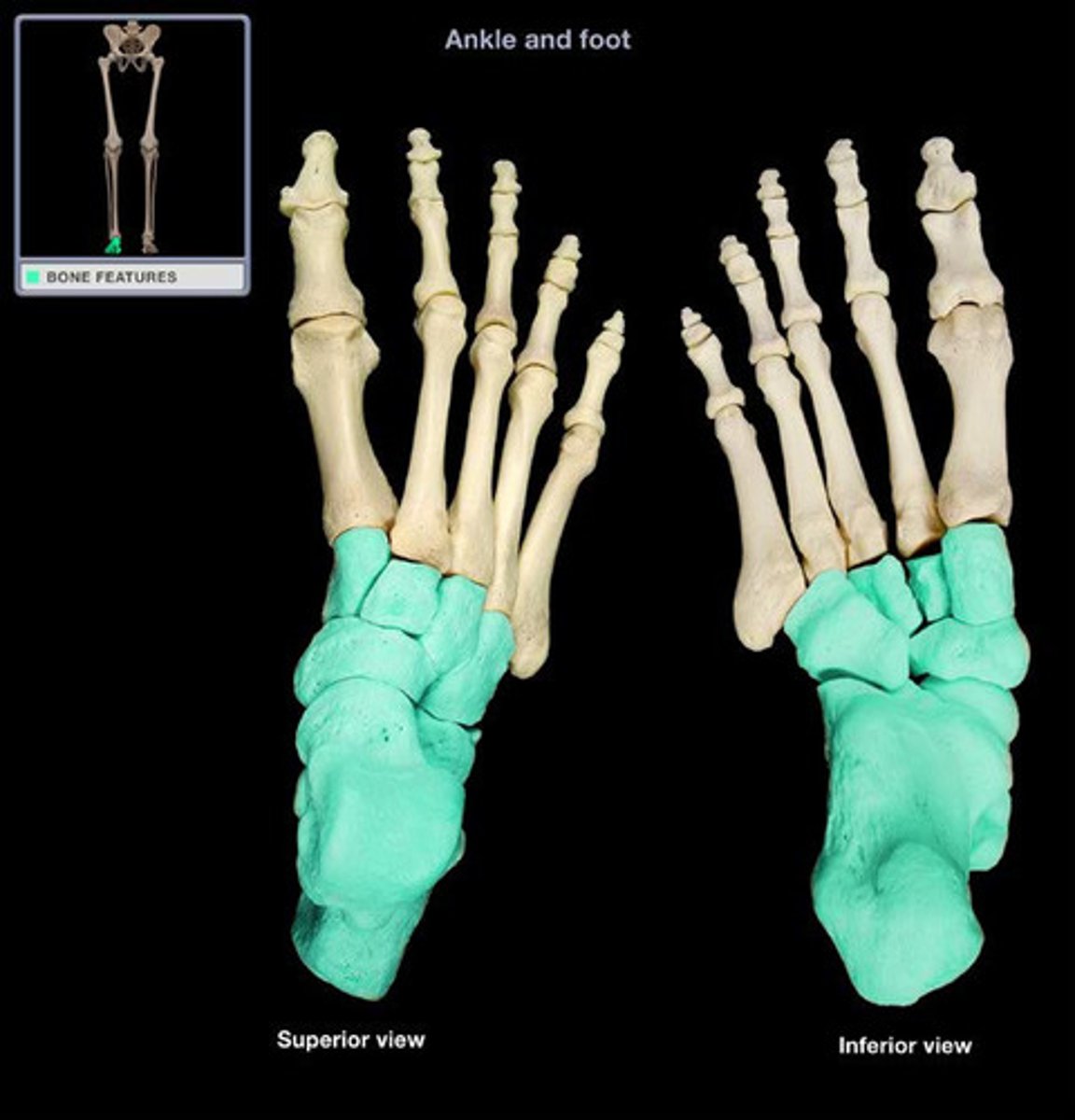
Tarsal bones
one of the seven bones that make up the posterior foot; includes the calcaneus, talus, navicular, cuboid, medial cuneiform, intermediate cuneiform, and lateral cuneiform bones
How many bones are in the appendicular skeleton
126 bones
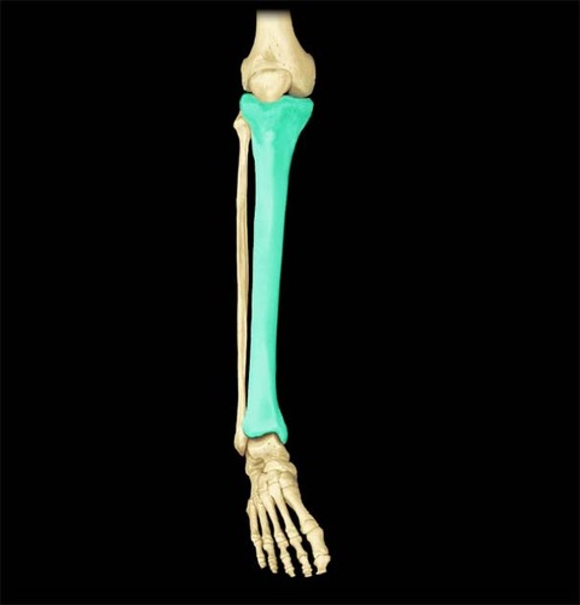
Tibia
the medial and larger bone of the lower leg
Tibial tuberosity
attachment of patellar ligament
Tibiofibular joint
pertaining to the joint between the tibia and fibula
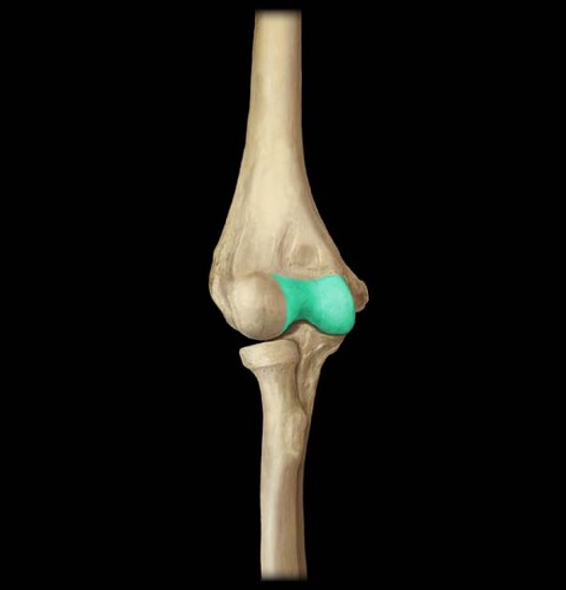
Trochlea of humerus
articulates with trochlear notch of ulna
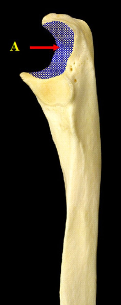
Trochlear notch
articulation between ulna and trochlea of the humerus at the elbow joint, found on ulna
trochlea
pulley-shaped region located medially at the distal end of the humerus; articulates at the elbow with the trochlear notch of the ulna
Ulna
medial bone of the forearm

Ulnar notch of the radius
shallow, smooth area located on the medial side of the distal radius; articulates with the head of the ulna at the distal radioulnar joint
Sternoclavicular joint
Articulation between the clavicle and the sternum
obturator foramen
opening in hip bone formed by the pubic and ischial rami