Chapter 28: Scrotal and Penile Sonography
1/15
There's no tags or description
Looks like no tags are added yet.
Name | Mastery | Learn | Test | Matching | Spaced | Call with Kai |
|---|
No analytics yet
Send a link to your students to track their progress
16 Terms
Scrotum Anatomy
Suspended from male pelvis between perineum and penis; contains: testis, testicular appendages, epididymis, proximal portion of vas deferens, and spermatic cord; sac of cutaneous tissue that supports testicles is divided internally and externally:
Externally - into lateral portions by median ridge called the median raphe
Internally - scrotum is divided into sacs by a septum called the dartos or tunica dartos (dartos contains superficial fascia and contractile tissue)

Cremaster Muscle
Surrounds each testicle and extends into abdomen over spermatic cord; covered by cremaster fascia; contraction of cremaster muscle performs the important function of regulating temperature of testicles
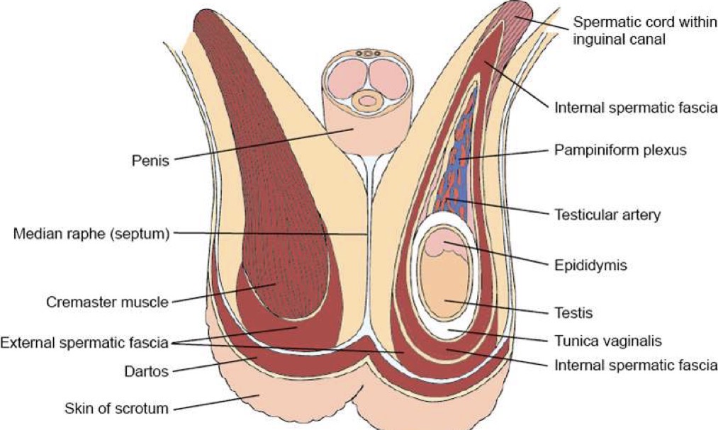
Tunica Vaginalis
Peritoneal sac composed of three layers; covers and surrounds testis and epididymis; outer parietal layer is closely attached to internal spermatic fascia; cavum vaginale is potential space between visceral and parietal layer; inner visceral layer that is closely attached to testicle

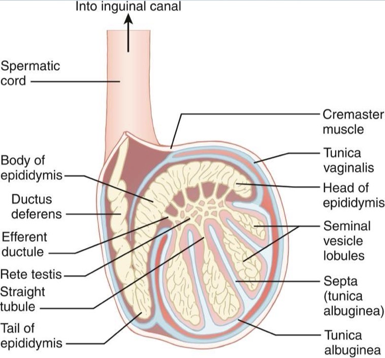
Testis Anatomy
Covered by dense, white fibrous tissue called tunica albuginea
Tunica albuginea extends into posterior wall of testicle; forms mediastinum testis and interlobar septa; septa of mediastinum radiate into testicle and separates into 200-300 lobules; each lobule contains one to three convoluted seminiferous tubules; seminiferous tubules are connected to straight tubules, which lead to the rete testis; rete testis is located within mediastinum testis; exits mediastinum as coiled efferent ducts
Normal Measurements (adult): 3-5cm length, 2-3cm width, 2-3cm height)
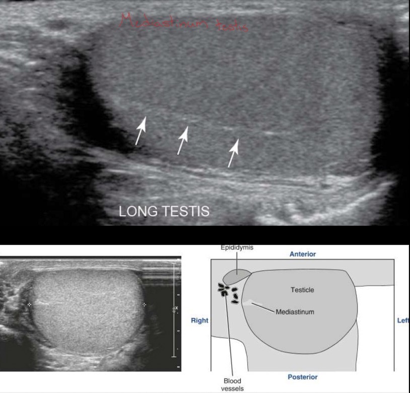
Sonographic Appearance of Testes
Homogeneous; medium-level echoes; hyperechoic linear structure is the mediastinum testis; low-resistance arterial blood flow; mediastinum testis – hyperechoic band running in cephalocaudad orientation in longitudinal plane and ovoid structure in transverse
Epididymis Anatomy
Posterolateral to testis; composed mostly of single convoluted tube, ductus epididymis; encapsulated by serosal layer; divided into the:
Head (globus major) – large superior portion, superolateral to testis (most common portion viewed on ultrasound)
Body – adjacent to posterolateral margin of testis
Tail (globus minor) – inferolateral to testis
Efferent ducts of the head empty into ductus deferens
Normal Measurements (adult): Head is 10-12mm AP and 5-12mm length, body is 2-4mm AP, and tail is 2-5mm AP
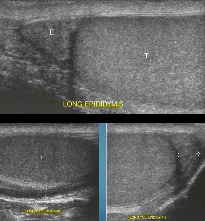
Sonographic Appearance of Epididymis
Iso/hypoechoic compared with normal testicle; coarse in appearance compared to normal testicle; head is superior and posterior to testicle, body courses posterior, and tail lies at inferior aspect
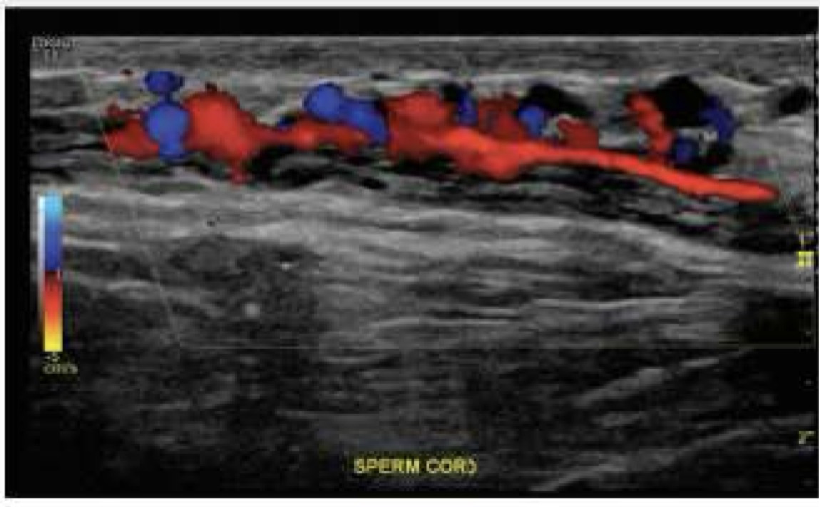
Spermatic Cord
Bilateral; extend from inguinal canal and internal inguinal ring into pelvis; each contains:
ductus deferens
testicular arteries (on image)
venous pampiniform plexus (on image)
lymphatics
autonomic nerves
fibers of cremaster muscle
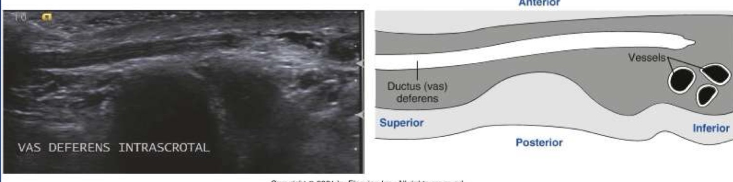
Ductus/Vas Deferens
thicker, less convoluted continuation of the ductus epididymis; three smooth muscle layers contribute to this duct’s increased thickness; runs in the spermatic cord through scrotum and inguinal canal; unites with seminal vesicle posterior to bladder to form ejaculatory duct; at terminal portion near seminal vesicles, the ductus deferens dilates; divided into four segments:
Scrotal – inferior
Suprascrotal – spermatic cord
Prepubic – inguinal region
Pelvic – posterior urinary bladder
Normal Measurements: 45cm length
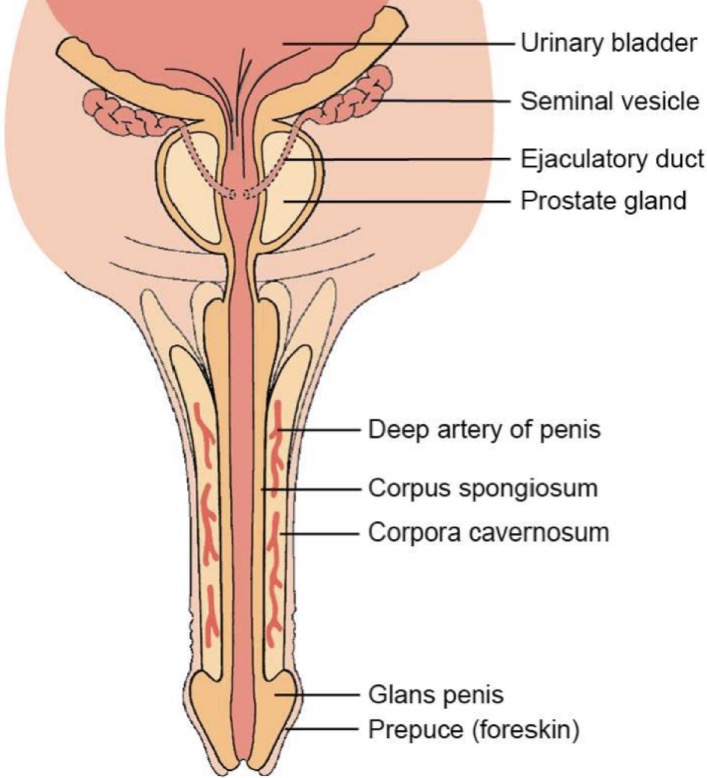
Penis Anatomy
Composed of 3 cylindrical masses of tussue:
• Two corpora cavernosa situated dorsolaterally
• Single corpus spongiosum in midventral region, which contains the spongy urethra
Bound and separated by tunica albuginea
Buck’s fascia: superficial to tunica albuginea and is a thick, fibrous, loosely applied covering of skin that envelops penis
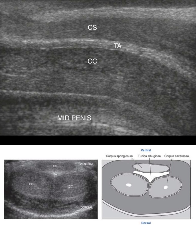
Sonographic Appearance of Penis
Corpus Spongiosum: lies anterior to the paired corpus cavernosa; homogeneous texture composed of medium-level echoes
Tunica Albuginea: highly echogenic covering of the corpus cavernosa
Septum Penis: echogenic plane dividing the two corpora cavernosa; extension of the tunica albuginea
Paired Corpora Cavernosa: Lie posterior to the corpus spongiosum and appear symmetrical, round or oval, with a medium-level homogeneous echo texture
Testis Vasculature
Arterial
Testicular arteries provide primary blood supply; arise from anterior AO; in spermatic cord, testicular artery accompanied by deferential artery and cremasteric artery
Deferential – supplies epididymis and vas deferens
Cremasteric – supplies peritesticular tissue
Superior epididymal artery supplies epididymis
Venous
Via pampiniform plexus; begins in scrotum with veins arising from mediastinum testis; plexus reduces to single vein – testicular vein as it ascends through inguinal canal; RIGHT testicular vein drains into IVC and LEFT testicular veins drains into LRV
Physiology
Testicles are classified as both endocrine and exocrine glands; testes produce testosterone at puberty which causes growth of male sex organs and secondary male sex characteristics; testes also produce spermatozoa which are transported through ducts (exocrine) that store and transport them; production of sperm one of most important functions
Testis Variants
Cryptorchidism is failure of testicles to descend into scrotum; common locations of undescended testes include: Inguinal canal, External inguinal ring, and Abdomen
Sonographic Application
Scrotum exams indicated for testicular size, inflammatory processes (epididymitis, orchitis), presence and composition of masses, detection of peritesticular fluid collections (hydrocele), evaluation of scrotal trauma, doppler evaluation to rule out testictesticular torsion, evaluation of scrotal pain, or location of undescended testicles
Penis exams indicated for detection of fibrosis (Peyronie’s disease) or scar tissue and plaques, evaluation of tumors or periurethral disease, penile hematoma, or doppler evaluation of vasculogenic impotence
Associated Tests
MRI, CT, or nuclear medicine