Bio II Exam 3
1/103
There's no tags or description
Looks like no tags are added yet.
Name | Mastery | Learn | Test | Matching | Spaced | Call with Kai |
|---|
No analytics yet
Send a link to your students to track their progress
104 Terms
Two types of symmetry
Radial and bilateral
Radial Symmetry
Body parts arranged around central axis
Can be bisected into 2 equal halves at any 2D plane
Bilateral Symmetry
Body has right and left halves that are mirror images
Only the sagittal plane bisects the animal into 2 equal halves
Sagittal Plane
Divides the body into right and left portions
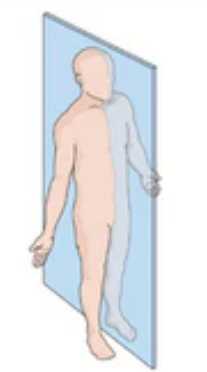
Midsagittal Plane
Divides the body into EQUAL right and left portions
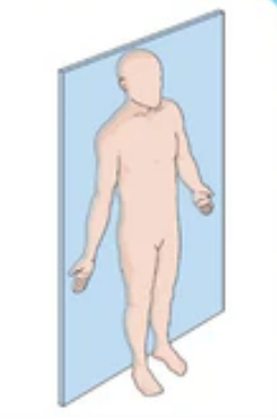
Frontal Plane (AKA Coronal Plane)
Separates the front from the back
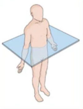
Transverse plane (horizontal plane)
Divides into upper and lower portions
AKA cross-section
Called oblique plane if cut at an angle
Basal Metabolic Rate
Average amount of energy used by an organism in a non-active state
Smaller animals have a higher BMR to compensate for heat lost from a higher surface-area-to-volume ratio
4 types of animal tissue
Epithelia
Connective Tissue
Muscles
Neurons
Epithelial Tissue
Lines cavities, open spaces and surfaces
Secretion
Classified by number of layer and shape of the cell
Types of Epithelial Tissue
Squamous, cuboidal, columnar, transitional
Connective Tissue
Connects tissue together, provides support
Arises from the mesoderm
Consists of cells (fibroblasts) embedded in a non-cellular matrix
Matrix usually composed of a ground substance
Ground Substance
Composed of collagen, elastic, and reticular fibers
Cartilage
Connective Tissue
Chondrocytes and chondroblasts
Makes up fetal bones, intervertebral discs, joint spaces
Bone
Connective Tissue
Osteoblasts, osteocytes, osteoclasts
Makes up vertebrates’ skeletons
Adipose
Connective Tissue
Adipocytes
Energy storage and insulation
Blood
Connective Tissue
red blood cells (erythrocytes) and white blood cells (leukocytes) and platelets (thrombocytes)
Immune response and oxygen transport
Muscle Tissue
Generates movement
Types of Muscle Tissue
Smooth, skeletal, cardiac
Incomplete digestive tract
Single opening = gastrovascular cavity
Food enters through mouth and muscular pharynx
Waste exits through mouth and muscular pharynx
Lacks specialized parts
Complete digestive tract
Two openings = alimentary canal
Food enters through mouth
Wastes exit through anus
Monogastric Digestive Systems
Humans and herbivores
Rabbits have a distended cecum
Ruminant digestive system
Four stomachs
Rumen, reticulum, omasum, and abomasum
Rumen and reticulum
Two of four stomachs of ruminants
Contains prokaryotes and protists to digest cellulose fiber
Omasum
Third ruminant stomach
Cud is regurgitated, chewed and swallowed
Removes water
Abomasum
Stomach where enzymes are produced by animal (most like monogastric stomachs)
Bird digestive system
Crop to store food
Two stomachs:
Proventriculus (enzymes)
Gizzard (grinding)
One opening (cloaca) to excrete urine and feces
Human Digestive Tract
complete (alimentary canal)
tube-within-a-tube body plan
digestion is entirely extracellular
digestive enzymes secreted by walls of digestive tract and nearby glands
Mouth
Three major pairs of salivary glands
Salivary amylase initiates starch digestion
Tongue mixes chewed food with saliva and forms bolus
Pharynx
Where digestive and respiratory passages come together
Epiglottis covers opening into trachea and keeps food from air passages
Esophagus
Takes food to stomach by peristalsis
Peristalsis is rhythmical contraction to move contents in tubular organs
Human Stomach
Stomach wall has deep folds called rugae
Gastric pits drain gastric glands
Gastric glands produce pepsin, a hydrolytic enzyme that acts on protein to produce peptides
Junction between stomach and small intestine controlled by pyloric sphincter
Chyme
Food (bolus) mixing with gastric juices
Small intestine
First segment is duodenum which chemically breaks down food (chyme) from the stomach
Liver
Produces bile which is stored in gallbladder
bile contains bile salts which break up fat into droplets via emulsification
helps maintain glucose concentration in blood by converting excess into glycogen
Pancreas
Exocrine gland
Produces pancreatic juice and digestive enzymes into the duodenum
Pancreatic amylase digests starch to maltose
Trypsin digests protein to peptides
Lipase digests fat droplets to glycerol and fatty acids
Large Intestine
Includes cecum, colon, rectum and anal canal
Larger in diameter but shorter in length than small intestine
Absorbs water, salts and some vitamins to dehydrate chyme
Essential amino acids for adults
Methionine, valine, threonine, phenylalanine, leucine, isoleucine, tryptophan, lysine
Fat-soluble vitamins
A, D, E, K
Vitamin A
Fat soluble
promotes eye health, helps form and maintain healthy skin, teeth and bones
Vitamin D
Fat soluble
Helps the body absorb calcium, maintains strong bones
Vitamin E
fat soluble
Antioxidant, boosts immune function
Vitamin K
fat soluble
aids in blood clotting
B vitamins
Water soluble
help the body produce energy, influence growth/development
Vitamin C
water soluble
boosts immune function, fights skin aging, antioxidant
Osmoregulation
Process of maintenance of salt and water balance across membranes within the body’s fluids
Kidneys
Located on either side of vertebral column, each connected to a ureter which connects urine from the kidney to the bladder
Tubular nephrons
Functional unit of the kidney
Composed of glomerular capsule, glomerulus, proximal convoluted tubule, loop of the nephron/Henle, distal convoluted tube, and collecting duct
Renal cortex
Outer region of the kidney, granular appearance
Renal medulla
Cone-shaped renal pyramids of kidney
Renal pelvis
Hollow-chambered innermost part of the kidney, receptive for collecting ducts
Processes of Urine Production
Glomerular filtration in glomerular capsule, tubular reabsorption (water) in proximal convoluted tubule, tubular secretion (and removal of sodium and chloride) at distal convoluted tube.
Excretion of hypertonic urine
Dependent on reabsorption of water from the loop of nephron and collecting duct
Antidiuretic hormone
Plays a role in water reabsorption, and released by posterior lobe of the pituitary gland in the brain
Contraction of EXTERNAL INTERCOSTAL MUSCLES
Expands the rib cage
Contraction of DIAPHRAGM
Expands the volume of thorax and lungs
Diaphragm is primary muscle of inspiration
Vital capacity
Maximum amount of air that can be expired after a forceful inspiration (you’ll almost always have a little left over)
Hemoglobin
Consists of four polypeptide chains, two alpha and two beta
Each chain is associated with a heme group that has a central iron atom that can bind a molecule of O2, forming oxyhemoglobin
Bohr shift
Increased CO2 in blood increases H2, lowering pH and reducing hemoglobin’s affinity for O2
High levels of CO2 mean oxygen is needed so the hemoglobin lets go of the oxygen
Central Nervous System
Includes the brain and spinal cord
Lies in the midline of the body
Peripheral Nervous System
Contains cranial nerves and spinal nerves that gather info from sensors and conduct decisions to effectors
Lies outside the CNS
Functions of the Nervous System
Receiving sensory input, performing integration, and generating motor output
Neurons
Cell body contains nucleus
Dendrites receive signals from sensory receptors
Axon conducts nerve impulses and is covered by myelin sheath that assists with action potential
Motor Neurons
Accept nerve impulses from the CNS (primary motor cortex in the frontal lobe) and transmits them to muscles or glands
Efferent (starts in brain and leaves through nervous system)
Sensory Neurons
Accepts impulses from sensory receptors and transmits them to the CNS
Afferent (starts in PNS and travels toward CNS then brain)
Four types of neurons
Unipolar neurons, bipolar neurons, multipolar neurons, and pseudounipolar neurons
Glial Cells
Support, protect, and nourish neurons
Outnumber neurons 10 to 1 in the brain
Fulfills immunity, making myelin sheaths, and nutritional support
Most brain tumors are caused by mutations in glia
Oligodendrocytes
CNS
Form myelin sheaths around axons
Astrocytes
CNS
Provides nutrients and structural support
Ependymal cells
CNS
Produce cerebrospinal fluid that cushions the neurons
Microglia
CNS
Scavenge pathogens and dead cells and immune support
Schwann cells
PNS
Forms the myelin sheath
Satellite cells
PNS
Provides nutrients and structural support to neurons