Bio 011 Lab Exam 3
1/107
There's no tags or description
Looks like no tags are added yet.
Name | Mastery | Learn | Test | Matching | Spaced | Call with Kai |
|---|
No analytics yet
Send a link to your students to track their progress
108 Terms

Identify A
Parietal bone

Identify B
Frontal bone

Identify C
Sphenoid bone

Identify D
Nasal bone

Identify E
Ethmoid bone

Identify F
Lacrimal bone

Identify G
Temporal bone
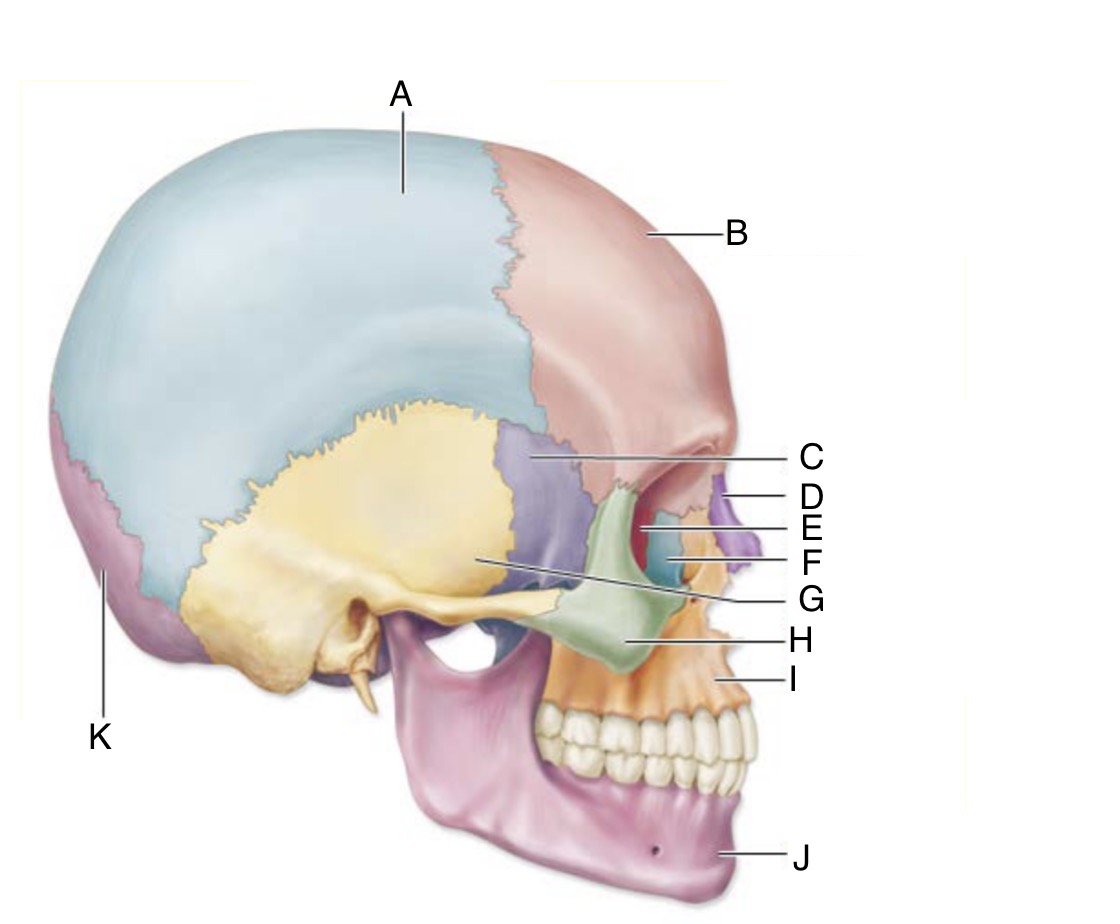
Identify H
Zygomatic bone

Identify I
Maxilla

Identify J
Mandible

Identify K
Occipital bone

Identify A
Seven cervical vertebrae (breakfast at 7a)

Identify B
Twelve thoracic vertebrae (lunch at noon)

Identify C
Five lumbar vertebrae (dinner at 5p)

Identify D
Five fused lumbar vertebrae
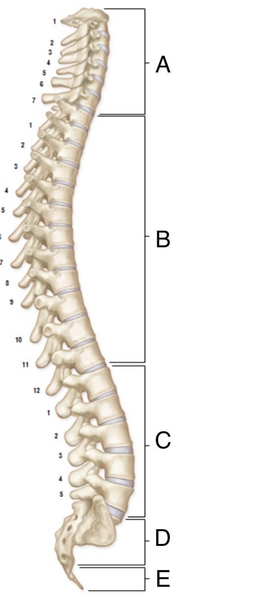
Identify E
Four fused coccyx vertebrae (usually 3-5)
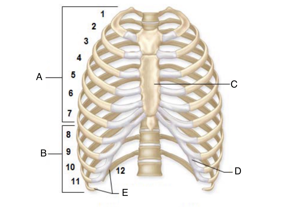
Identify A
True ribs (1-7), which are directly connected to the sternum in the front of the rib cage by costal cartilage

Identify B
False ribs (8-12), which either have an indirect connection to sternum or none at all. 8-10 are connected indirectly by way of costal cartilage belonging to the rib above. 11-12 are not connected to the sternum and are referred to as floating ribs.

Identify C
Sternum

Identify D
Costal cartilage

Identify E
Floating ribs

Identify A
Clavicle/collarbone

Identify B
Scapula/shoulder blade

Identify C
Humerus (arm bone)
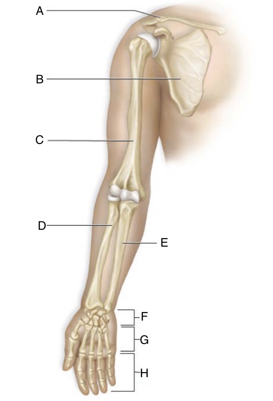
Identify D
Radius (forearm bone)

Identify E
Ulna (forearm bone)

Identify F
8 carpals (wrist bones)

Identify G
5 metacarpals (palm bones)

Identify H
14 phalanges (finger bones)

Identify A
Coxal bone, which is a fusion of three bones: ilium, ischium and pubis

Identify B
Ilium, part of the coxal bone
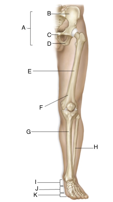
Identify C
Pubis, part of the coxal bone

Identify D
Ischium, part of the coxal bone

Identify E
Femur (thigh bone)

Identify F
Patella/kneecap

Identify G
Tibia (leg bone)

Identify H
Fibula (leg bone)

Identify I
7 tarsals (ankle bones)

Identify J
5 metatarsals (feet bones)

Identify K
14 phalanges (toe bones)
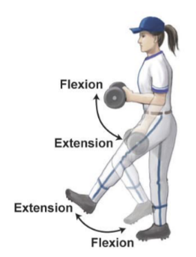
Flexion and extension
Decreasing the angle of a joint and increasing the angle of a joint
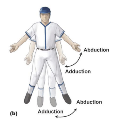
Abduction and adduction
Moving a body part away from the body and moving the body part toward the body
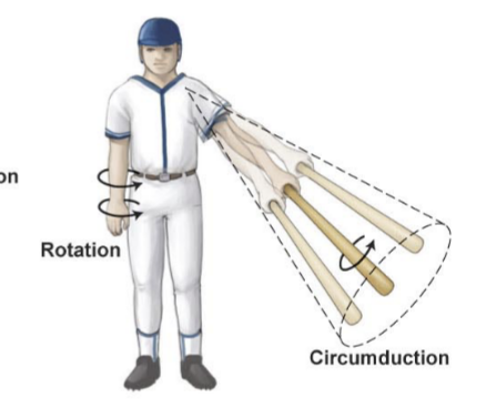
Lateral and medial rotation
Rotating a body part from front to side of the body, and rotating it back to the front of the body
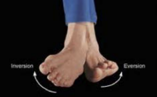
Inversion and eversion
Inverting feet so the bottoms face each other, and everting so the bottoms face away from each other.

Identify A and describe function
Orbicularis oculi: responsible for closing the eye and crow’s feet

Identify B and describe function
Closes the lips (as in kissing)

Identify C and describe function
Pectoralis major, medially rotates arm

Identify D and describe function
Serratus anterior: abducts scapula

Identify E and describe function
External oblique: compresses abdomen and flexes spine

Identify F and describe function
Quadriceps femoris: flexes thigh and extends leg

Identify G and describe function
Tibialis anterior: flexes foot toward shin (turns foot upward)

Identify H and describe function
Masseter: elevates the jaw and clenches teeth for chewing

Identify I and describe function
Deltoid: abducts, flexes and extends arm
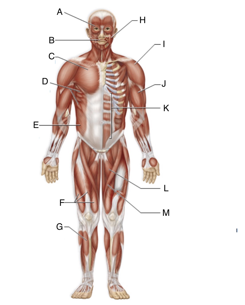
Identify J and describe function
Biceps brachii: flexes forearm and bends forearm at elbow

Identify K and describe function
Rectus abdominis: compresses abdomen and flexes spine

Identify L and describe function
Adductor longus: moves thigh toward midline, flexes and laterally rotates thigh

Identify A and describe function
Trapezius: raises scapula and extends neck

Identify B and describe function
Latissimus dorsi: extends, adducts and medially rotates arm

Identify C and describe function
Triceps brachii: straightens forearm at elbow to extend the arm

Identify D and describe function
Gluteus maximus: extends thigh back and laterally rotates it
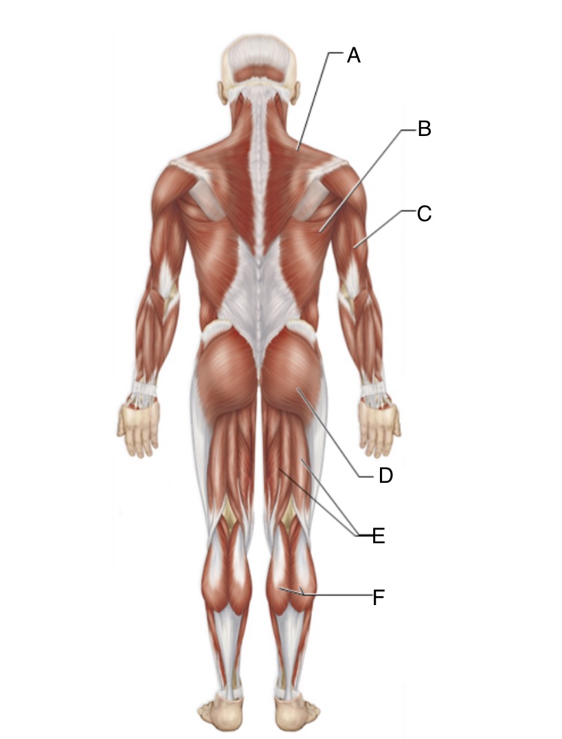
Identify E and describe function
Hamstrings: bends leg at knee (extends thigh) and flexes leg

Identify F and describe function
Gastrocnemius: turns foot downward and bends leg at knee
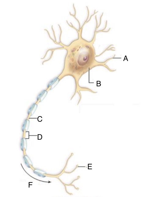
Identify A and describe function
Dendrites, where signals are inputted.

Identify B and describe function
Cell body, where the nucleus is

Identify C and describe function
Axon (propagates/moves the signal down the cell)

Identify E and describe function
Axon terminal with terminal boutons (the output region)
What forms the myelin sheath?
Glial cells called Schwann cells make up the sheath.
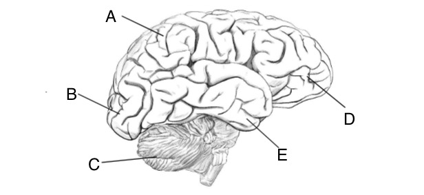
Identify A and describe function
The parietal lobe, involved in somatosensation: interpreting sensations coming from the skin and joints.

Identify B and describe function
Occipital lobe: visual processing area

Identify C and describe function
Cerebellum, a smaller version of the cerebrum which receives information from eyes/ears about body position and movement, and maintains balance and equilibrium.

Identify D and describe function
Frontal lobe, involved in motor control

Identify E and describe function
Temporal lobe, interprets hearing signals
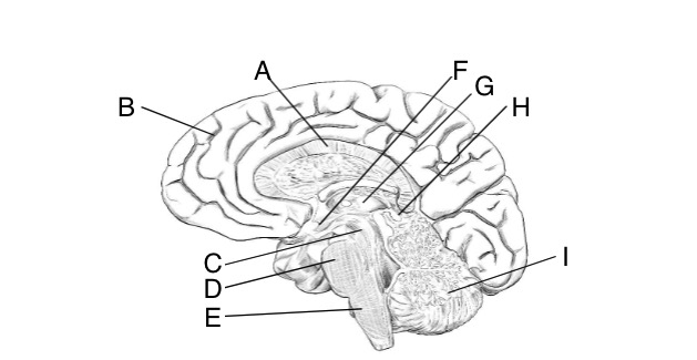
Identify A and describe function
Corpus callosum, which connects the two halves of the cerebrum and allows for communication between the them, transporting nerve signals for multiple senses, movement, and cognitive function.

Identify B and describe function
Cerebellum, maintains balance and equilibrium.

Identify C and describe function
Midbrain, responsible for auditory and visual reflexes.

Identify D and describe function
Pons, which is a sensory and motor relay area. Transmits sensory signals up to the brain and motor signals down to the spinal cord.

Identify E and describe function
Medulla oblongata, with cardiovascular and respiratory control centers which modulate how the heart, blood vessels, and lungs function.

Identify F and describe function
Hypothalamus, which contains clusters of cells involved in body functions that keep us alive. Tells us when to eat, drink, maintains body temp, controls s*xual behavior/aggression.

Identify G and describe function
Thalamus, a sensory relay area transmitting sensory signals to respective parts of the cerebrum, except for smell.

Identify H and describe function
Epithalamus, which has a functional gland called the pineal gland. This secretes the hormone melatonin, which makes us sleepy when it is dark outside.

Identify I and describe function
Also the cerebellum, balance and equilibrium.

Identify A and describe function
Gray matter, where synapses form.

Identify B and describe function
Dorsal horn, where sensory information is processed

Identify C and describe function
Ventral horn, where motor information is processed

Identify D and describe function
White matter, made of myelinated axons and allows for fast movement of nerve impulses up and down the cord
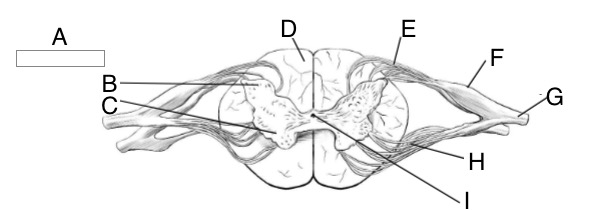
Identify E and describe function
Dorsal nerve root, which carries sensory information to the spinal cord.

Identify F and describe function
Dorsal root ganglion, where cell bodies of sensory neurons are.

Identify G and describe function
Spinal nerves, formed by dorsal and ventral nerve roots. Function is to leave the vertebral column and innervate the organs of the body.

Identify H and describe function
Ventral nerve root, carrying motor information from the cord.

Identify I and describe function
Central canal, a hole containing cerebrospinal fluid which bathes the brain and spinal cord.

Identify A and describe function
Ciliary body, surrounds the iris and is made of smooth muscle. The muscle is attached to the lens and can alter the lens shape.

Identify B and describe function
Cornea, which has a curvature that bends the light rays when they enter eye.

Identify C and describe function
Aqueous humor, fluid sitting in front of the lens. Keeps eye inflated with proper ocular pressure, in addition to helping focus light as it passes through the layer.

Identify D and describe function
Pupil, which constricts to limit light entering the eye, and dilates (opens) to increase the amount of light entering the eye.

Identify E and describe function
Iris, made of smooth muscle and con contract/relax when necessary to open or close the hole in the center, called the pupil.

Identify F and describe function
Lens, attached to the ciliary body smooth muscle. It is the protein-based focuser of the eye.

Identify G and describe function
Sclera, the white of the eye made of tough, protective tissue holding the eye together. It protects the delicate structures within the eye.

Identify H and describe function
Vitreous humor, a clear gel-like substance between the lens and retina. Function is to help keep the eye’s shape and provide nutrients. It sticks to your retina at the back of your eye and lets light in. Your retina is the part of your eye that communicates with your brain so you can see.
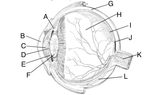
Identify I and describe function
Retina, the innermost layer and also called the neural tunic. It has the photoreceptors of the eye: cones and rods which convert light signals into electrical signals that are conveyed to the brain. The second area is the optic disc/blind spot. This is where the optic nerve leaves the eye, and there are no photoreceptors here. Light rays focused on this part of the retina cannot be seen, but we don’t realize the blind spot exists because our brain makes us think it isn’t there.

Identify J and describe function
The fovea centralis on the retina has mostly cones, processing our sharpest, color filled visual signals.