2 - Deep back and spinal cord
1/34
There's no tags or description
Looks like no tags are added yet.
Name | Mastery | Learn | Test | Matching | Spaced | Call with Kai |
|---|
No analytics yet
Send a link to your students to track their progress
35 Terms
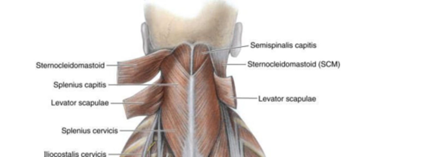
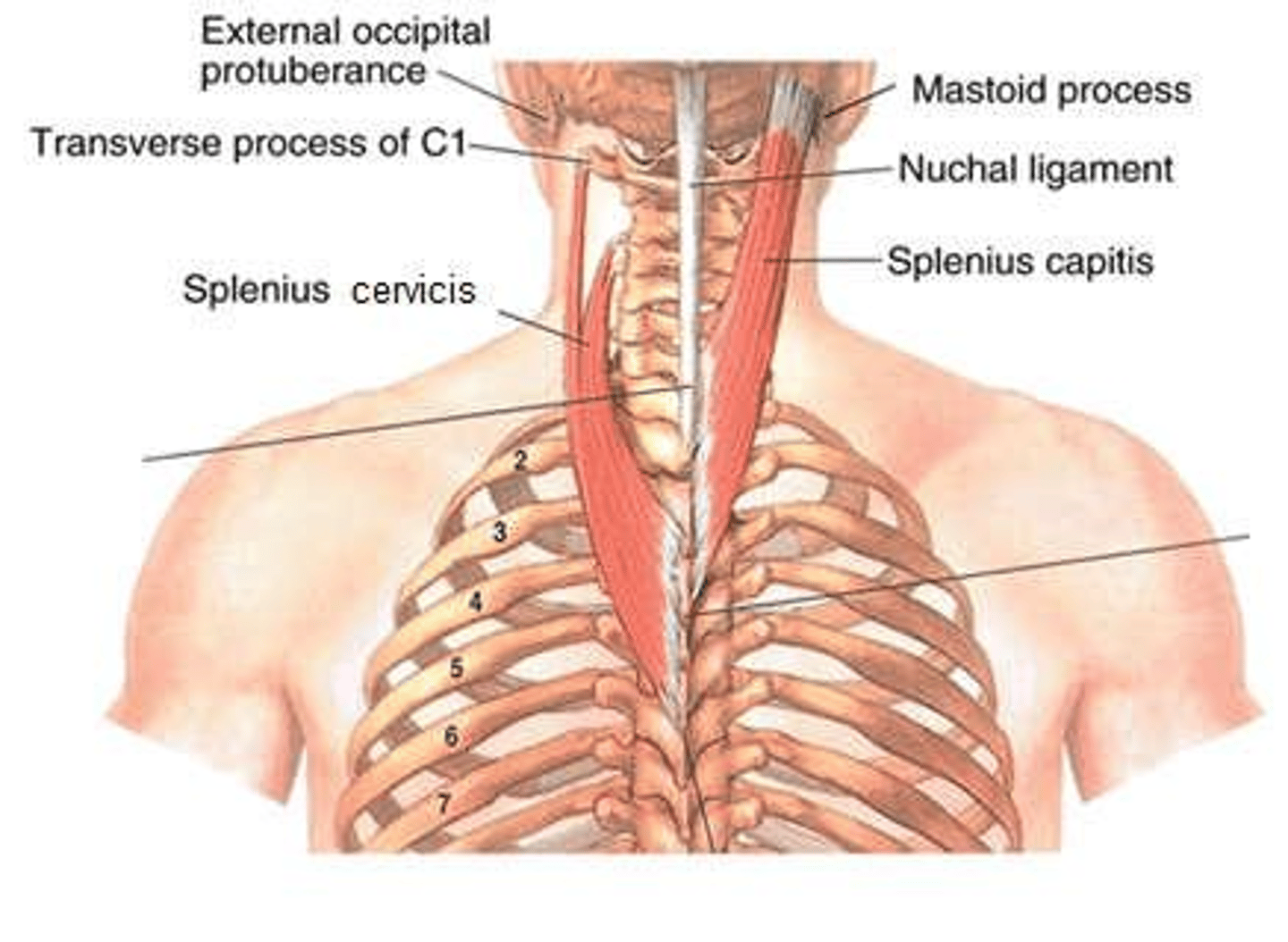
Splenius Muscle: Attachments, Action, and Innervation
Attachments: Spinous processes (C/T) to mastoid process/transverse processes (C). Action: Bilaterally extends; unilaterally laterally bends/rotates head/neck. Innervation: Posterior rami of C1-C8.

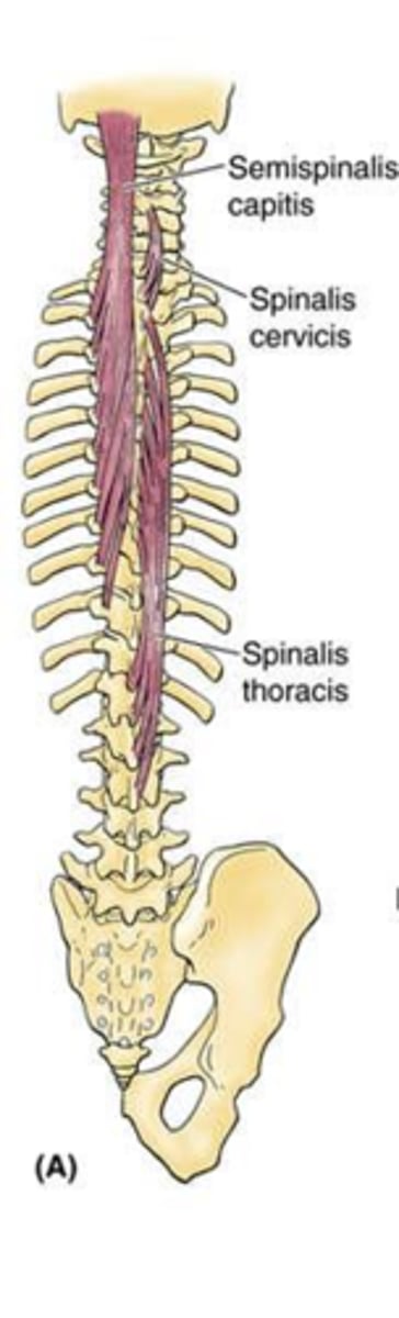
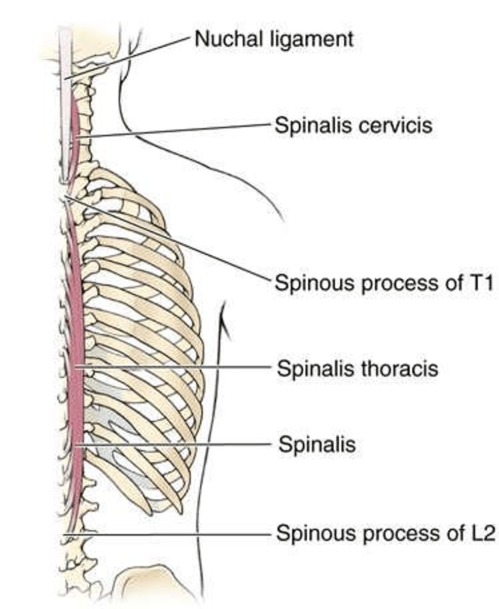
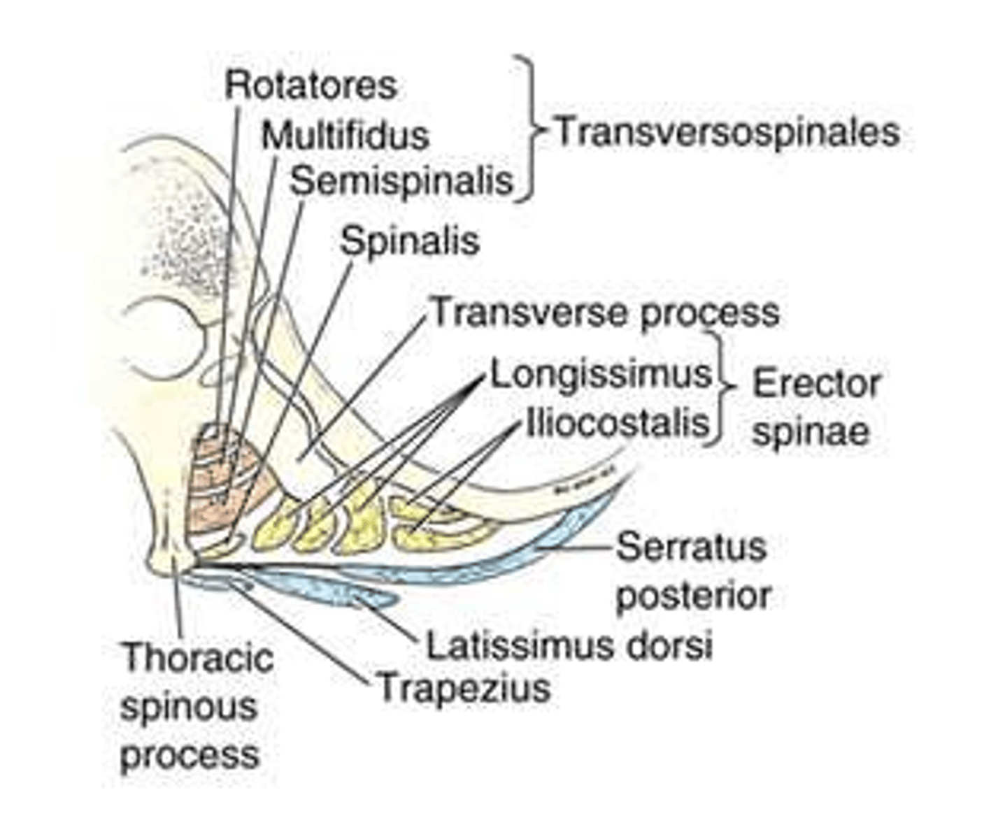
Spinalis: Attachments, Action, and Innervation
Attachments: Spinous process to spinous process. Action: Bilaterally extends; unilaterally laterally bends vertebral column. Innervation: Posterior rami of C1-L5.

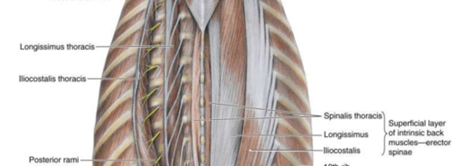
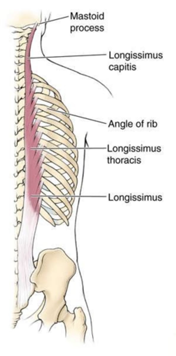
Longissimus: Attachments, Action, and Innervation
Attachments: Sacrum/lumbar vertebrae/iliac crest to medial ribs/mastoid process. Action: Bilaterally extends; unilaterally laterally bends vertebral column. Innervation: Posterior rami of C1-L5.

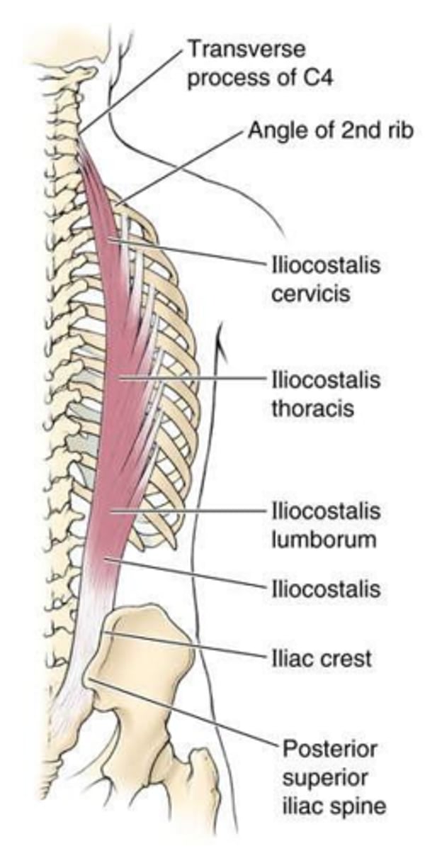
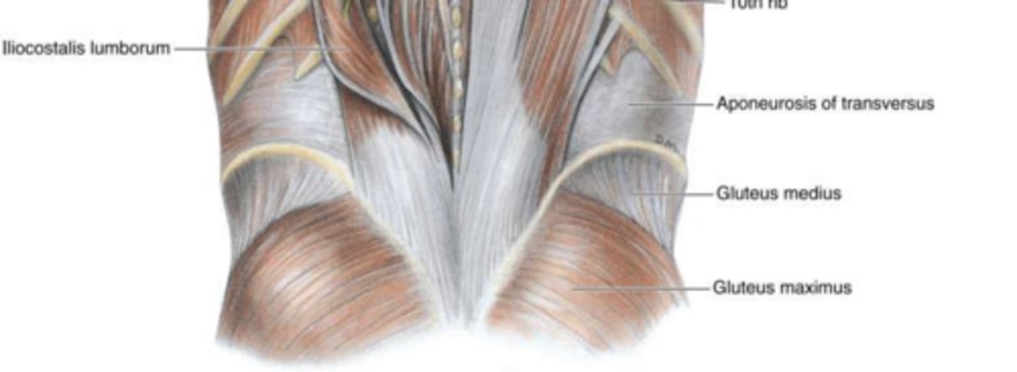
Iliocostalis: Attachments, Action, and Innervation
Attachments: Iliac crest/sacrum to lateral ribs. Action: Bilaterally extends; unilaterally laterally bends vertebral column. Innervation: Posterior rami of C1-L5.
Semispinalis: Attachments, Action, and Innervation
Attachments: Transverse processes (C/T) to occipital bone (capitis)/spinous processes (cervicis). Action: Bilaterally extends; unilaterally rotates head/vertebral column. Innervation: Posterior rami of C1-T12.
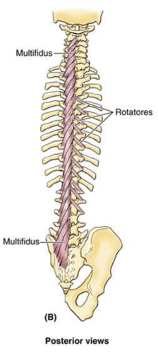
Multifidus: Attachments, Action, and Innervation
Attachments: Sacrum/ilium/transverse processes to spinous processes of C, T, L. Action: Extends and rotates vertebral column. Innervation: Posterior rami of C1-L5.

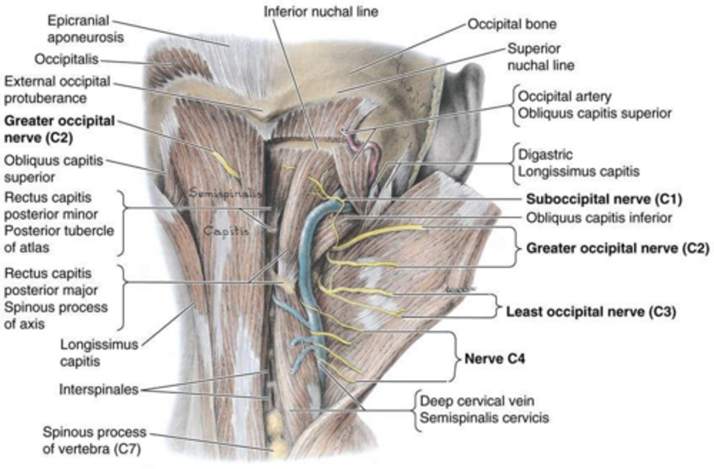
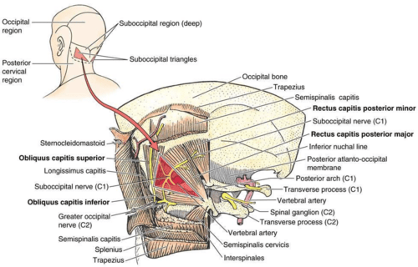
Rectus Capitis Posterior Major: Attachments, Action, and Innervation
Attachments: Spinous process of C2 to occipital bone. Action: Extends head. Innervation: Suboccipital nerve (posterior ramus of C1).
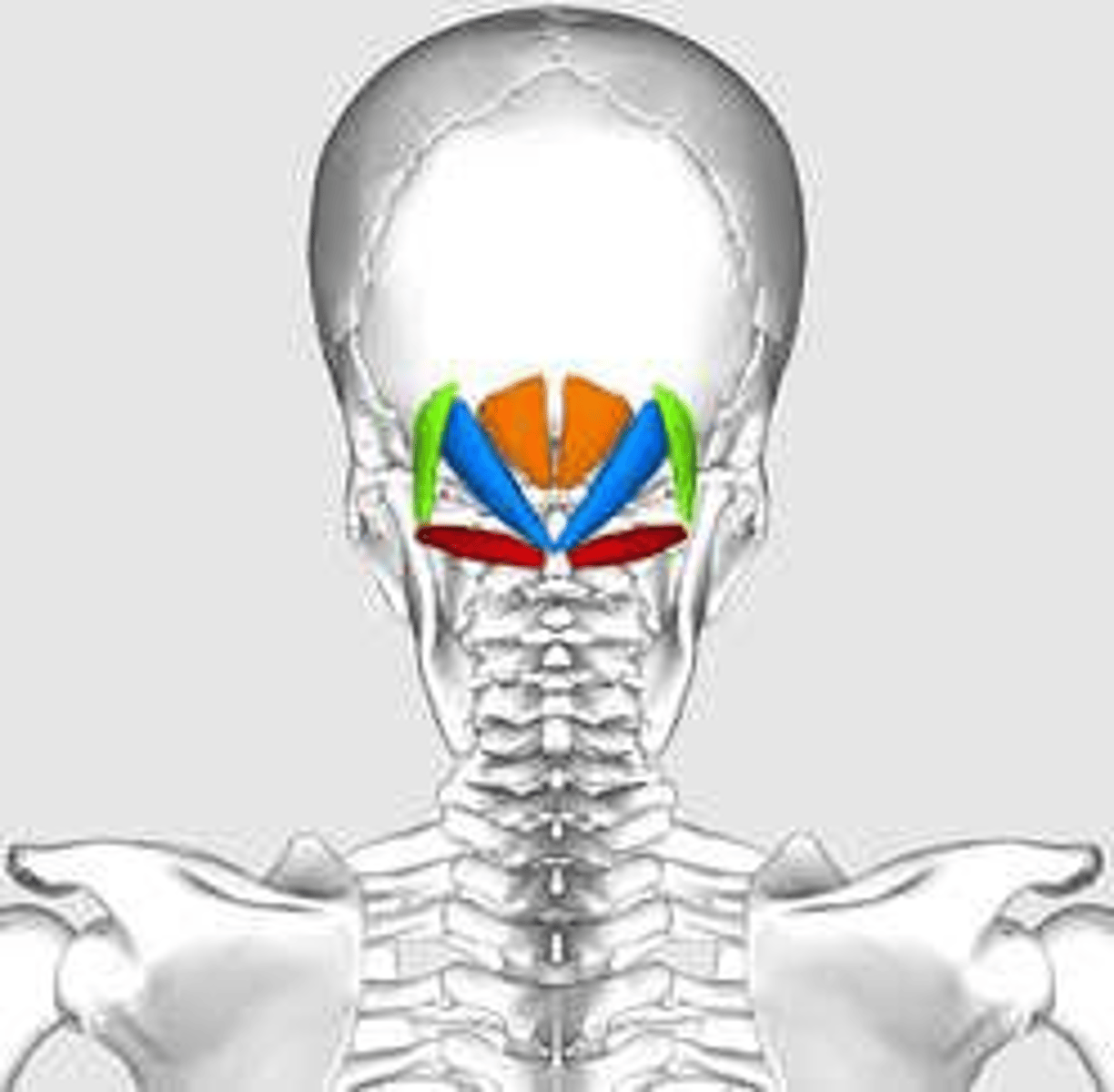
Rectus Capitis Posterior Minor: Attachments, Action, and Innervation
Attachments: Spinous process of C1 to occipital bone. Action: Extends head. Innervation: Suboccipital nerve (posterior ramus of C1).
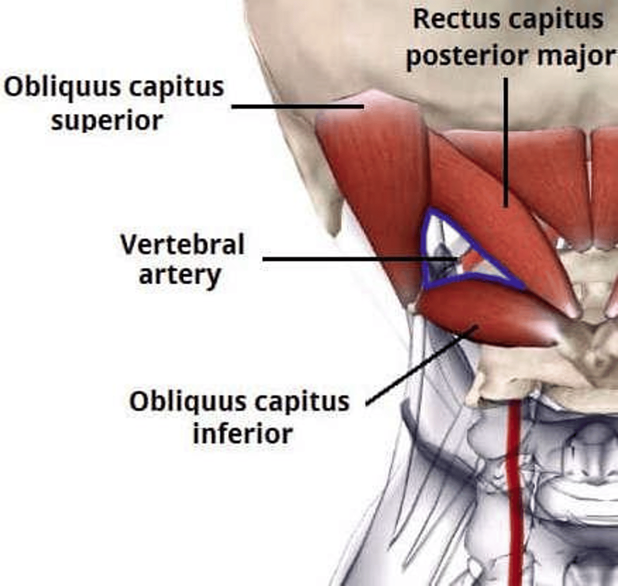
Obliquus Capitis Superior: Attachments, Action, and Innervation
Attachments: Transverse process of C1 to occipital bone. Action: Extends head. Innervation: Suboccipital nerve (posterior ramus of C1).
Obliquus Capitis Inferior: Attachments, Action, and Innervation
Attachments: Spinous process of C2 to transverse process of C1. Action: Rotates head. Innervation: Suboccipital nerve (posterior ramus of C1).
Anterior Longitudinal Ligament
Joins vertebral bodies anteriorly; resists hyperextension of the vertebral column.
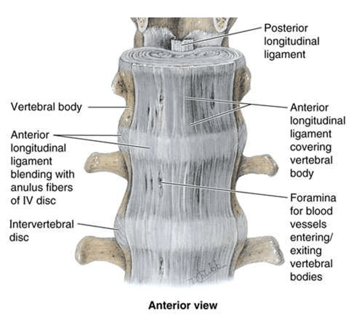
Posterior Longitudinal Ligament
Joins vertebral bodies posteriorly; resists hyperflexion and prevents posterior disc herniation.
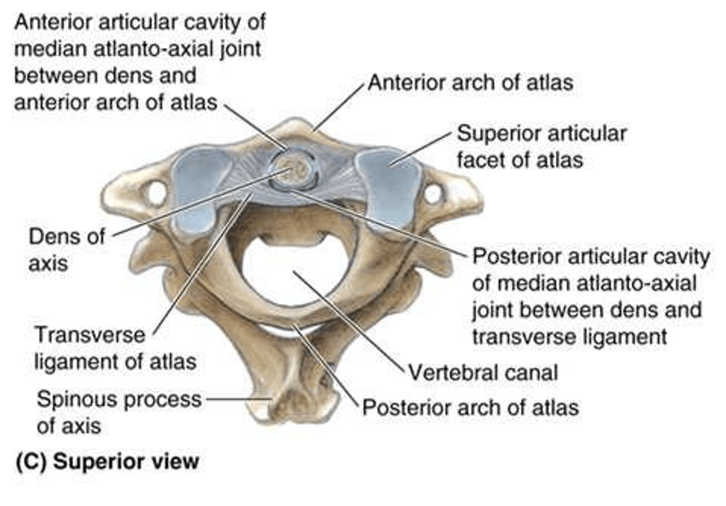
Transverse Ligament of the Atlas
Holds the dens of C2 against the anterior arch of C1; facilitates rotation.
Cruciate Ligament
Cross-shaped ligament connecting the dens to lateral masses of the atlas, foramen magnum, and C2 body.
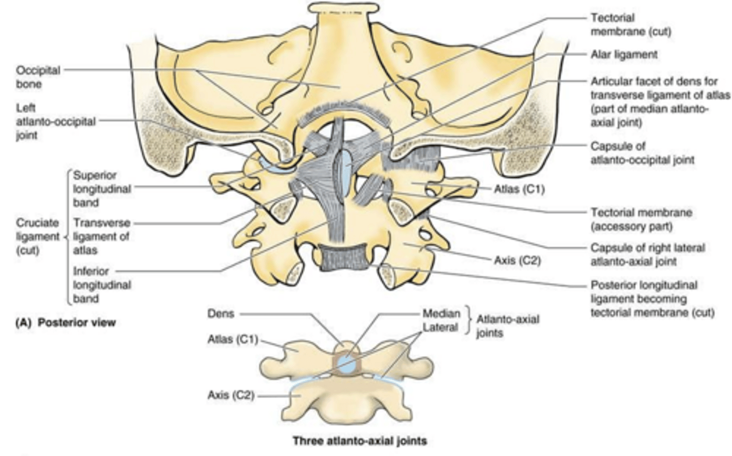
Alar Ligament
Connects the dens to anterolateral margins of the foramen magnum; prevents excessive rotation.
Tectorial Membrane
Extension of the posterior longitudinal ligament from the posterior body of C2 to the internal foramen magnum.
Nuchal Ligament
Connects external occipital protuberance and crest to all cervical spinous processes.
Supraspinous Ligament
Continuation of the nuchal ligament; connects tips of spinous processes from thoracic region to sacrum.
Intertransverse Ligament
Connects adjacent transverse processes of the vertebrae.
Interspinous Ligament
Connects adjacent spinous processes of the vertebrae.
Ligamentum Flavum
Connects adjacent laminae of the vertebrae.
Arterial Supply of the Back: Cervical Region
Vertebral and ascending cervical arteries (from the subclavian artery).
Arterial Supply of the Back: Thoracic Region
Posterior intercostal arteries (branches of the thoracic aorta).
Arterial Supply of the Back: Lumbar Region
Lumbar arteries (branches of the abdominal aorta).
Arterial Supply of the Back: Sacral Region
Iliolumbar, lateral, and medial sacral arteries (from the internal iliac artery).
Spinal Branches of the Back
Enter intervertebral foramina, continuing as radicular or segmental medullary arteries.
Venous Drainage of the Back: Plexuses
Internal and external vertebral venous plexuses, which drain into intervertebral veins.

Destination of Intervertebral Veins
Vertebral (neck), posterior intercostal (thorax), lumbar (abdomen), and sacral veins (sacral region).
Clinical Significance of Vertebral Venous Plexuses
Allows cancer metastasis to vertebrae/brain and bypasses obstructed inferior vena cava.
Lymphatic Drainage of the Upper Back
Middle/inferior trunks drain to thoracic duct; superior trunks to bronchomediastinal trunks.
Lymphatic Drainage of the Lower Back
Drains to lumbar trunks, then to chyle cistern at thoracic duct origin.

Arterial Supply of the Spinal Cord
One anterior spinal artery and two posterior spinal arteries running its length.
Radicular vs. Segmental Medullary Arteries
Segmental medullary arteries anastomose with spinal arteries; radicular arteries do not.

Great Anterior Segmental Medullary Artery (of Adamkiewicz)
Arises on left at T9-T12; supplies spinal cord from T8 to conus medullaris.
Venous Drainage of the Spinal Cord
Three anterior and three posterior spinal veins draining to external vertebral venous plexus.
