TAPP Exam UTK
1/383
There's no tags or description
Looks like no tags are added yet.
Name | Mastery | Learn | Test | Matching | Spaced | Call with Kai |
|---|
No analytics yet
Send a link to your students to track their progress
384 Terms
somatic and autonomic
two kinds of peripheral nerves
somatic nerve
voluntary functions
somatic nerve
one continuous nerve from the CNS to target
somatic nerve
move skeletal muscle & transmit sensation from body walls (e.g., skin, mucous membranes)
autonomic nerves
involuntary functions
autonomic nerve
two nerves, linked by a synapse, occur between CNS & target
autonomic nerve
activate organs, transmit sensation from organs, glands, and internal structures
afferent
sensory
efferent
motor
synapses
electrochemical communication between nerve cells and with tissues
sympathetic & parasympathetic
two types of autonomic nerves
sympathetic nerve
fight or flight
parasympathetic nerve
rest & digest
sympathetic synapse
synapse before reaching their target, in sympathetic trunk, in pre-vertebral ganglia
parasympathetic synapse
synapse in or near the walls of their targets
vagus nerve
maintains rhythm of heart & ability to pump heart
parasympathetic nerves
originate from cranial nerves or from the sacrum
T1-L2
sympathetic trunk runs
ganglion
where synapse occurs in autonomic nerve
parasympathetic
is vagus nerve parasympathetic or sympathetic
thorax region
ribs 1-12
protect heart & lungs
rib cage function
right pleural cavity, left pleural cavity, mediastinum
divisions of thorax
bronchi tubes & lungs
pleural cavity contains
heart & aortas
mediastinum contains
heart, esophagus, trachea, aorta
mediastinum components
xiphoid process
can break off
costal cartilage
connects rib to sternum
true ribs
attach directly
false ribs
do not attach directly
floating ribs
does not attach to sternum or another rib
2
how many floating ribs
inferior to 4th rib
nipple placement younger female
inferior to 6th rib
nipple placement developed female
pectoral muscles
breast lays over what
suspensory ligaments
connective tissue that maintains shape of tissue region in breast
breast region
large amount of blood going to what region
retromammary space
layer of loose connective tissue that separates the breast from the deep fascia overlying pectoralis major and serratus anterior muscles
lactiferous ducts
carries breast milk from breast lobules to nipple
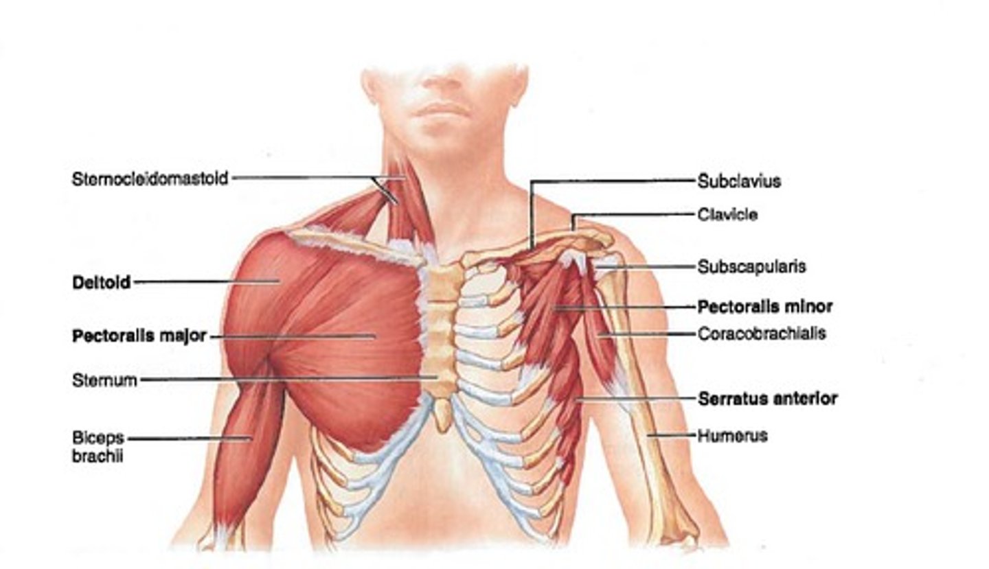
pectoralis major, pectoralis minor, serratus anterior
anterior thorax superficial muscles
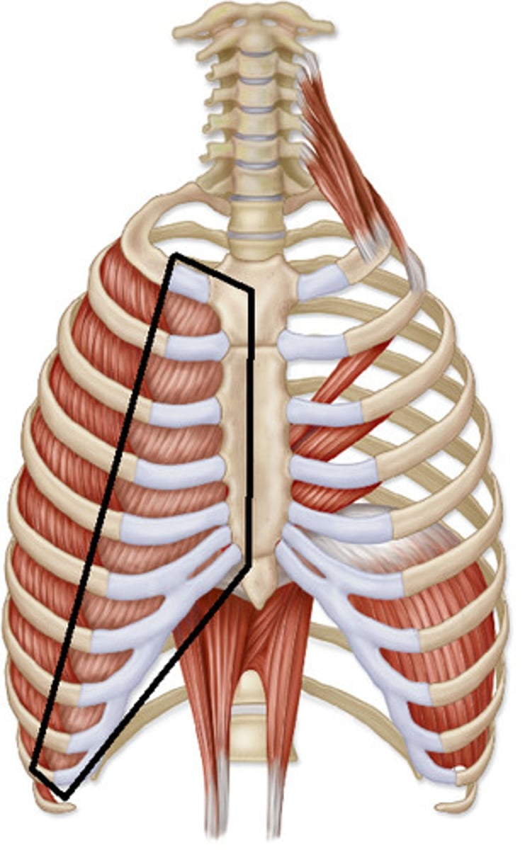
external intercostal muscles, internal intercostal muscles, innermost intercostal muscles
intercostal muscles
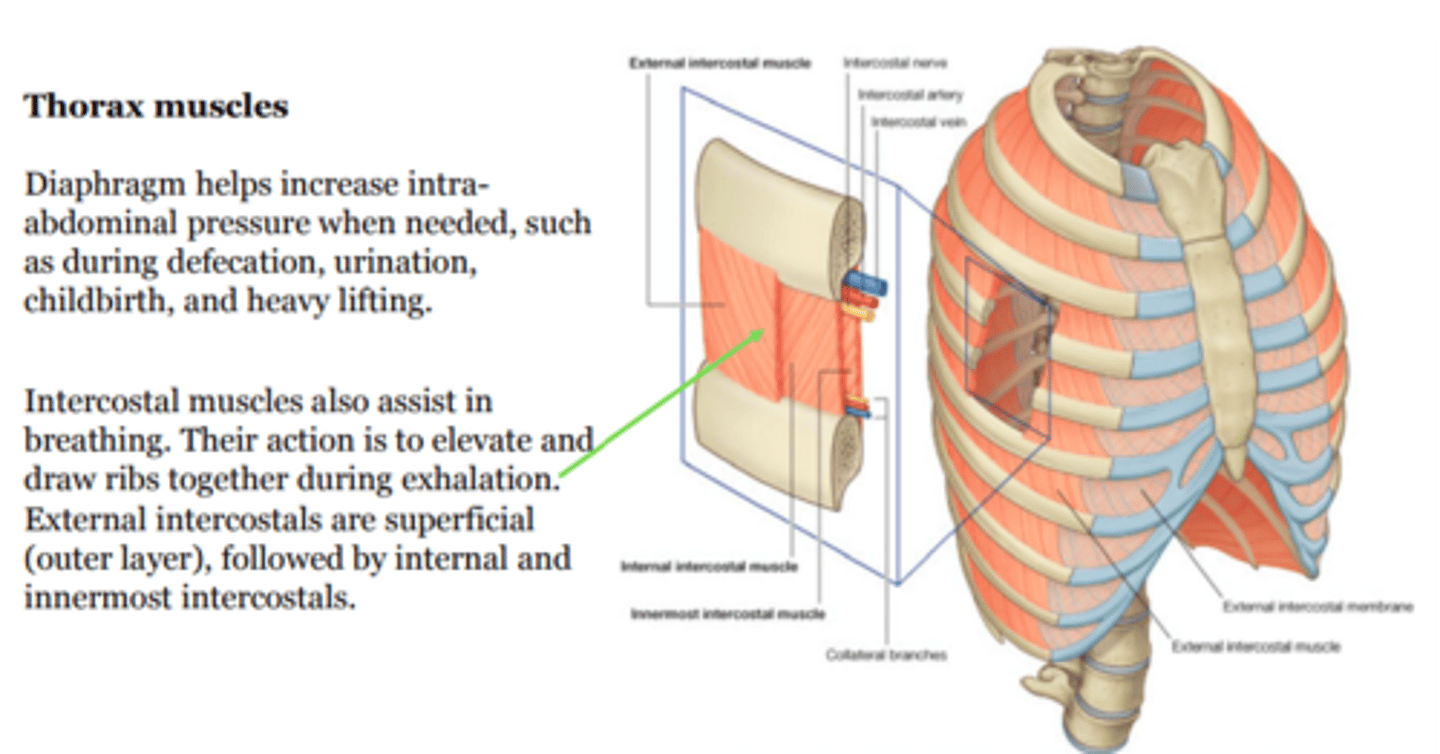
external intercostal muscles
run from sternum to vertebrae
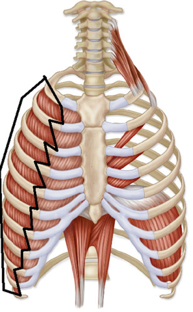
intercostal muscles
wrap completely around thoracic wall
internal intercostal muscles
start at sternum but do not go to vertebrae
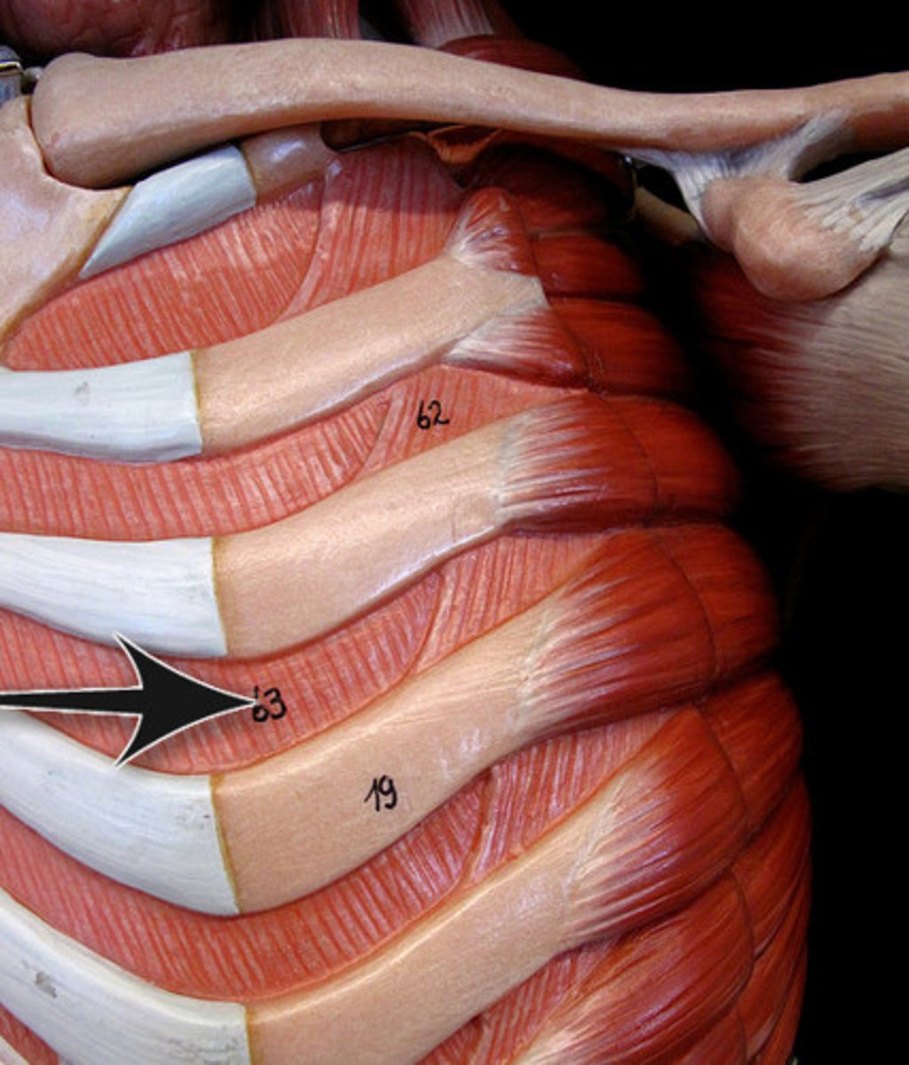
innermost intercostal muscles
deep to internal & external intercostal muscles
when inhaling or exhaling, either maintain the position of ribs to each other or let ribs return to natural position when relaxing
intercostal muscles function
endothoracic fascia
loose connective tissue that lines entire thoracic cavity
external intercostal aponeurosis
covers where external intercostal muscles stop
intercostal veins, arteries, and nerves
intercostal v.a.n.
diaphragm
muscle mainly responsible for respiration
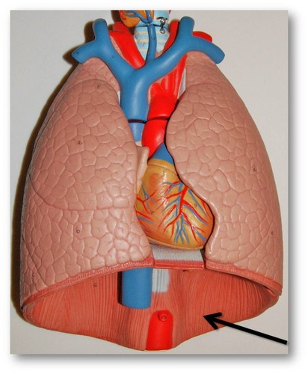
diaphragm
most inferior border of thoracic cavity
hiccups
spasm of diaphragm
liver
shape of what organ causes asymmetry to diaphragm
right dome of diaphragm
more superior than left dome of diaphragm
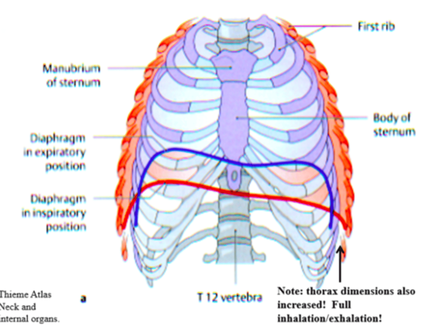
diaphragm contracts & flattens out, increases space in chest cavity, lungs expand into chest cavity, pressure is lower
inspiration
diaphragm relaxes and returns to starting point, reducing space in chest cavity, lungs deflate, pressure is higher
expiration
phrenic nerve
innervation of diaphragm
C3, C4, C5
what specific nerves innervate diaphragm
somatic
is phrenic nerve somatic or autonomic
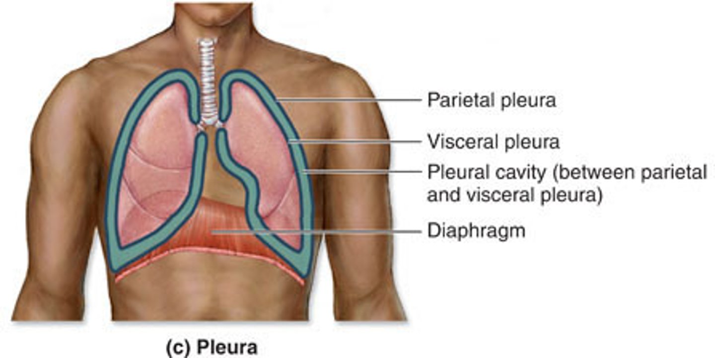
pleural cavity of lungs
space between visceral and parietal pleura
costodiaphragmatic recess
allow lungs to expand into openings
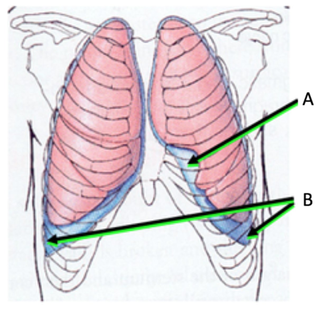
parietal pleura
does not directly touch lung
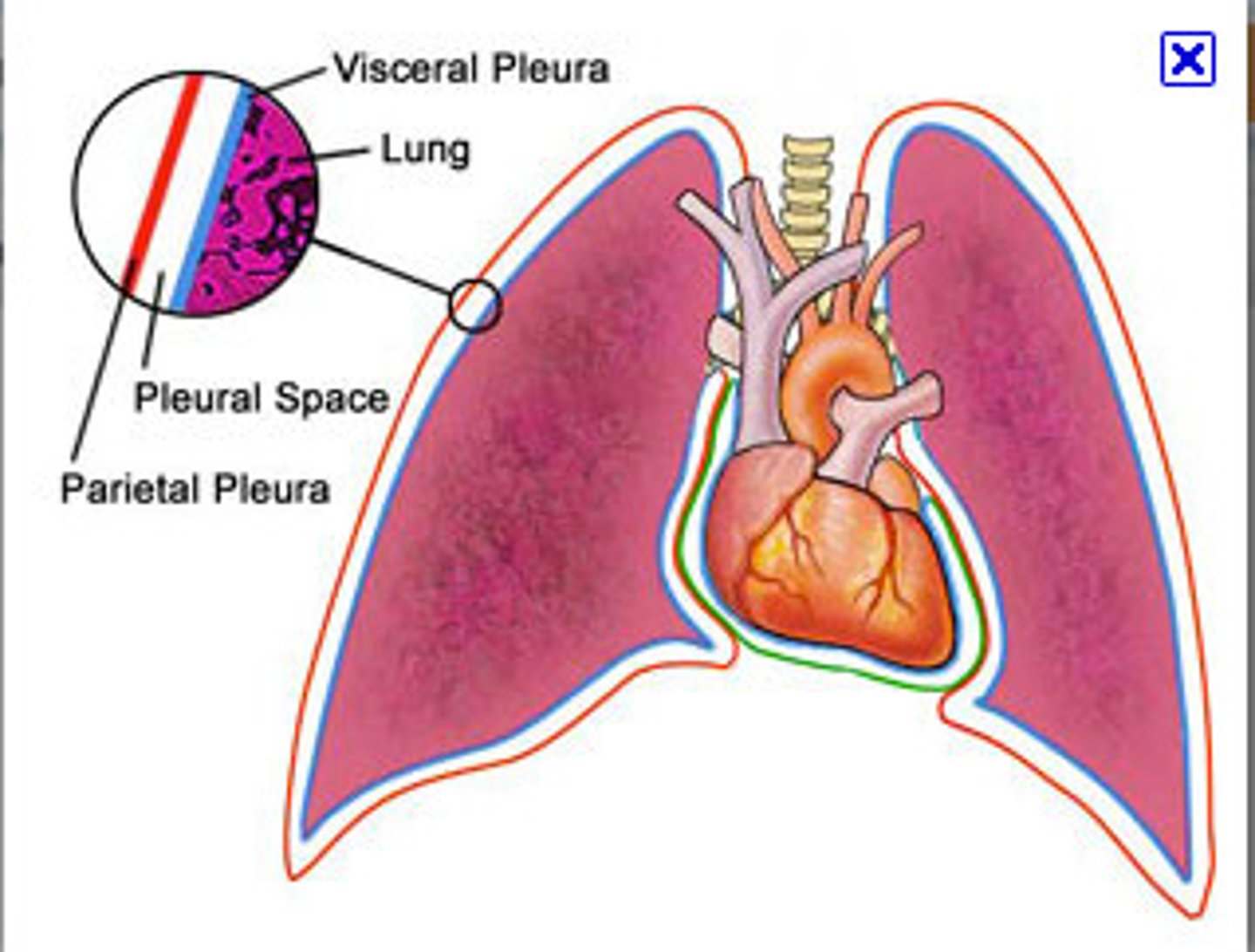
visceral pleura
directly touches lung
serous fluid in pleural cavity
keeps 2 layers of pleura as close as possible
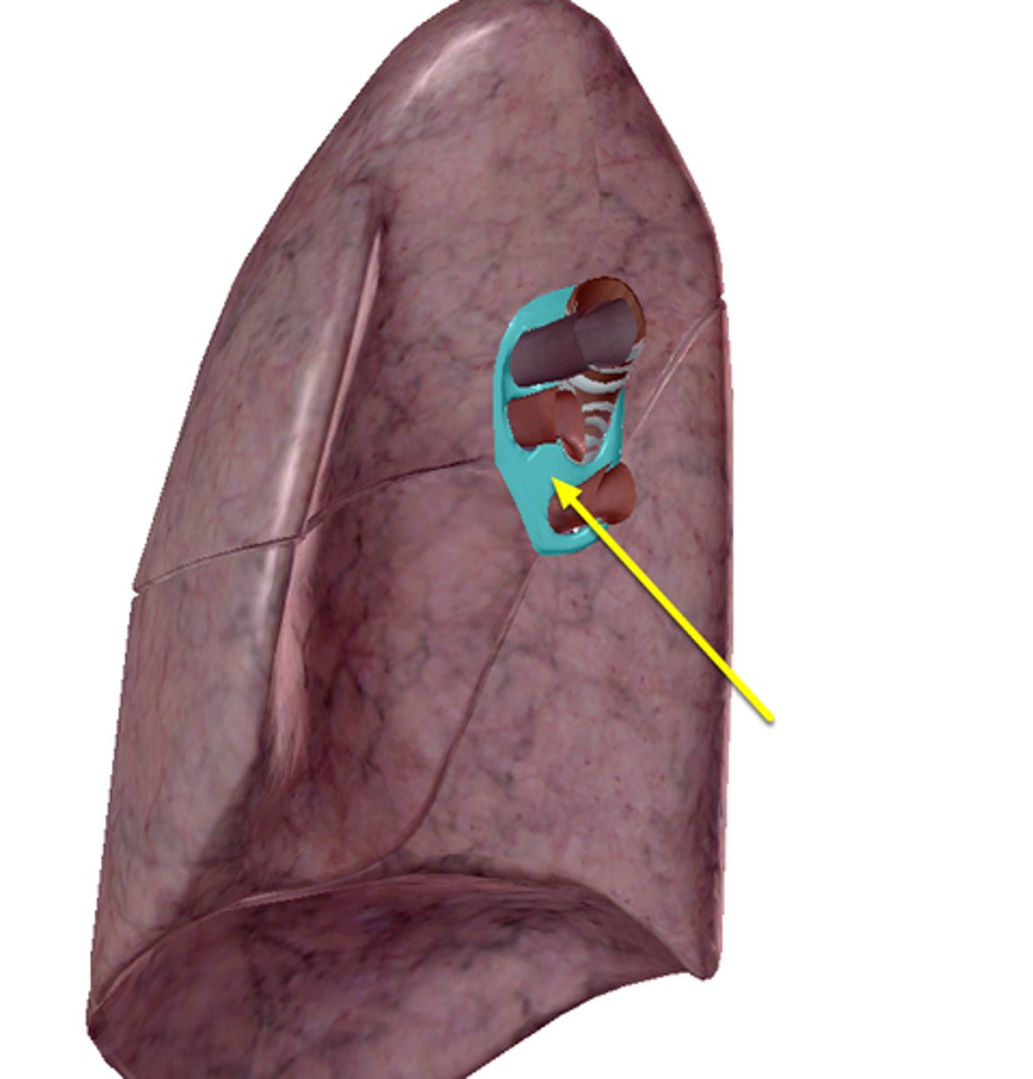
hilum of lung
contains pulmonary vessels & right and left main bronchus
right lung
horizontal fissure is only on
upper, middle , lower
lobes of right lung
upper and lower
lobes of left lung
upper & middle lobe of right lung
horizontal fissure separates what
left lung - upper & lower lobe
right lung - middle & lower lobe
oblique fissure separates what
force air into trachea, goes into right/left main bronchus, travel to lobar bronchi, go to different lobes
air travel to trachea
bronchopulmonary units
can operate without all units working properly
lobar bronchi
goes into individual functional units
main bronchus to lobar bronchus to tertiary/segmental bronchus to bronchioles to respiratory bronchioles
air flow of bronchopulmonary units
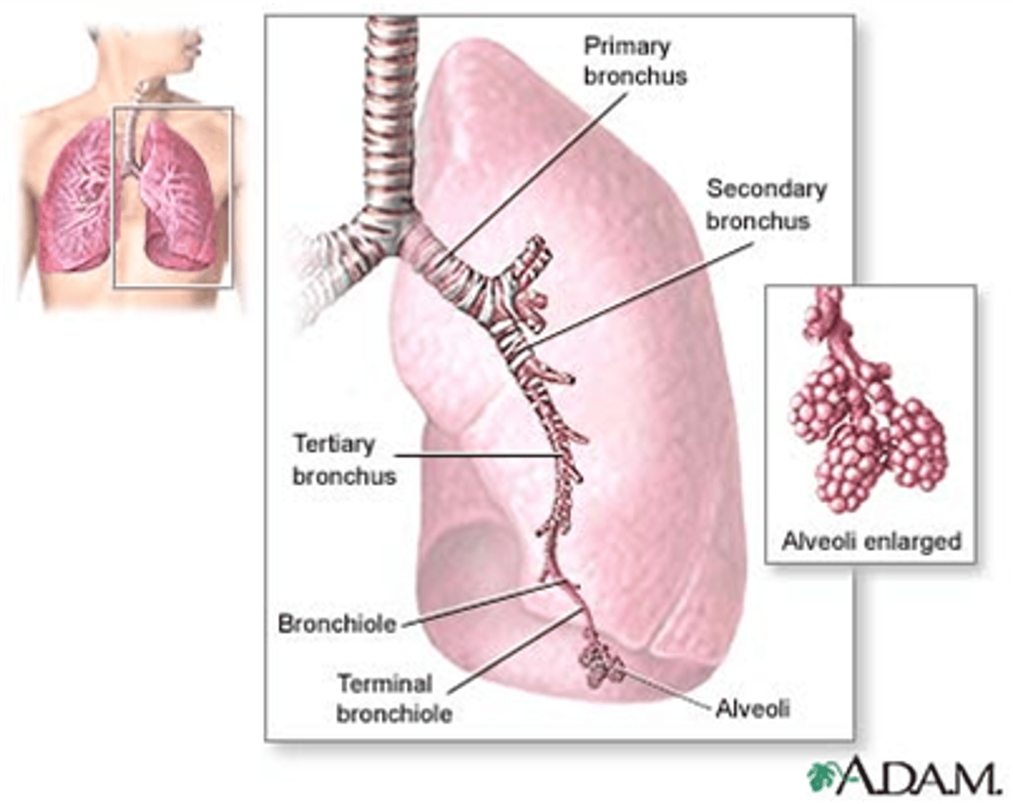
alveolus
site of gas exchange
branches of pulmonary vein
take direct root to hilum of lung
respiratory bronchiole
where aveoli are located
branch of pulmonary artery
follow branching of airways (bronchi & bronchioles)
allows for movement as expansion/flexion, attracts visceral pleura to parietal pleura, lowers friction between parietal layers
serous fluid in respiration
pulls against central tendon, lowers diaphragm, expands pleural cavity & creates negative pressure
diaphragm contraction in respiration
life and expand
ribs in respiration
passively fill expanded space
lungs in respiration
increases vertically
surface area in inspiration
decrease vertically
surface area in expiration
helps to increase volume in three dimensions
rib cage in respiration
exercise & controlled breathing activities
forced respiration happens
scalenes, pectoralis minor, serratus anterior, rectus abdominis
forced respiration uses what muscles
pleural cavity in expiration in pneumothorax
air in cavity pushes & deflates lung
collapsed lung in pneumothorax
lung cannot respond to change of pressure
superior mediastinum & inferior mediastinum
mediastinum divisions
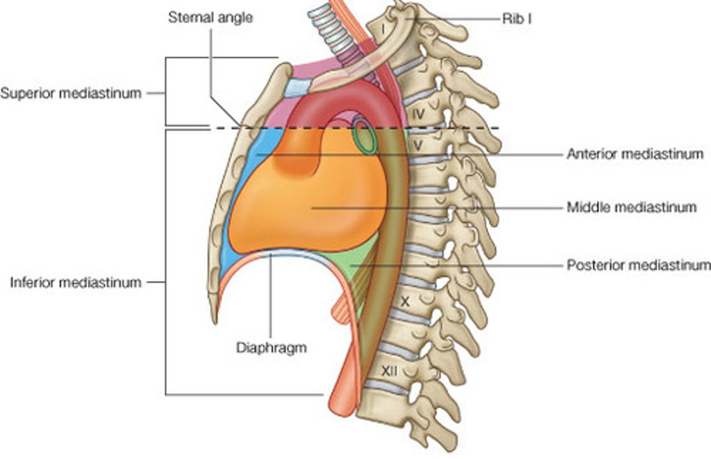
anterior mediastinum, middle mediastinum, posterior mediastinum
inferior mediastinum divisions
organs: thymus, trachea, esophagus,
arteries: aortic arch, brachiocephalic trunk, left common carotid artery, left subclavian artery,
veins: superior vena cava, brachiocephalic veins, arch of azygos, thoracic duct,
nerves: left and right vagus nerve, recurrent laryngeal nerve, cardiac nerve, left and right phrenic nerves
superior mediastinum contents
organs: thymus
arteries: internal thoracic branches
veins: internal thoracic branches
anterior mediastinum contents
organs: heart & great vessel roots, trachea, main bronchi
arteries: ascending aorta, pulmonary trunk, pericardiacophrenic arteries
veins: superior vena cava, pulmonary veins, pericardiacophrenic veins
nerves: phrenic, vagus, sympathetics
middle mediastinum contents
organs: esophagus
arteries: descending thoracic aorta
veins: azygos hemiazygos veins, thoracic duct
nerves: vagus, splanchnic, sympathetic chain
posterior mediastinum contents
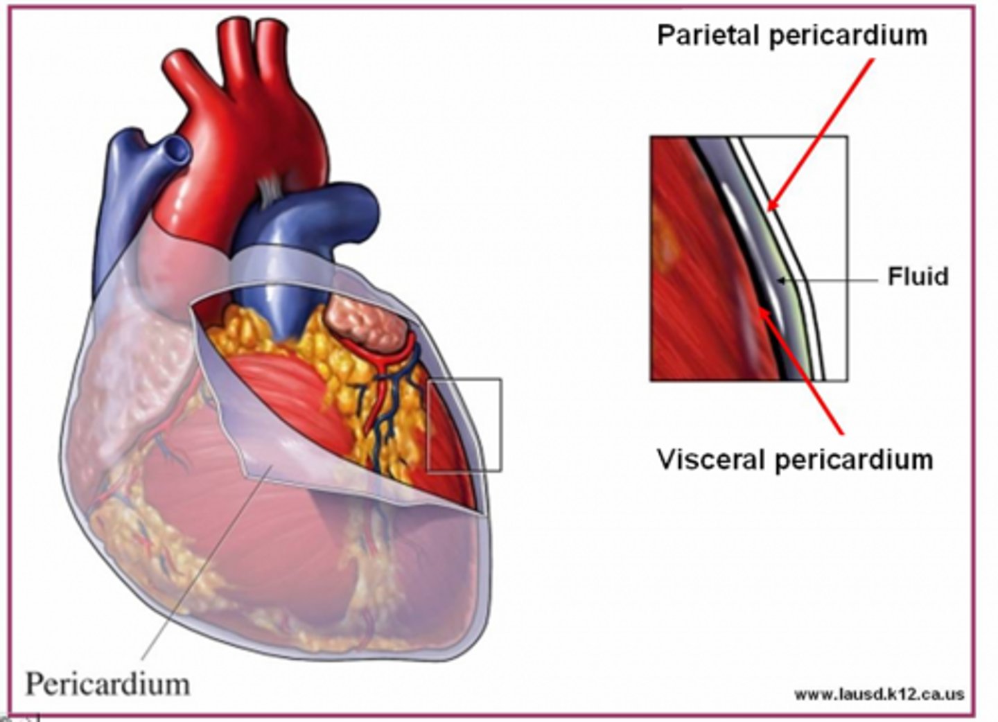
pericardium
double - layered connective tissue fibrous membrane that protects and holds heart
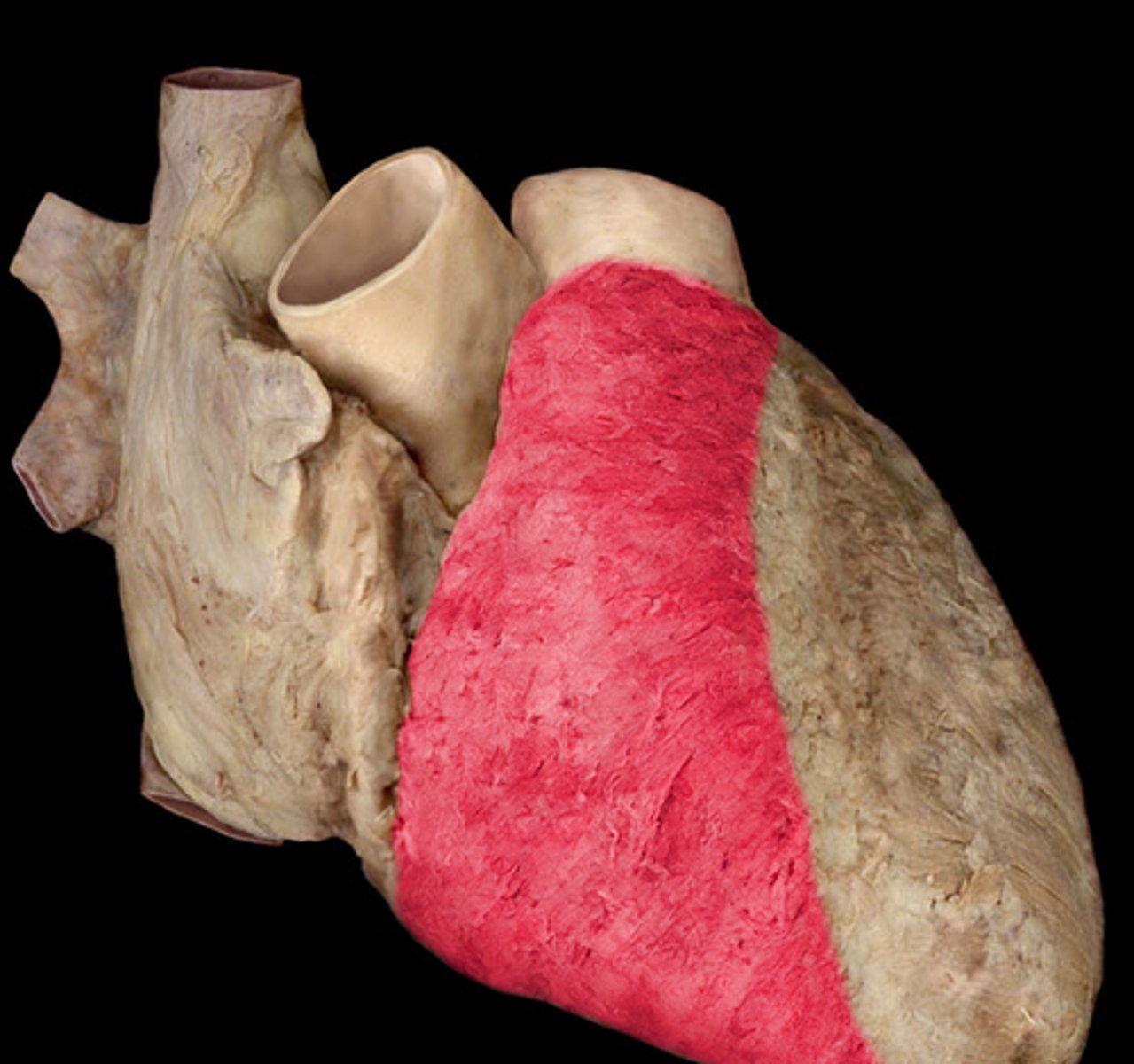
right ventricle of heart
majority of heart we see in situ
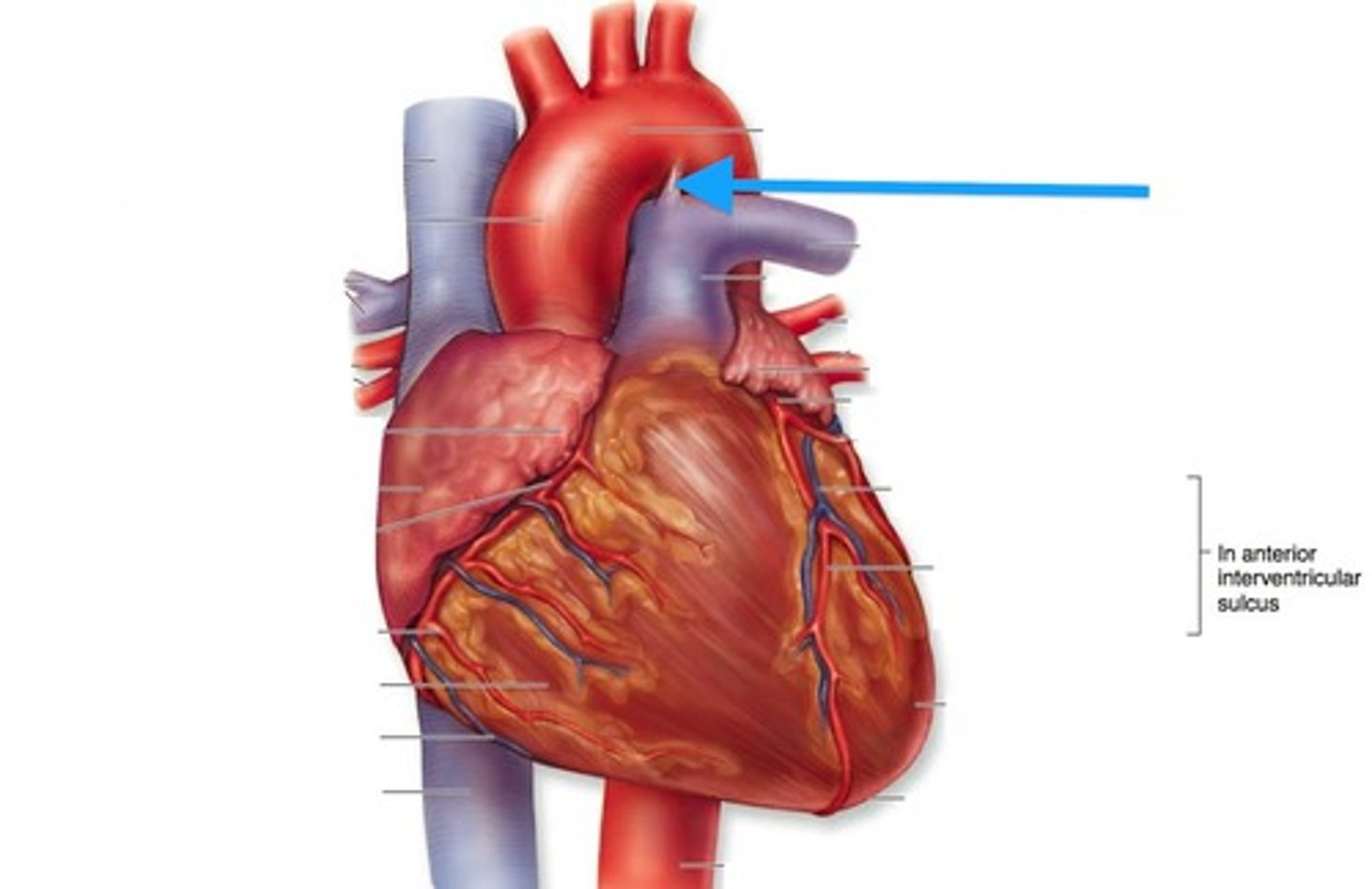
ligamentum arteriosum
hold parts of great vessels in place & connects arch of aorta and pulmonary trunk
fetal circulation
bypass taking blood to and from lungs
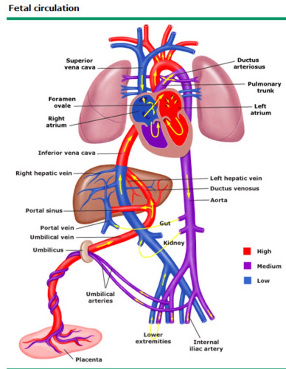
ductus arteriosus
shunts blood from pulmonary arteries to the arch of aorta