exam 1 anat 102 everything 😭
1/126
There's no tags or description
Looks like no tags are added yet.
Name | Mastery | Learn | Test | Matching | Spaced | Call with Kai |
|---|
No analytics yet
Send a link to your students to track their progress
127 Terms
Meissner Corpuscle
General: Somatic:
Tactile aka Corpuscles of Touch; found in dermal papillae of the dermis; its capsule detects the onset of touch; some detection of low-frequency vibration; abundant in fingertips, hands, eyelids tip of tongue, lips, nipples, soles,
clitoris, tip of penis
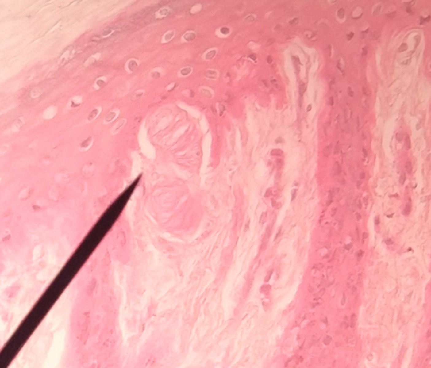
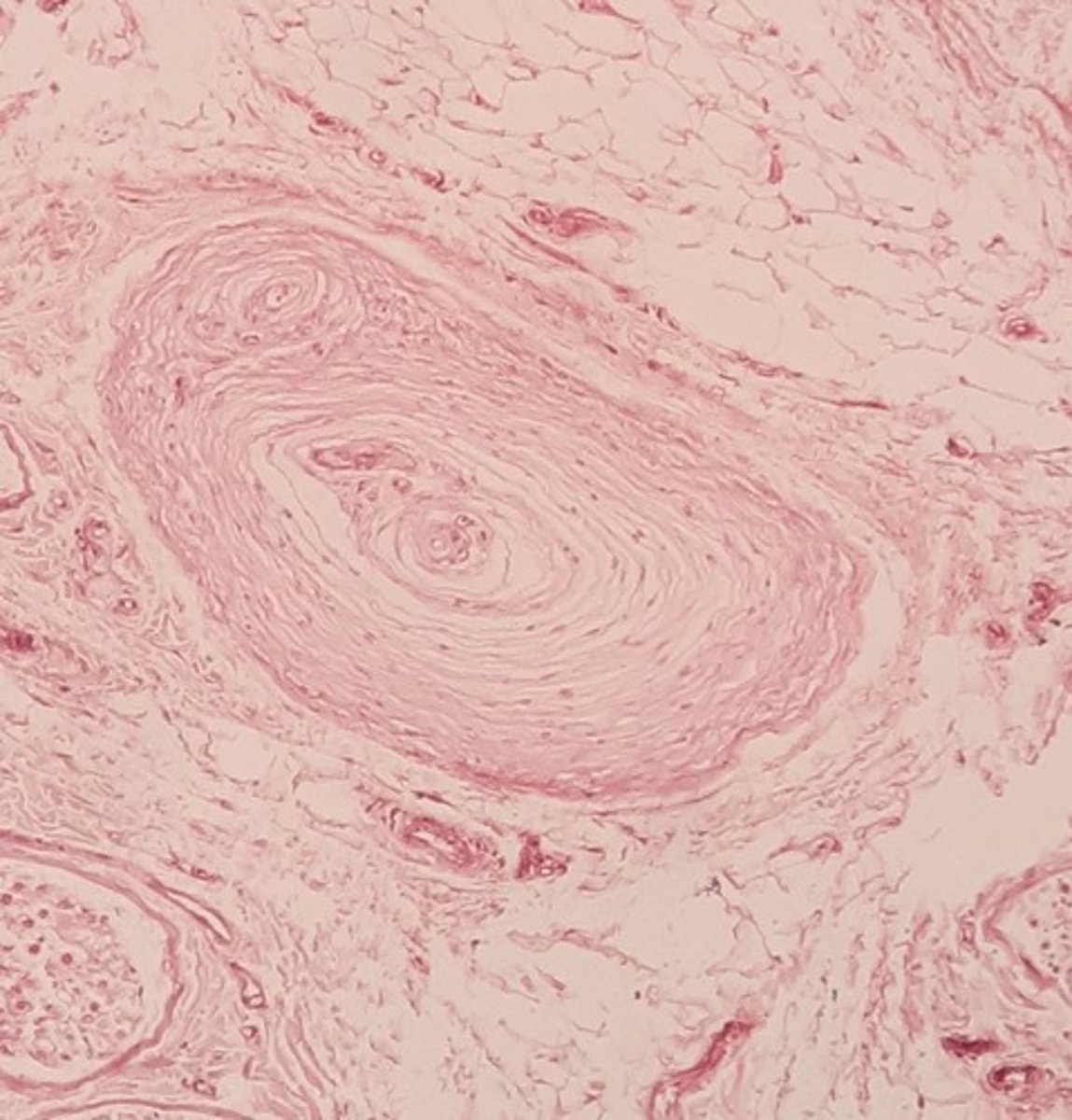
Pacinian Corpuscle
General: Somatic:
Tactile aka Lamellated Corpuscle; found in the dermis, hypodermis and other body tissues; its capsule resembles
an onion and detects high-frequency vibration
Vallate Papillae
Special: Taste elevations on the tongue's surface that house 100-300 taste buds per papillae; there are roughly 12 of them; they
form an inverted "v" on the posterior aspect of the tongue
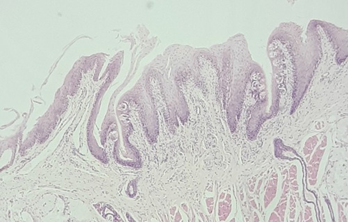
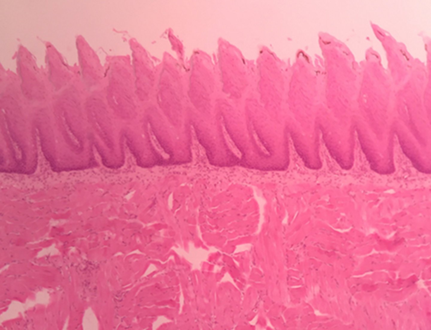
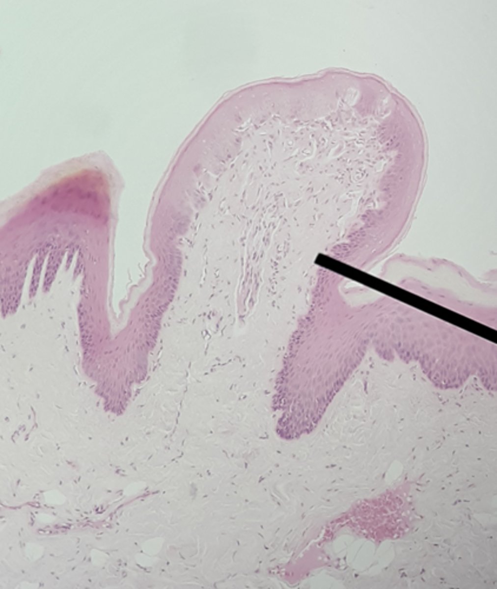
Filiform Papillae
Special: Taste
pointed, threadlike elevations on the entire surface of the tongue; contain tactile receptors; do NOT contain taste buds; increase friction of food in the mouth to enhance mechanical breakdown
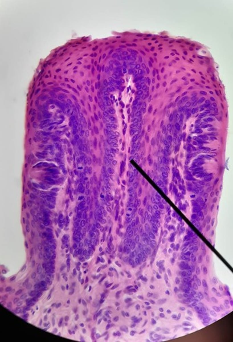
Fungiform Papillae
Special: Taste
mushroom-shaped elevations, scattered over entire surface of the tongue; contain about 5 taste buds each.
Foliate Papillae
Special: Taste
elevations found on the lateral aspect of the tongue of children
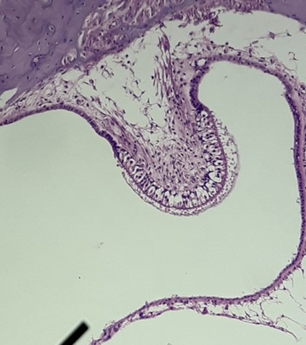
Crista Ampullaris
Special: Hearing
an elevation at the base of each of the semicircular ducts of the inner ear; contains hair cells that alter the movement of endolymph within the ampulla with rotational forces; involved in equilibrium and balance.
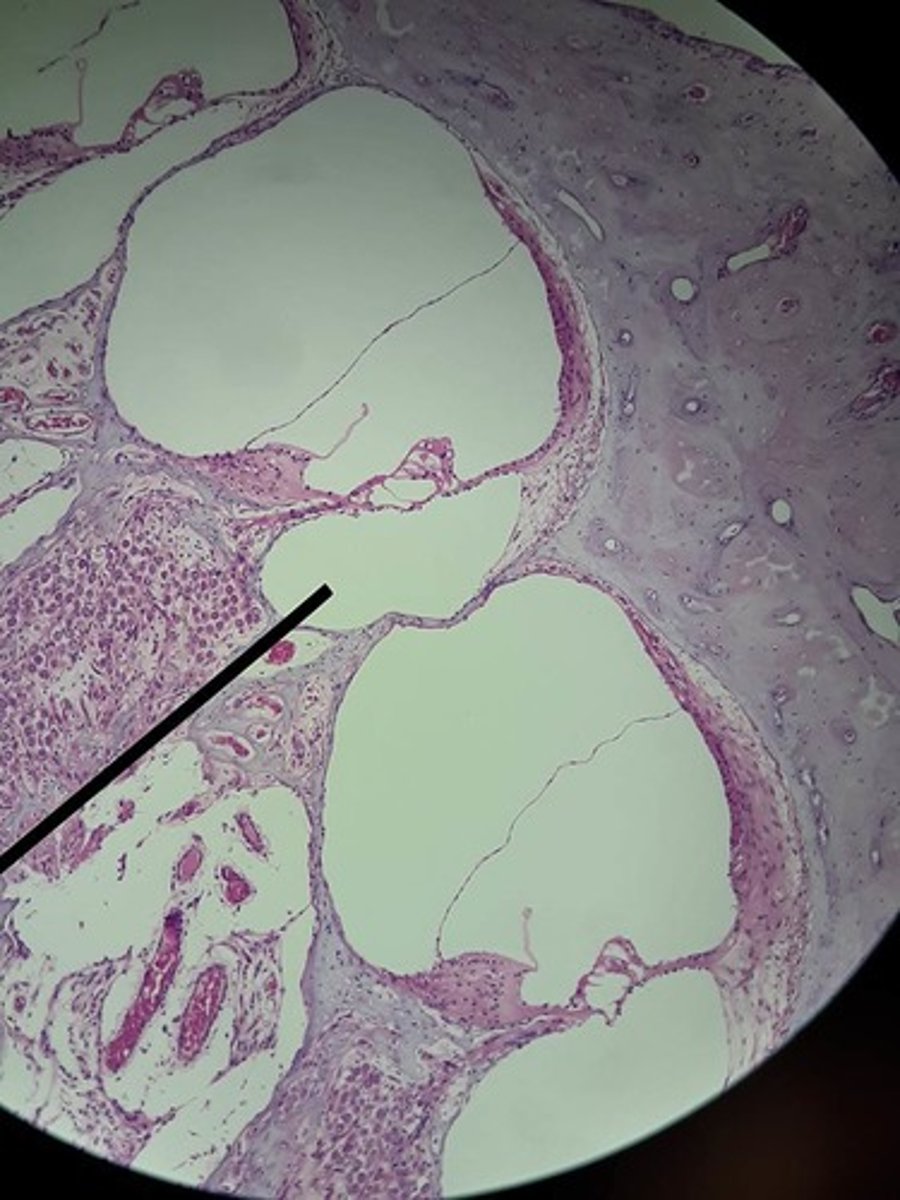
Cochlea
Special: Hearing
snail-shell spiral canal of the inner ear; contains fluid compartments (scala) and the Spiral Organ of Corti that contains the hair cells that are the receptors for hearing.
Inner hair cells and outer hair cells are found in a 1:3 ratio.
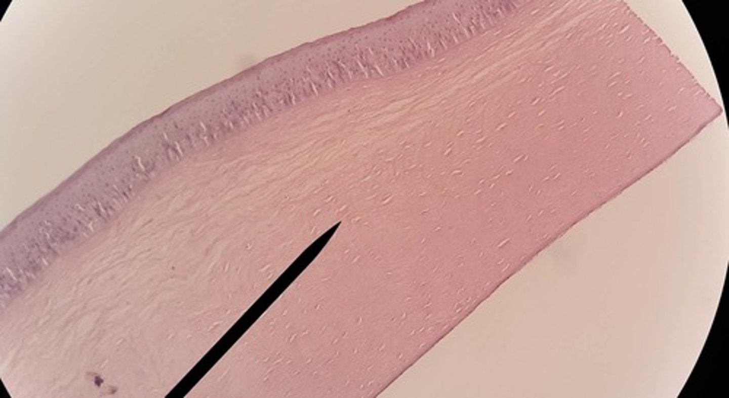
Cornea
Special: Vision
tissue structure located in the anterior aspect of the eyeball, anterior to the iris; its curved structure functions to focus light on the retina posteriorly; has three main layers: outer (nonkeratinized stratified squamous epithelium), middle "stroma" (collagen fibers and fibroblasts) and inner (simple squamous epithelium)
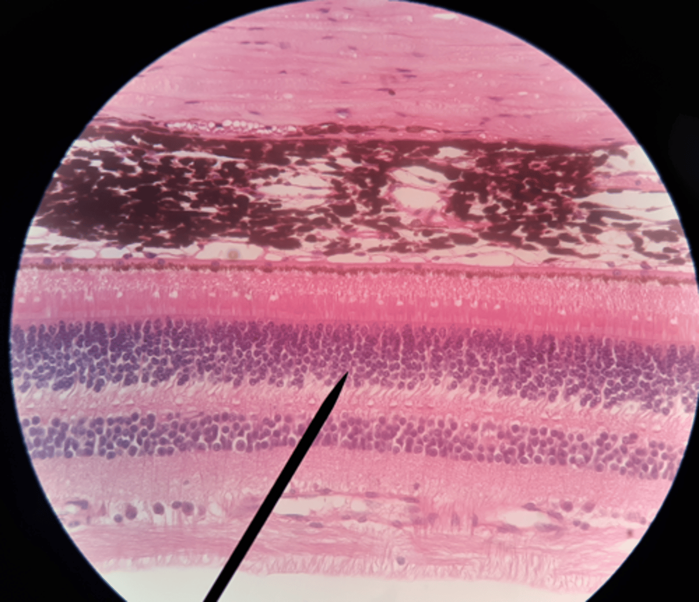
Retina
Special: Vision
tissue structure located on the inner part of the posterior eyeball, just anterior to the choroid; contains the cells involved in the visual pathway. Involves six main layers from posterior to anterior: pigmented layer (rods and cones), outer synaptic layer, bipolar layer, inner synaptic layer, ganglion cell layer, optic (II) nerve axons
General or Special Sense?-Meissner Corpuscle
General: Somatic-Touch
General or Special Sense?- Pacinian Corpuscle
General: Somatic-detects vibration
General or Special Sense?- Vallate Papillae
Special: Taste
General or Special Sense?- Filiform Papillae
Special: Taste
General or Special Sense?- Fungiform Papillae
Special: Taste
General or Special Sense?- Foliate Papillae
Special: Taste
General or Special Sense?- Crista Ampullaris
Special: Hearing
General or Special Sense?- Cochlea
Special: Hearing
General or Special Sense?- Cornea
Special: Vision
General or Special Sense?- Retina
Special: Vision
General senses include
• touch, pressure, vibration, itch, tickle
• thermal sensation:
warm/cold
• pain sensations
• proprioceptive sensations
• provide information about the internal organs
Visceral Senses, provide information about the internal organs (pressure, stretch, chemicals, nausea, hunger, temperature, are considered a general or special sense?
general sense
special senses include
• smell
• taste
• vision
• hearing
• equilibrium
• balance

1
Aqueous humor. Clear, watery fluid that fills the anterior chamber of the eye, between the cornea and lens.

2
Blind spot. Optic disk, which creates a small break in the visual field; point where the retina's receptor cell axons converge to form the optic nerve.

3
Choroid. Thin, pigmented layer behind the retina; in many animals a light-reflective portion, the tapetum lucidum, aids night vision.
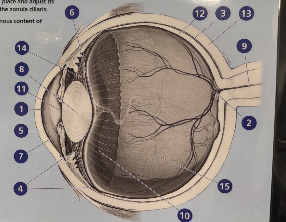
4
Ciliary body. Structure that encircles the eye opening posterior to the iris; connects the iris, suspensory ligaments, and ora serrata; includes muscles that operate the lens.

5
Cornea. Transparent covering over the front of the eye that allows light to enter; attached at its perimeter to the opaque sclera.

6
Hyaloid fossa. Depression on the anterior of the vitreous body that helps hold the lens in place.

7
Iris. Pigmented diaphragm that regulates the size of the pupil to control the amount of light entering the eye.

8
Lens. Biconvex transparent structure that focuses light onto the retina.

9
Optic nerve. Bundle of nerve fibers that transmit visual signals from the eye to the brain.

10
Ora serrata. The serrated disk of tissue behind the lens that attaches to the choroid at the anterior rim of the retina.

11
Pupil. The adjustable opening through the iris where light enters the eye.

12
Retina. The light-sensitive lining of the eye that contains receptor cells called rods and cones.

13
Sclera. Tough, opaque outer covering of the eyeball that helps maintain the eye's shape.

14
Suspensory ligaments. Fibrous strands that connect the lens and the ciliary body; they help hold the lens in place and adjust its thickness to change focus; also known as the zonula ciliaris.

15
Vitreous humor. The transparent gelatinous content of The posterior chamber of the eye.
Name of all the Cranial nerves?
Olfactory, Optic, Oculomotor, trochlear, trigeminal, abducens, facial, vestibulocochlear, glossopharyngeal, vagus, accessory, hypoglossal.
CN 1?
Olfactory
Innervates: Olfactory epithelium (nose)
Function: Smell (sensory)
CN 2?
Optic
Innervates: Retina
Function: Vision (sensory)
CN 3?
Oculomotor
Innervates: Most eye muscles, eyelid, pupil
Function: Eye movement, opens eyelid, constricts pupil (motor)
CN 4?
Trochlear
Innervates: Superior oblique muscle
Function: Moves eye down & in (motor)
CN 5?
Trigeminal
Innervates: Face, chewing muscles
Function: Facial sensation + chewing (both)
Exit: V1 (Ophthalmic) – Superior orbital fissure
V2 (Maxillary) – Foramen rotundum
V3 (Mandibular) – Foramen ovale
CN 6?
Abducens
Innervates: Lateral rectus muscle
Function: Moves eye laterally (motor)
CN 7?
Facial
Innervates: Facial expression muscles, lacrimal & salivary glands
Function: Facial expression, taste, tears & saliva (both)
CN 8?
Vestibulocochlear
Innervates: Inner ear
Function: Hearing & balance (sensory)
CN 9?
Glossopharyngeal
Innervates: pharynx, parotid gland
Function: Taste, swallowing, saliva (both)
CN 10?
Vagus
Innervates: Pharynx, larynx, thoracic & abdominal organs
Function: Swallowing, speech (both)
CN 11?
Accessory
Innervates: Sternocleidomastoid & trapezius
Function: Head turn & shoulder shrug (motor)
CN 12?
Hypoglossal
Innervates: Tongue muscles
Function: Tongue movement (motor)
Cerebrum
What is number 1? (including both hemispheres)

Cerebellum
What is number 2?

spinal cord
What is number 3?

frontal lobe
What is number 4?

parietal lobe
What is number 5?

temporal lobe
What is number 6?

occipital lobe
What is number 7?

left hemisphere
What is number 8?

Medial Longitudinal fissue
What is number 9?

Right Hemisphere
What is number 10?
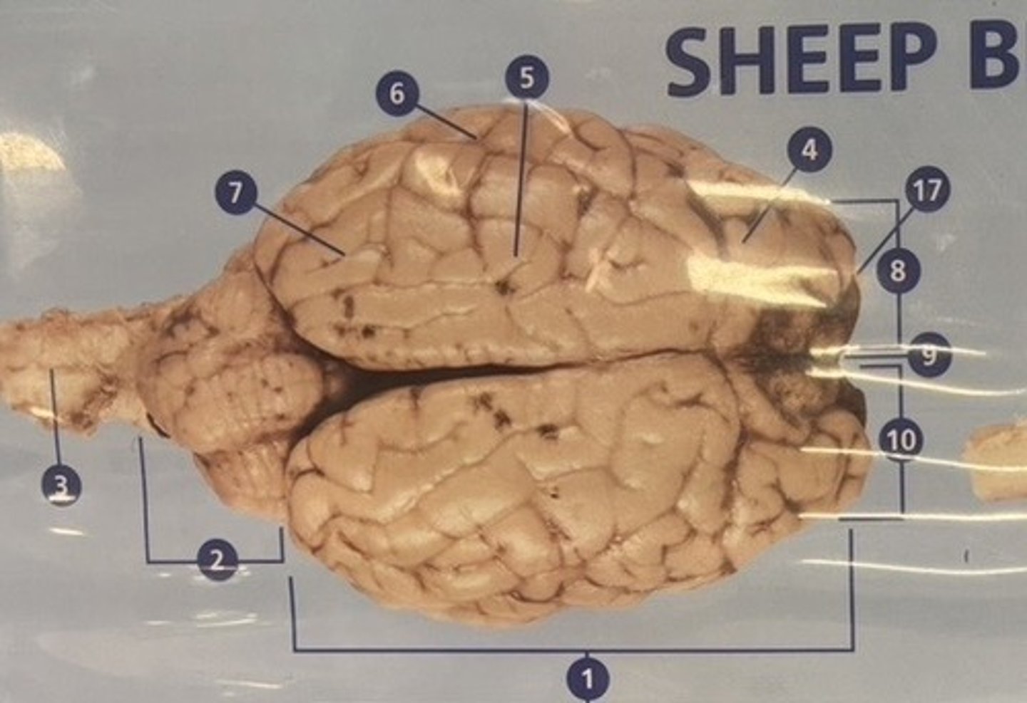
Brain Stem
What is number 11?

Medulla
What is number 12?

Pons
What is number 13?

Optic Tract
What is number 14?

Optic Chiasm
What is number 15?
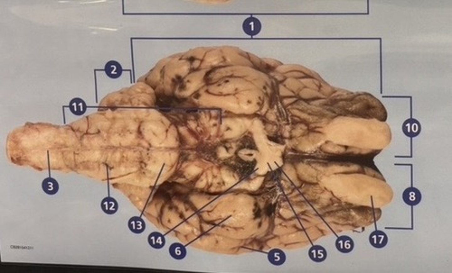
Optic Nerve
What is number 16?

Olfactory Bulb
What is number 17?

Midbrain
What is number 18?

Sulci
What is number 19?
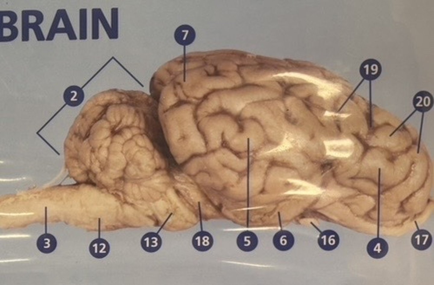
Gyri
What is number 20?

Septum Pellucidum
What is number 21?

Third Ventricle
What is number 22?

Left Lateral Ventricle
What is number 23?

Fourth Ventricle
What is number 24?
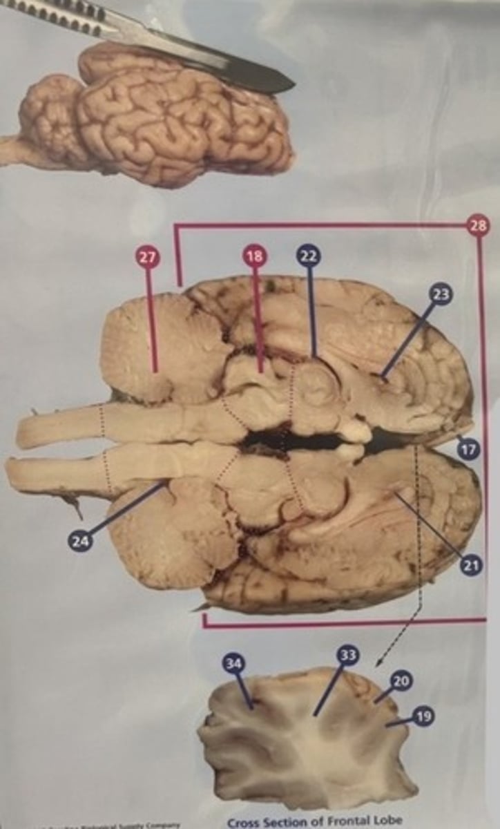
Arbor Vitae
What is number 25?

Superior Colliculus
What is number 26?

Hindbrain
What is number 27?

Forebrain
What is number 28?

Pineal Body
What is number 29?

Thalamus
What is number 30?

Corpus Calllosum
What is number 31?
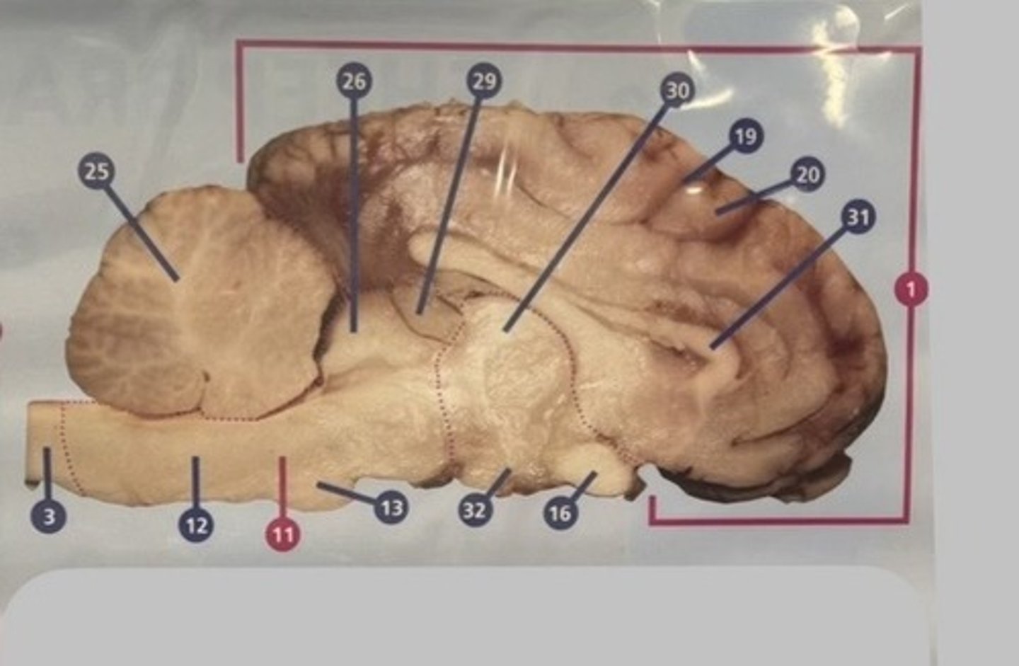
Hypothalumus
What is number 32?

White Matter
What is number 33?

Gray Matter
What is number 34?

Corpus callosum
White fiber tract connecting the left and right hemispheres
Olfactory Foramen
Cranial nerve I exit
Sulcus
A groove or depression on the cortex
Fourth ventricle
The cerebral aqueduct empties into this ventricle
Olive
Group of cell bodies of neurons and output to the cerebellum
Hypoglossal canal
This foramen outlets cranial nerve XII
Septum pellucidum
Divider between lateral ventricles
Hindbrain
Lower part of the brainstem that includes the cerebellum, pons, and medulla oblongata
Optic canal
Cranial nerve II exit
Diencephalon
Region that contains the thalamus and hypothalamus
Mammillary body
Bulge found at the hypothalamic floor
Cerebrum
Region that contains the frontal, parietal, occipital, and temporal lobes
Foramen ovale
Exit for the third trigeminal nerve (V3)
Arbor vitae
Branching myelinated fibers in the cerebellum
Inferior colliculus
Reflex movement of the head to auditory stimulus