biosci107 07 muscle
 Call Kai
 Learn
Learn Practice Test
 Spaced Repetition
 Match
Match Flashcards
 Knowt Play
1/16

There's no tags or description
Looks like no tags are added yet.
Name | Mastery | Learn | Test | Matching | Spaced | Call with Kai |
|---|
No analytics yet
Send a link to your students to track their progress
17 Terms
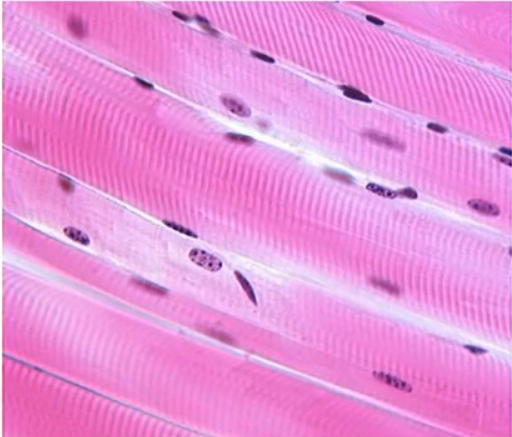
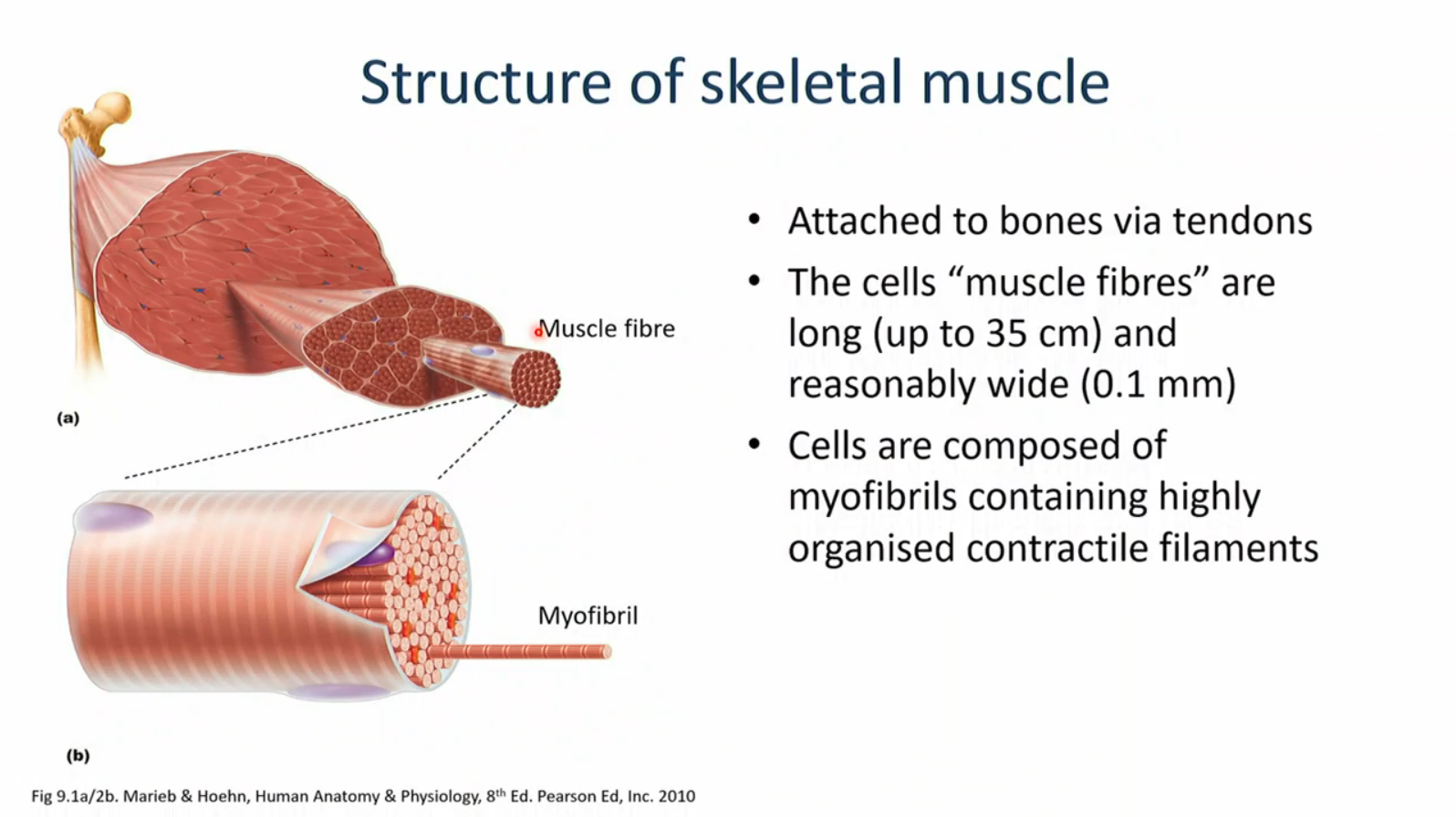
description of skeletal muscle
under voluntary control, striated, single long cylindrical cells, multiple peripheral nuclei
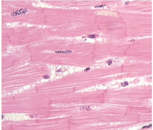
description of cardiac muscle
located only in the heart, striated, branched cells with 1-3 central nuclei, connected via intercalated discs, involuntary control
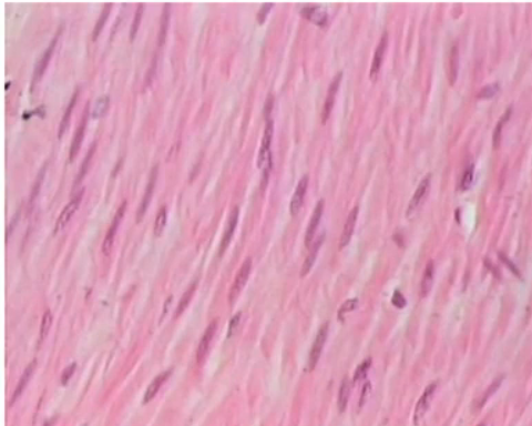
description of smooth muscle
involuntary, found in the wall of internal organs (gut, blood vessels), spindle shaped, uninucleated cells, not striated
structure of skeletal muscle
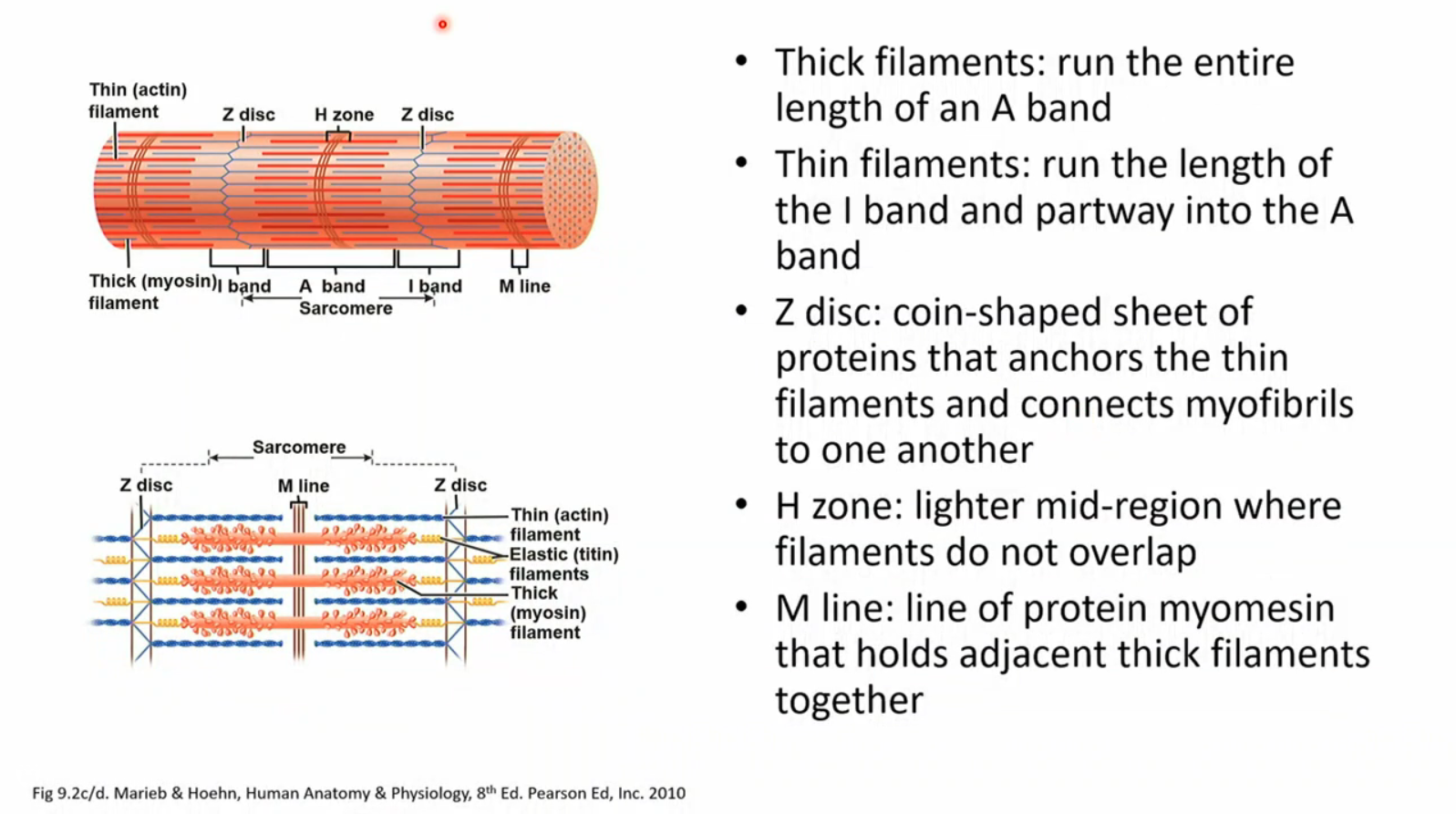
structure of a sarcomere
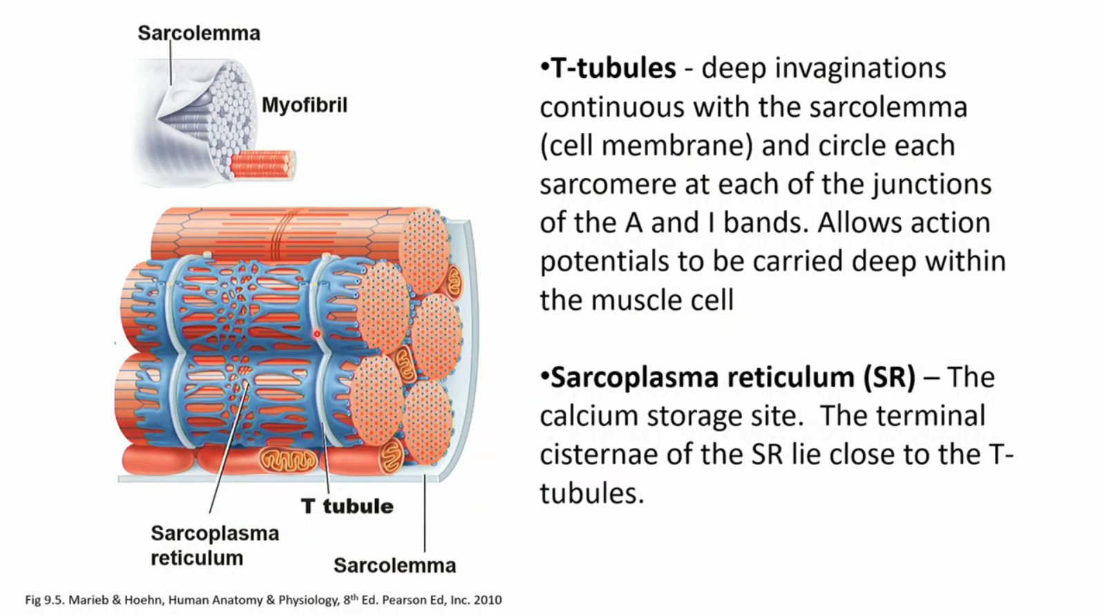
T-tubules and sarcoplasma reticulum
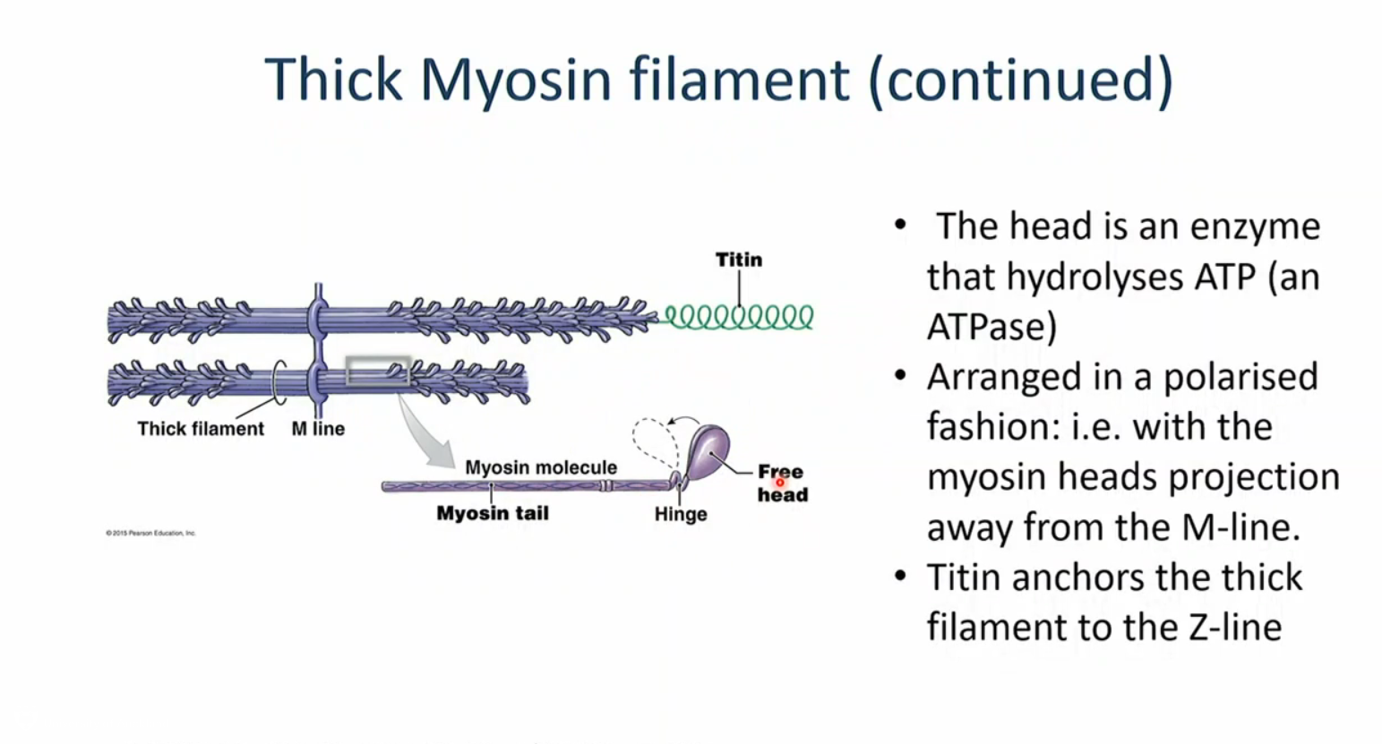
the thick filaments
composed of myosin, each myosin has 2 subunits each with a globular head and a tail, the two tails intertwine to form a helix, the heads have a binding site for actin. arranged in a polarised fashion so heads are sticking out in the ends and tails meet in the middle
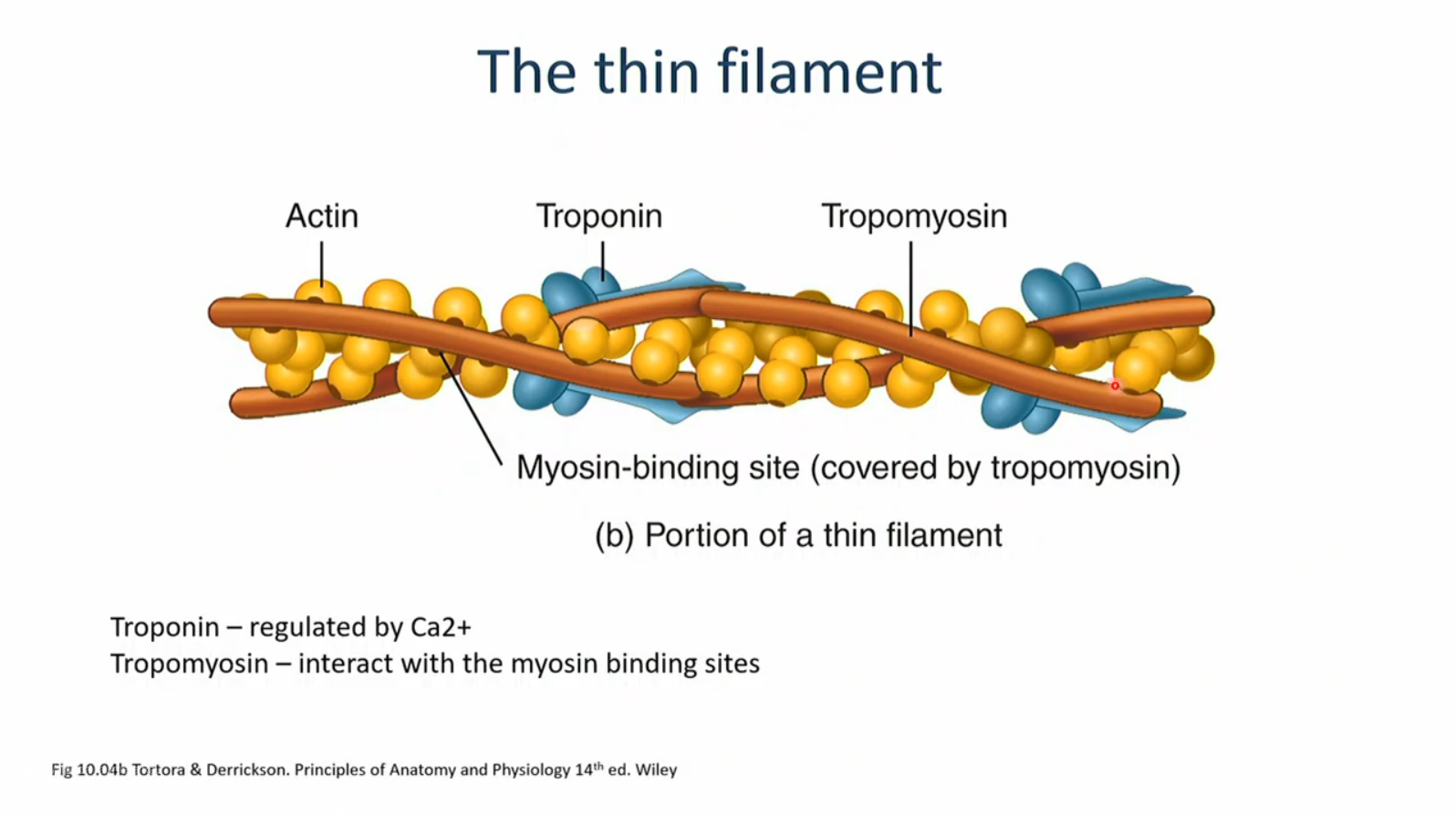
the thin filaments
composed primarily of globular actin proteins, the filaments are composed of a double stranded helical actin chain (polymers), troponin and tropomyosin are regulatory proteins associated with actin in skeletal and cardiac muscle
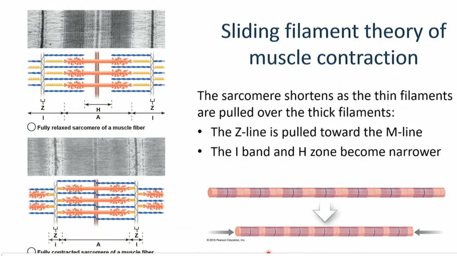
sliding filaments theory of muscle contraction
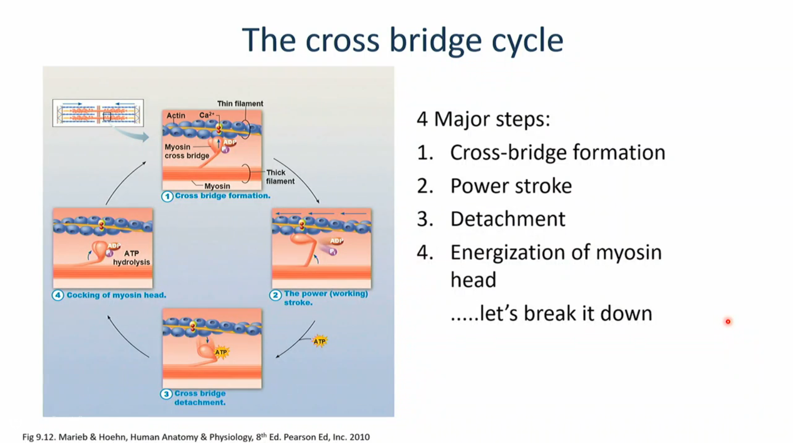
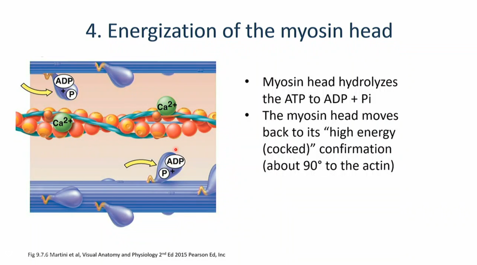
4 major steps of the cross bridge cycle
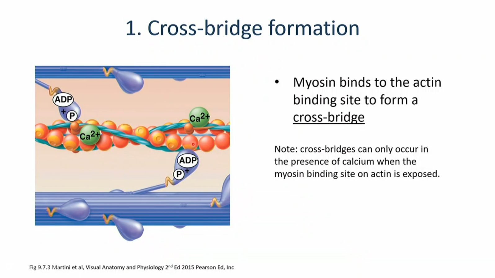
cross bride formation
the power stroke
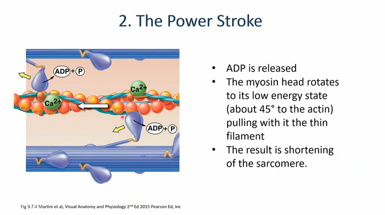
detachment
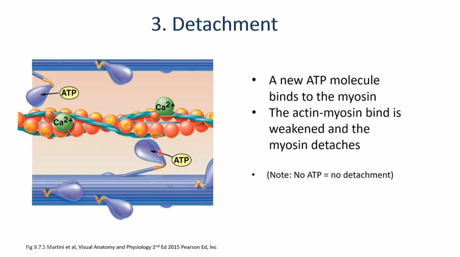
energisation of the myosin head
the importance of calcium in muscle contraction
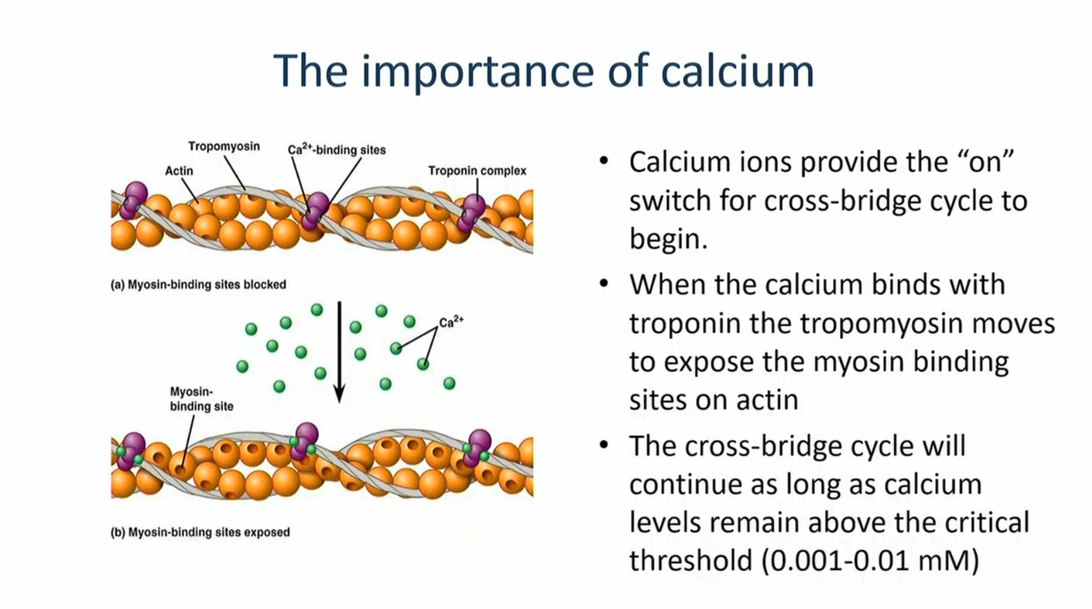
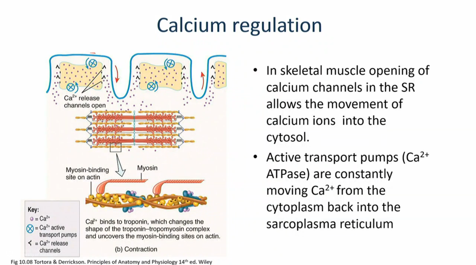
calcium regulation in skeletal muscle
action potential causes release of Ca2+
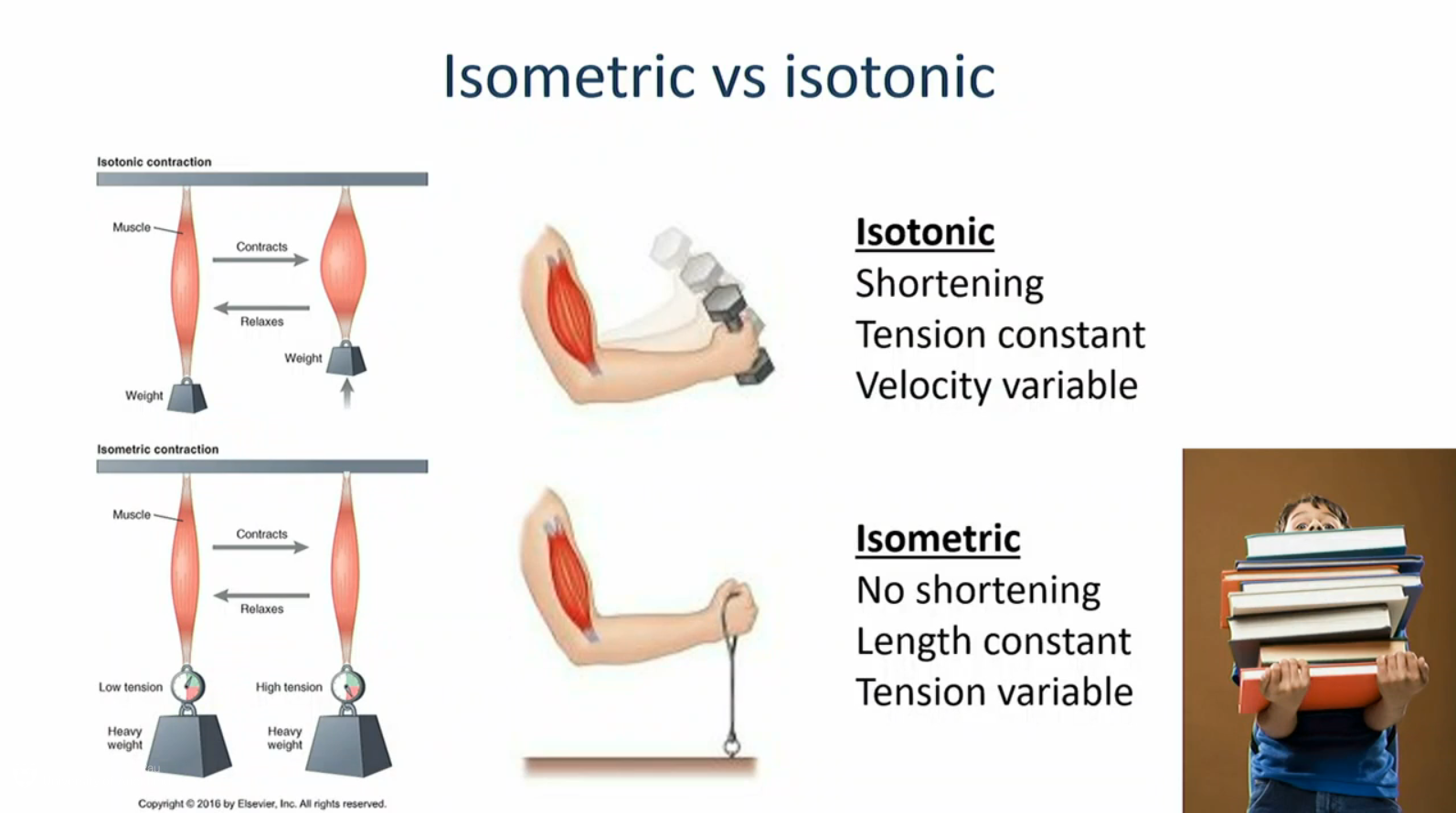
isometric vs isotonic