Sheep Eye Dissection Photos
1/29
There's no tags or description
Looks like no tags are added yet.
Name | Mastery | Learn | Test | Matching | Spaced | Call with Kai |
|---|
No analytics yet
Send a link to your students to track their progress
30 Terms
Sclera
Attachment site for eye muscle
Optic Nerve
transfer of visual information
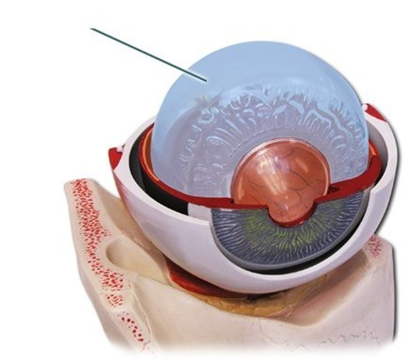
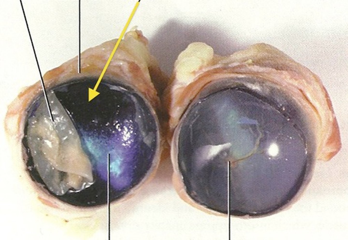
Cornea
to allow light through
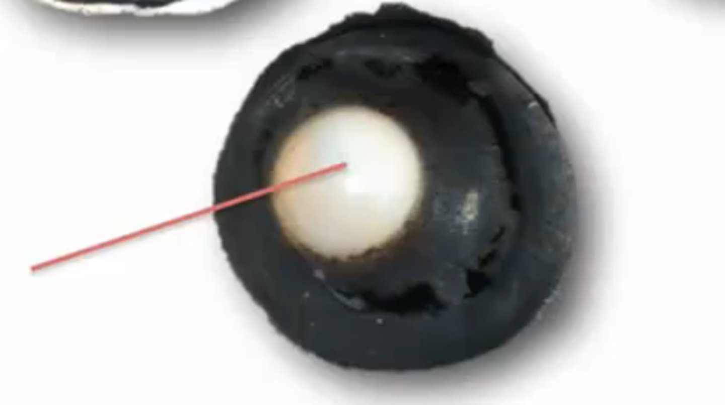
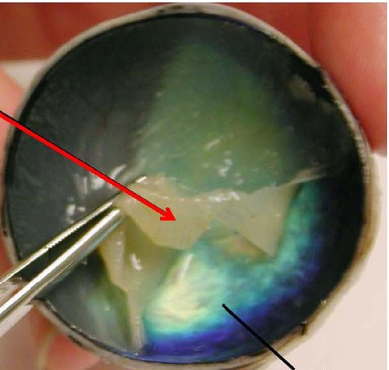
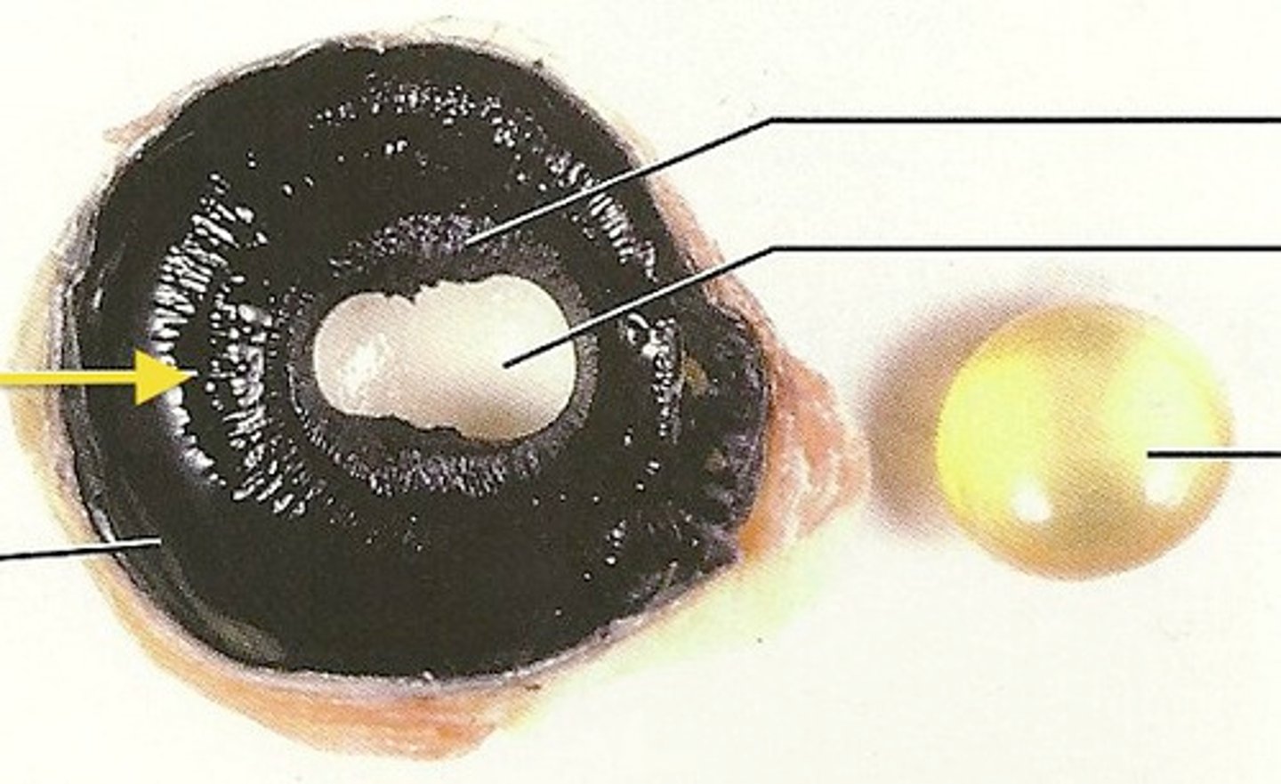
Lens
focusing the arriving image
Ciliary Body
adjust shape of lens
Choroid
Black/ delivers oxygen and nutrient to retina

Retina
receive light from lens
Optic Disk
places where axon exits
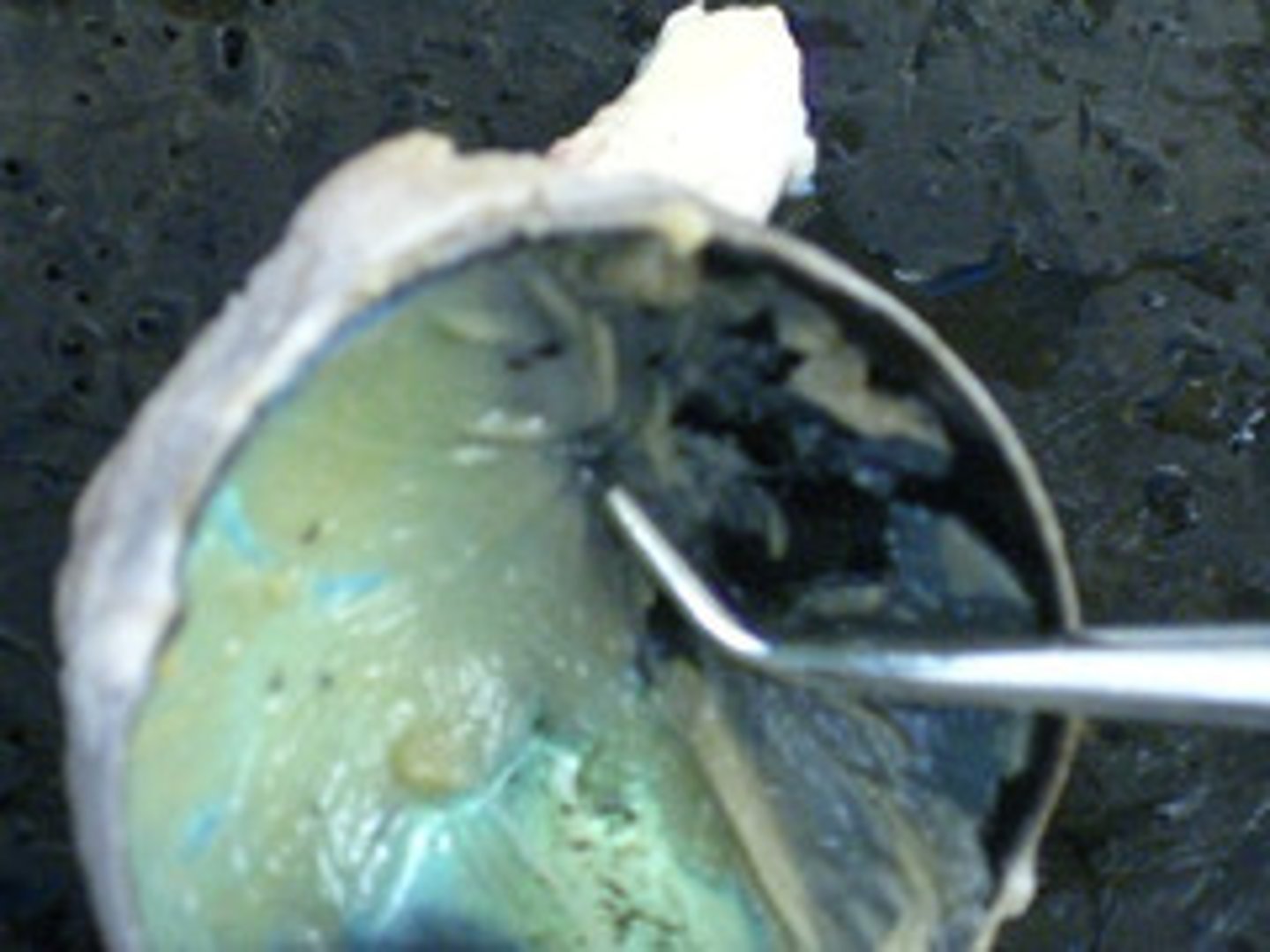
Vitreous Humor
helps maintain eye shape
Pupil Function
light into eye
Iris Function
controls the size of pupil
Choroid
thin black/brown film covering the eye.
Ciliary Body
black with "spokes"
anchors suspensory ligaments that flatten the lens for focusing your vision
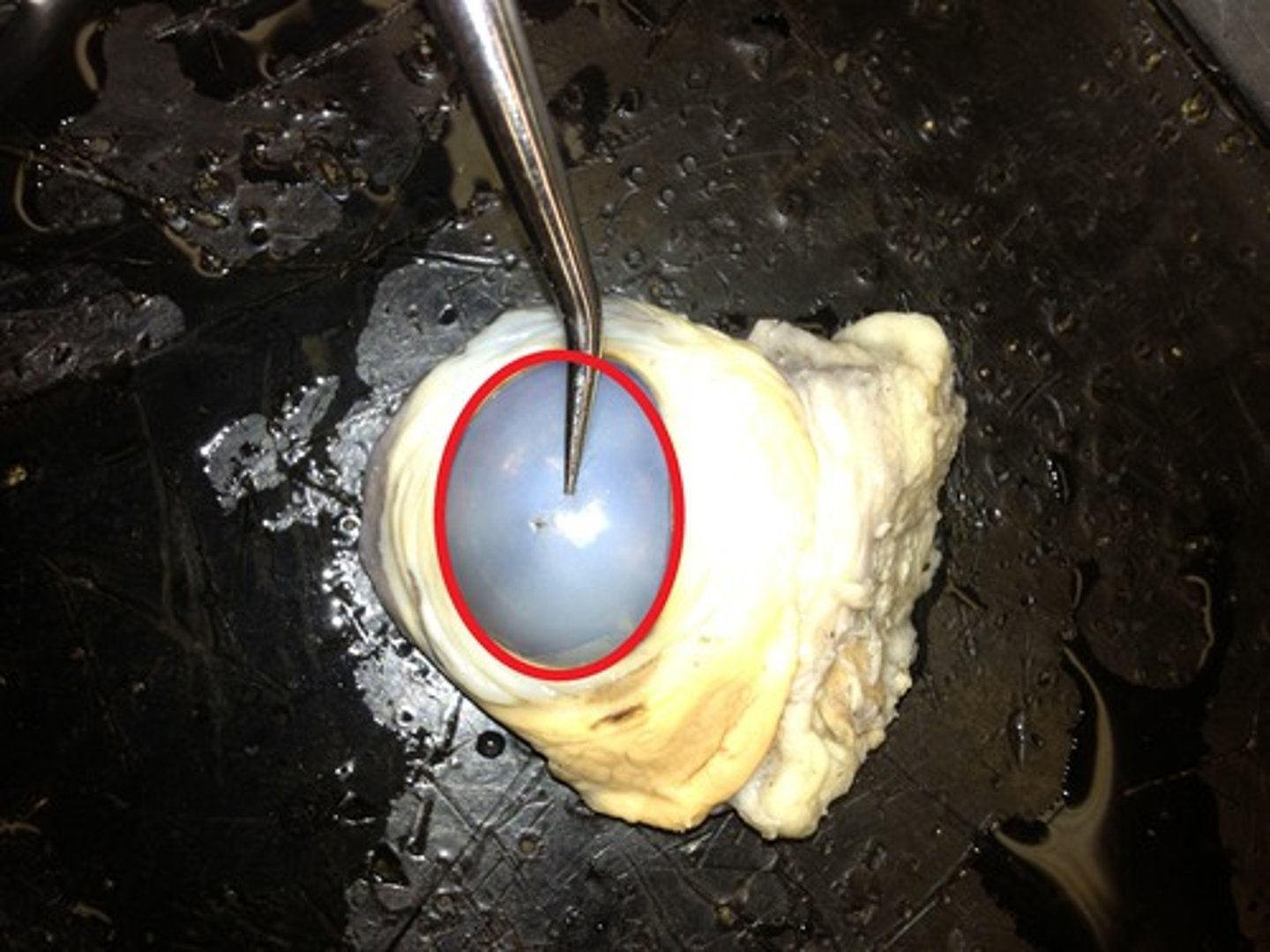
Cornea
clear "disk" covering the iris and pupil
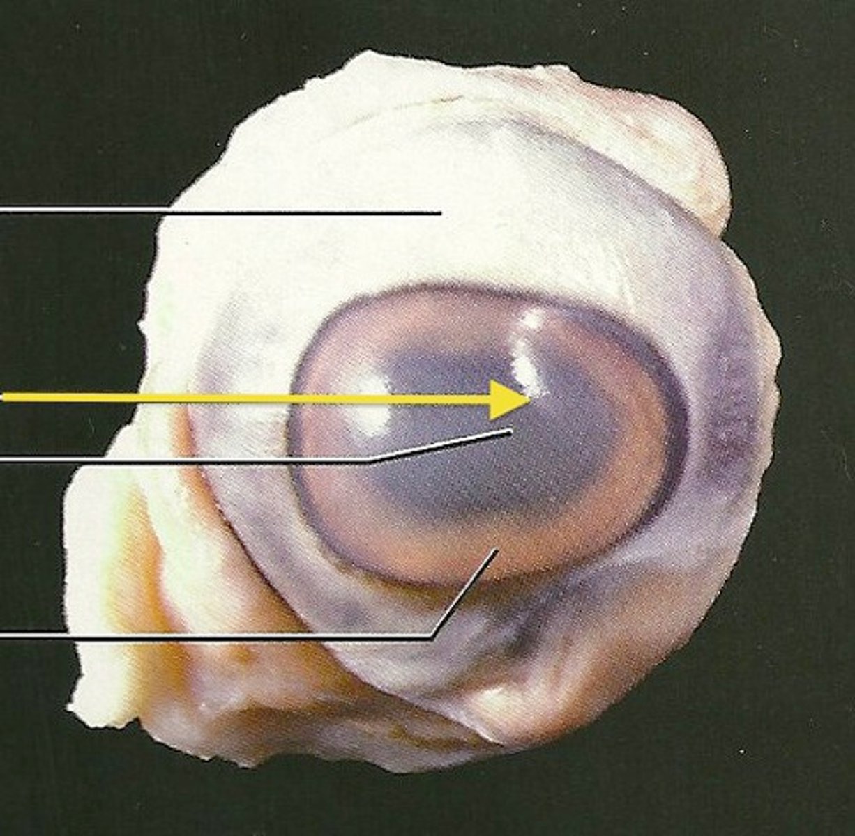
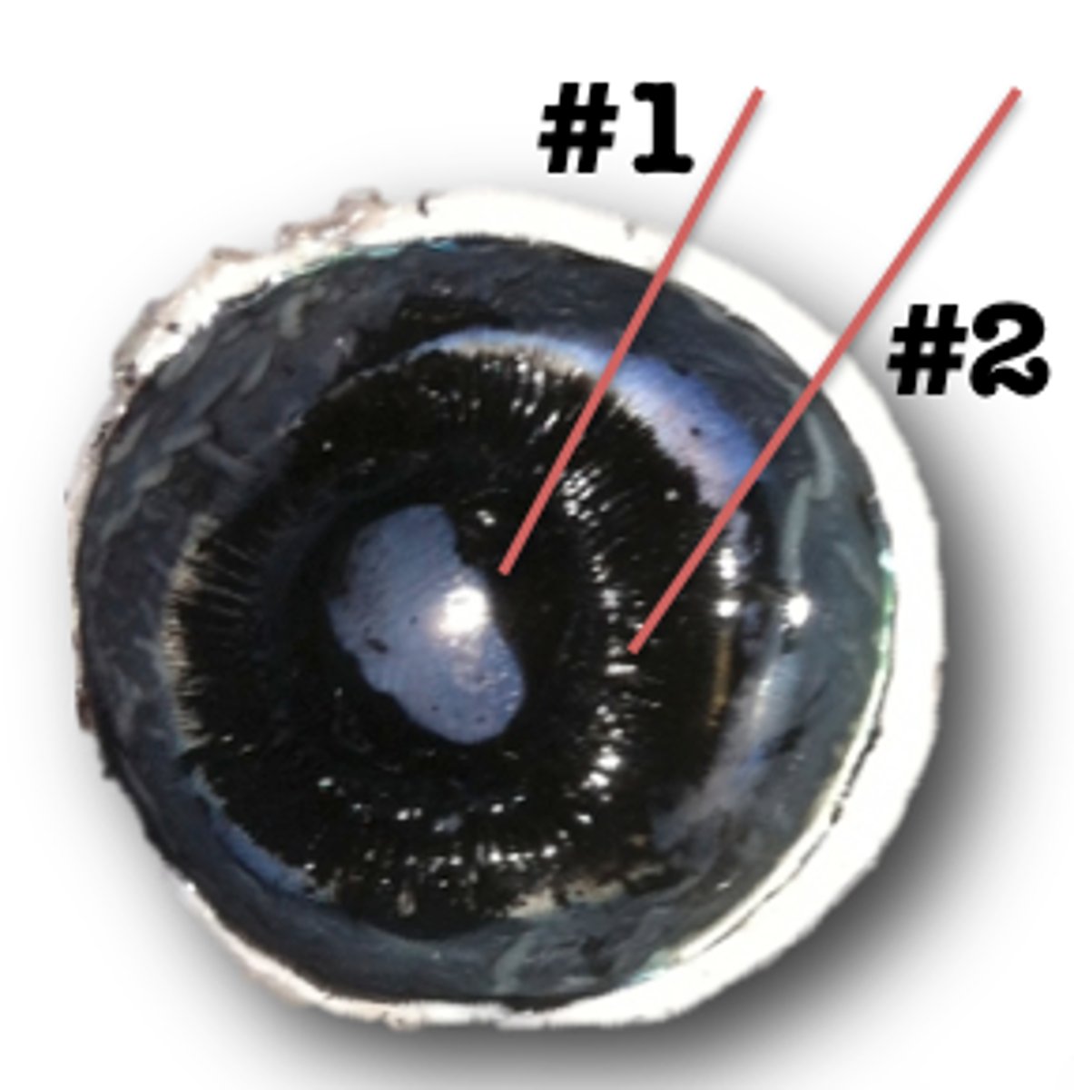
Iris
the colored part of the eye.

Optic Disk
the point where the optic nerve enters the retina. Blind spot.
Optic Nerve
the cranial nerve that serves the retina, appears as a white line or stem off center in the bacl of the eye.
Pupil
the opening through which light enters the eye
Retina
thin, translucent film covering the eye, beneath the choroid.
Sclera
Tough, white outer coat of the eyeball
Tapetum Lucidum
Iridescent layer, blue green irridescent sheen on the choroid

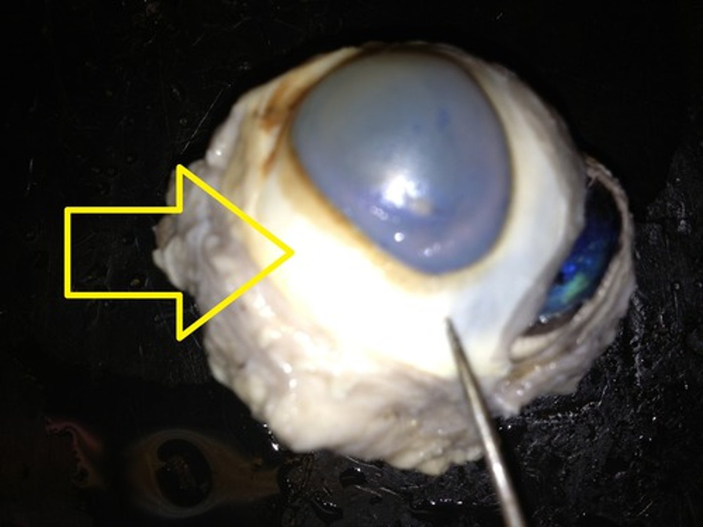
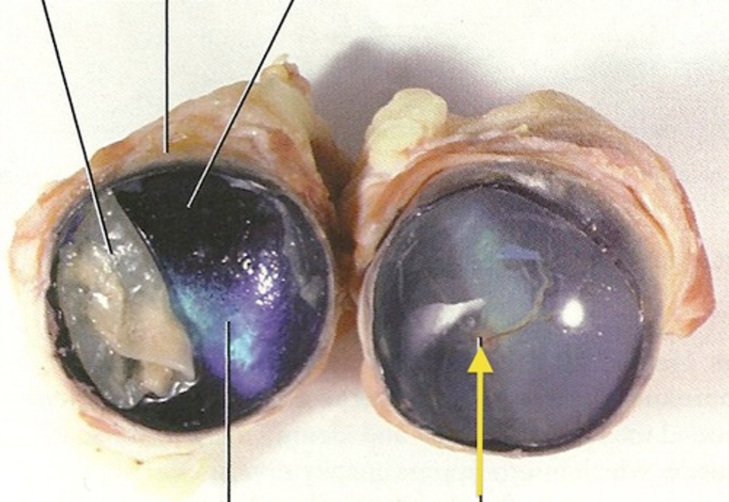
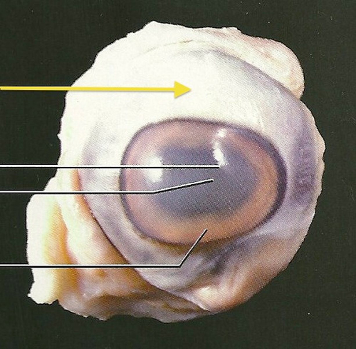
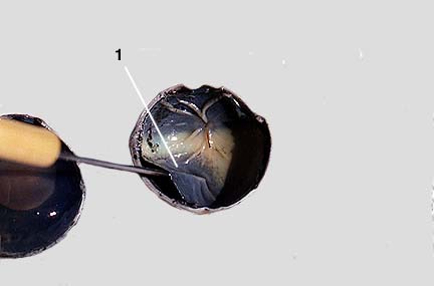
1) cornea 2) sclera
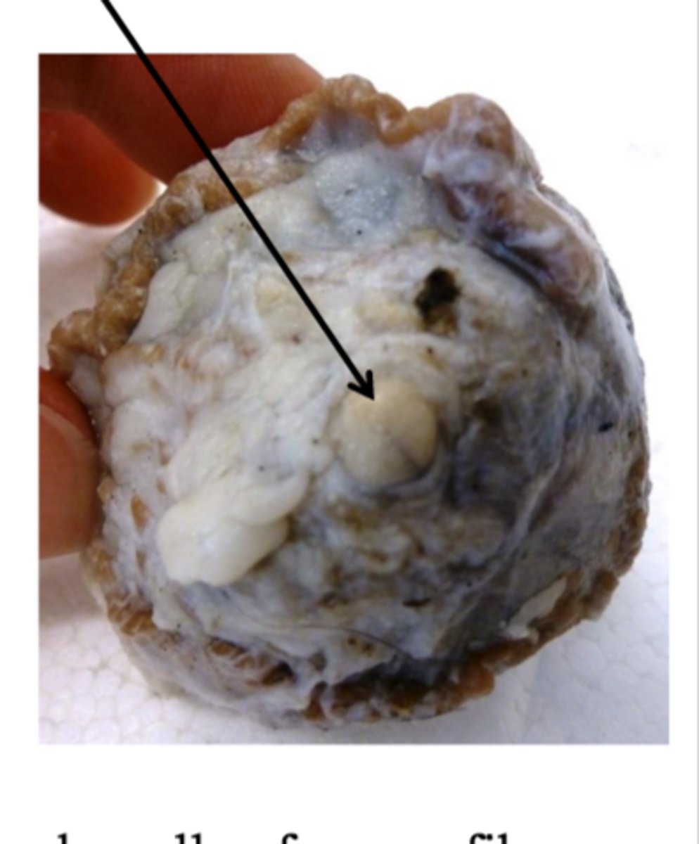
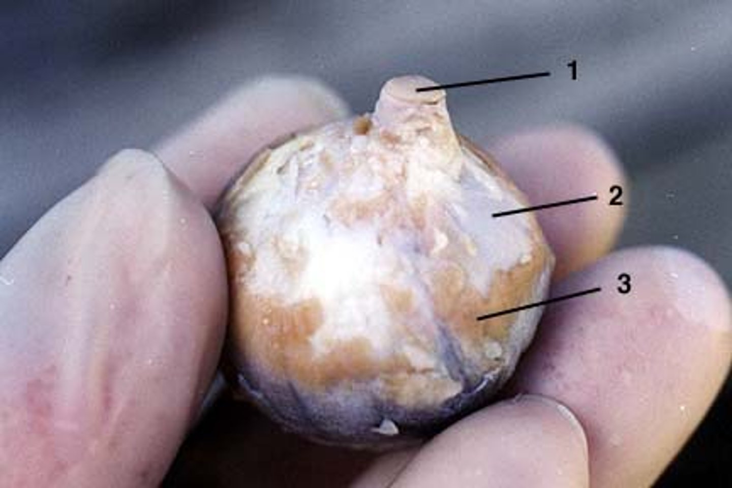
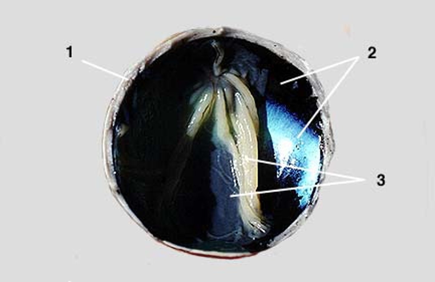
1) optic nerve 2) sclera 3) extrinsic muscles
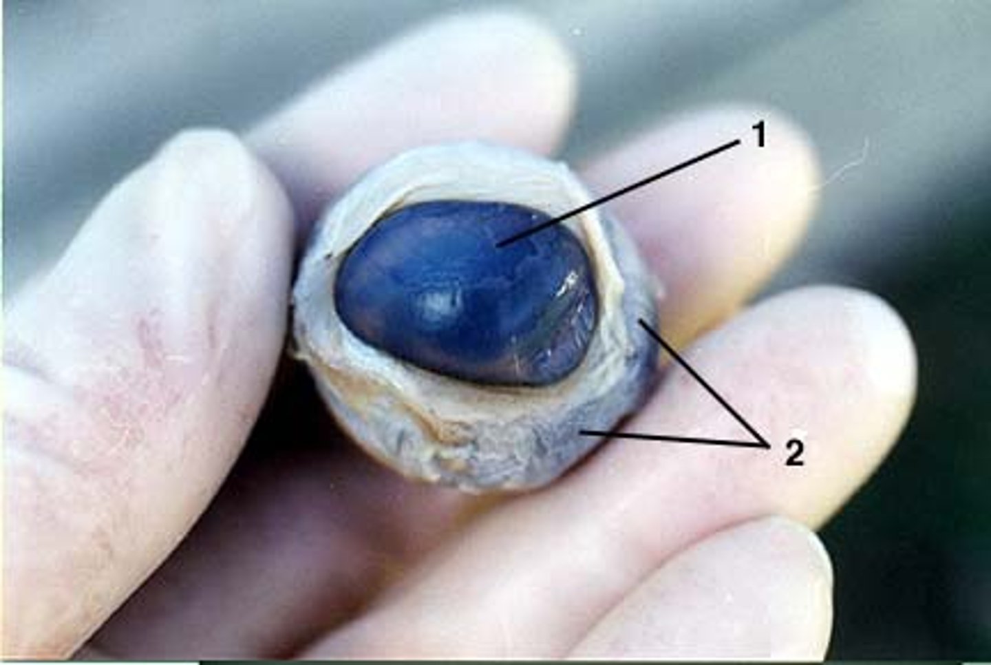
1) cornea 2) extrinsic muscle
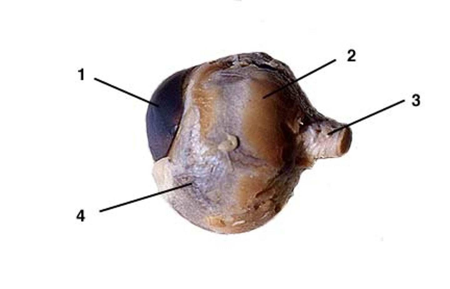
3) optic nerve 4) sclera

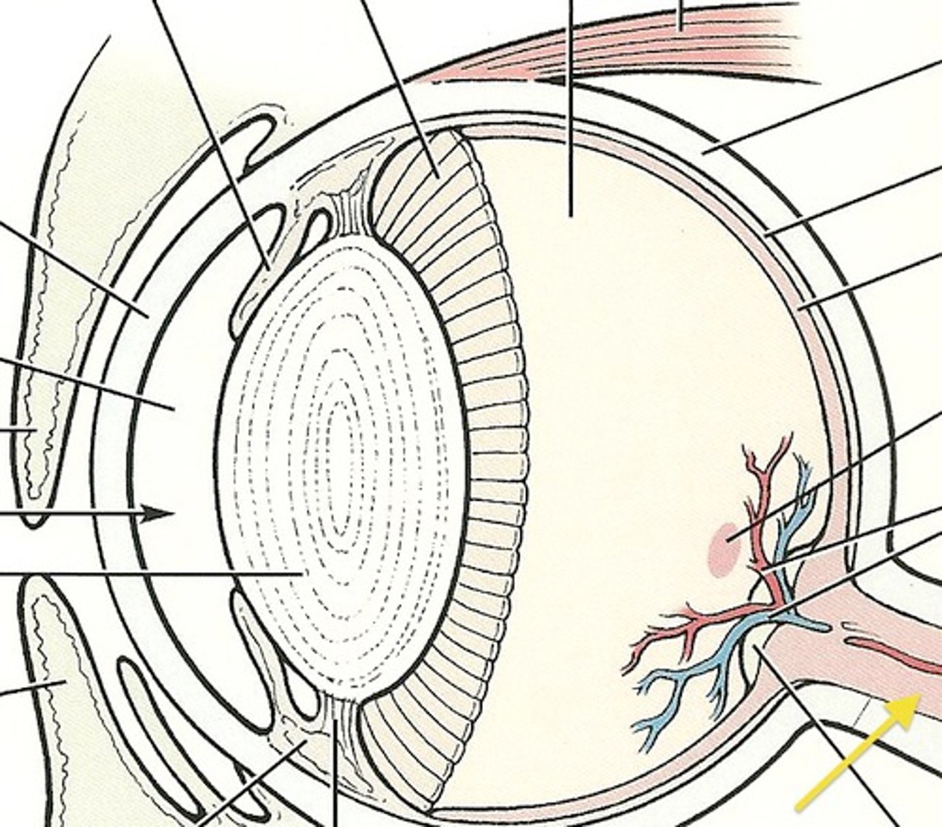
1) retina
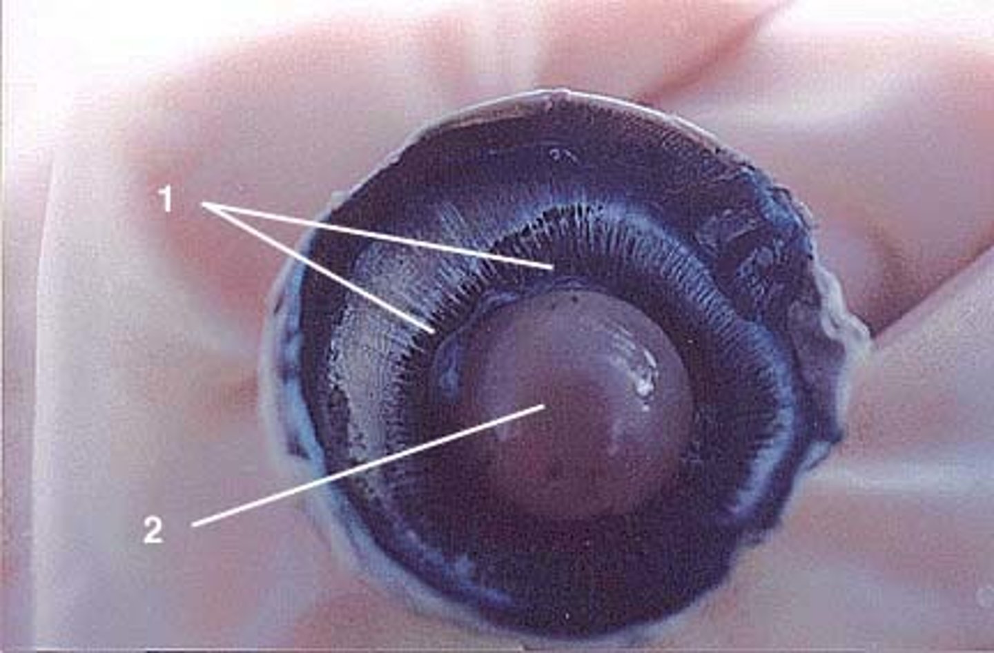
1) sclera 2) choroid 3) retina
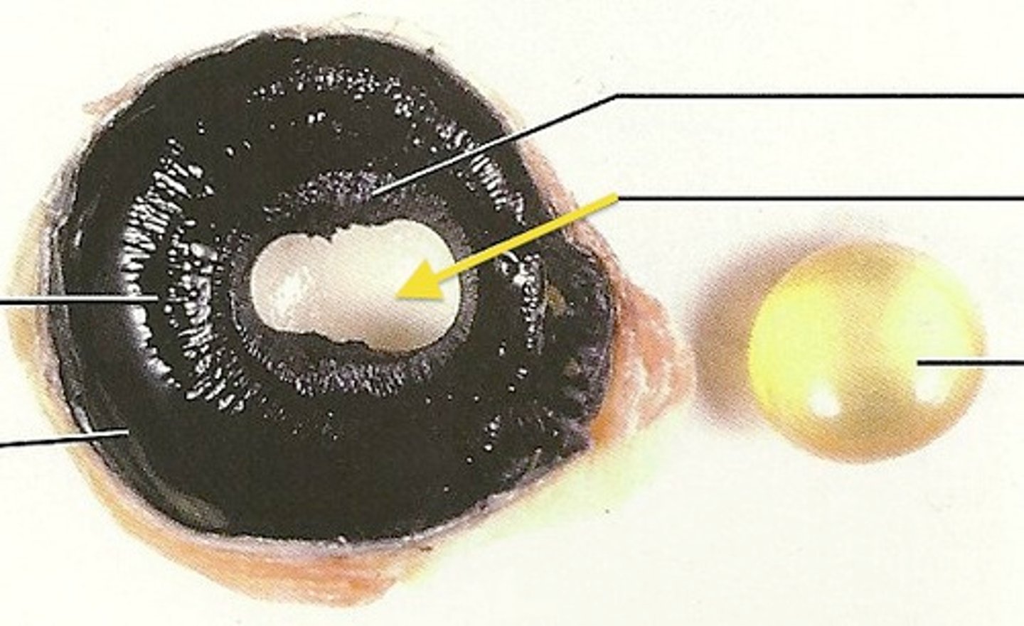
1) ciliary body 2) lens
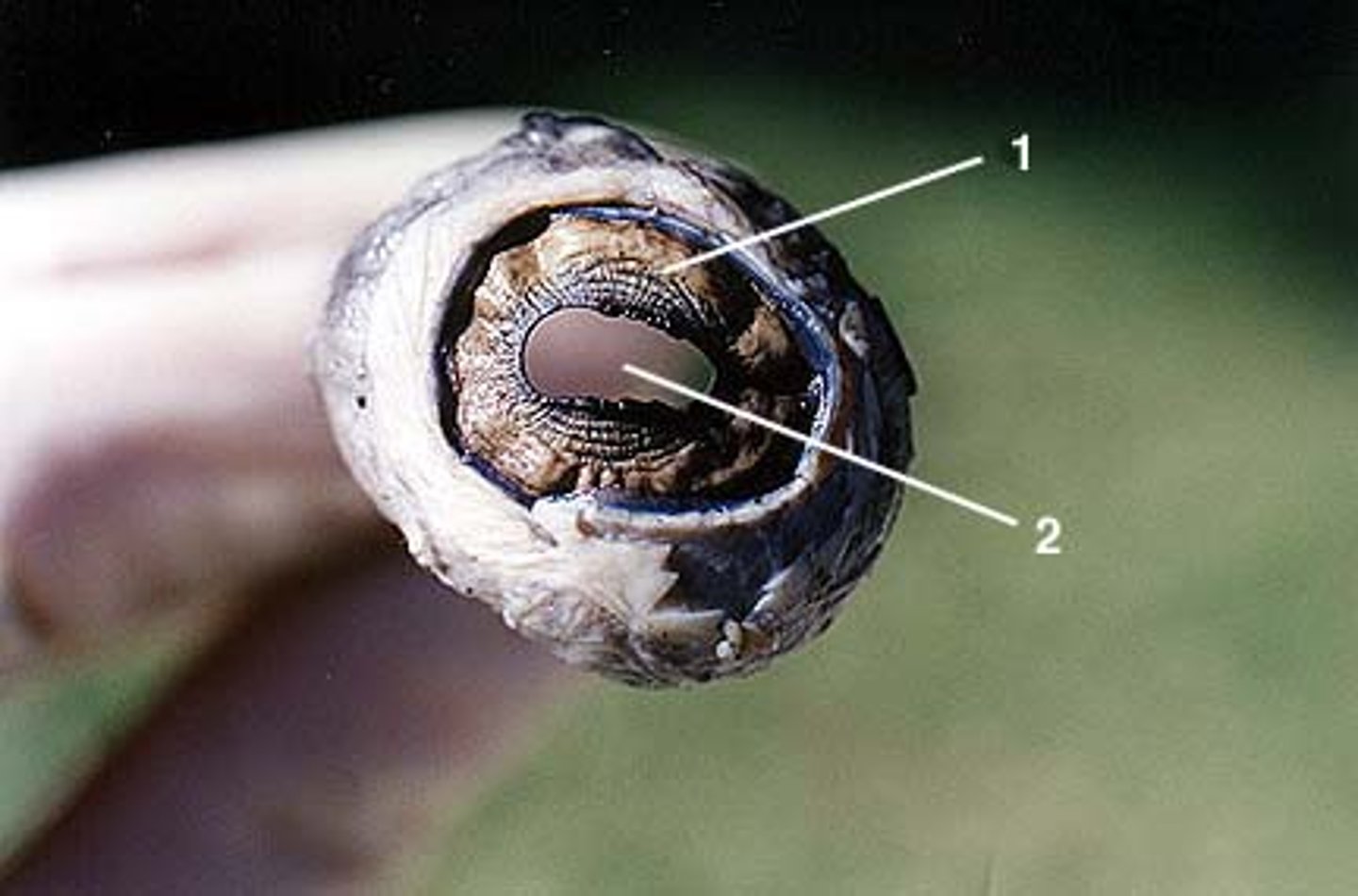
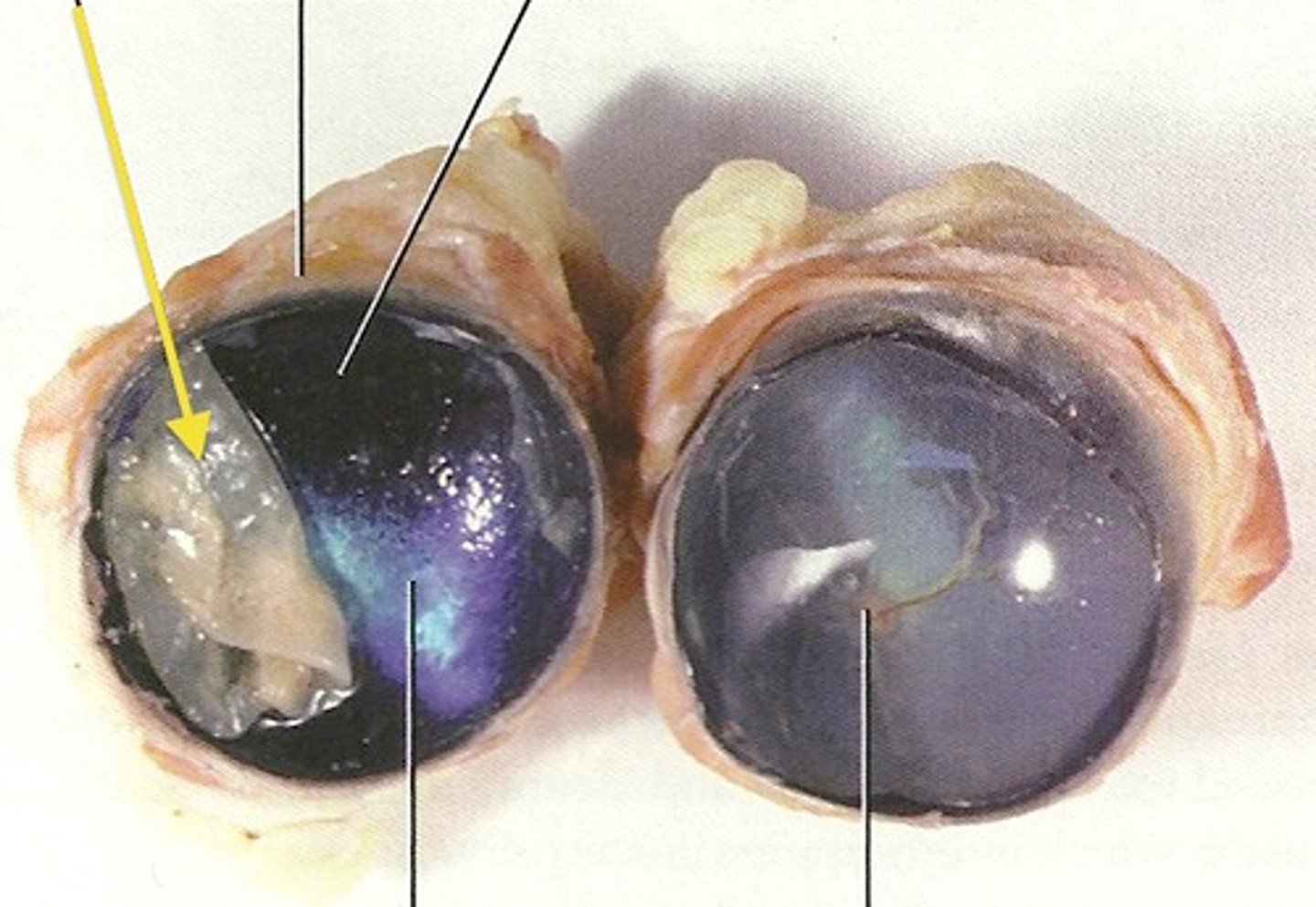
1) iris 2) pupil
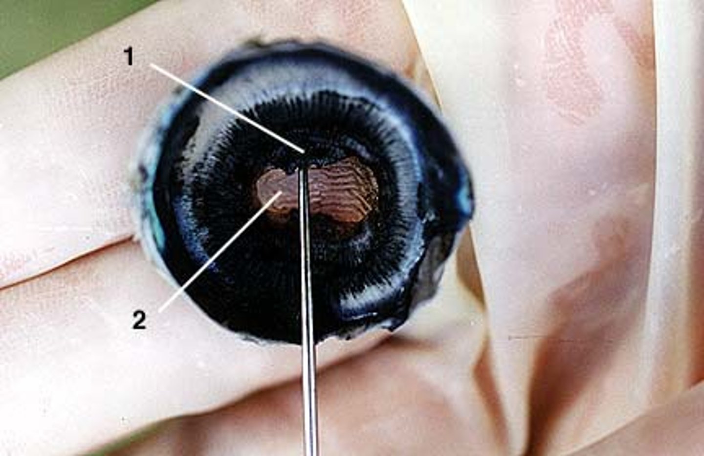
1) iris 2) pupil