Biomechanics WK2- DPT
 Call Kai
 Learn
Learn Practice Test
 Spaced Repetition
 Match
Match Flashcards
 Knowt Play
1/110

There's no tags or description
Looks like no tags are added yet.
Name | Mastery | Learn | Test | Matching | Spaced | Call with Kai |
|---|
No analytics yet
Send a link to your students to track their progress
111 Terms
types of joint surfaces
ovoid, planar, sellar
ovoid
most joints, convex and concave members
saddle
reciprocal surfaces, convex and concave members will switch depending on plane view
arthrokinematic movements
roll, glide, spin
roll movement
different points on the surface are in contact with different points on a second surface
glide movement
one point is in contact with different points on a second surface
rule for convex bone moving on concave
roll and glide occur in opposite direction
bone end rolls in direction bone moves
bone end glides in opposite direction
(shoulder abducting)
rule for concave bone moving on convex
roll and glide occur in same direction
bone end rolls in direction bone moves
bone end glides in same direction
osteokinematic and arthrokinematic movements
classical and accessory
classical movement is split into…
active ROM and passive ROM
accessory movement is split into…
component and joint play
component movements can be split into
adjunct rotation and conjunct rotation
adjunct rotation
can control
occurs because of muscle force
conjunct rotation
cannot be controlled
occurs because of joint geometry
joint play movements
occur in response to external force
medial/lateral glide
anterior/posterior glide
distraction/compression
* some joint play is healthy to absorb shock
movements in the planar joint surface
osteo= translatory
arthro= glide
movements in the ovoid joint surface
osteo= swing osteo= spin
arthro= roll & glide arthro= spin
movements in the sellar joint surface
osteo= swing
arthro= roll & glide
2 joint positions
close packed position (CPP) and loose packed position (LPP)
close packed position
max congruency between articular surfaces
greatest tension in capsule & ligaments
creates compression of joint
loose packed position
rest position, greatest amount of wiggle in joint
positioning of the hip joint in LPP and CPP
LPP: 30° flex, 30° abd, slight lat rotation
CPP: ext, med rotation, abd
positioning of the knee joint in LPP and CPP
LPP: 25° flex
CPP: full ext and lat rotation of tibia
problems with manual muscle testing (MMT)
spasticity and return to sport
key points for positioning a patient for MMT
free full ROM
against gravity/ movement away from the floor
minimize chance for muscle substitution
where and when to apply resistance during MMT
perpendicular pressure at the distal end of the moving segment
after seeing full ROM
types of resistance tests
break test
make test
strength through range
break test
PT applies max force to move body part out of end range
make test (active resistance test)
PT slowly builds resistance to match patient’s force
strength through range
PT applies force but allows motion through full ROM
fair (3/5)
move through full ROM against gravity
good (4/5)
move through full ROM and somewhat less than max resistance
normal (5/5)
full ROM and full max resistance
poor (2/5)
full ROM in gravity lessened position
trace (1/5)
palpable muscle contraction, no visible movement
zero (0/5)
no movement or contraction
3+ / 5
can take little resistance at end ROM against gravity
3- / 5
more than ½ but not full ROM against gravity
2+ / 5
less than ½ but some ROM against gravity OR can take little resistance at end ROM in gravity lessened position
2- / 5
more than ½ but not full ROM in gravity lessened position
1+ / 5
less than ½ but some ROM in gravity lessened position
factors of MMT
tester strength
patient understanding/cooperation
patient age
pain
limits in ROM
fatigue
muscle substitution
upper motor neuron dysfunction
how to improve reliability in MMT
proper positioning
adequate stabilization
non-painful contact
consistent pressure and position
avoid bias and change in patient instructions
acetabular labrum
cartilage ring around acetabulum, embraces femoral head
hip joint capsule
strong and dense
2 types of fibers
longitudinal and zona orbicularis
nearly complete socket for femoral head
blood vessels supply head and neck
what lines non-articular surfaces
synovial membrane
ligaments of hip joint
iliofemoral
ischiofemoral
pubofemoral
iliofemoral ligament
Y ligament of Bigelow
triangle shape
2 dense bands
pubofemoral ligament
from pubis to base of neck
ischiofemoral ligament
posterior and inferior acetabulum to inner greater trochanter
spiral course
weakest
ligament function - abduction
pubofemoral taut
ligament function - adduction
lateral band of iliofemoral taut
ligament function - ER
iliofemoral and pubofemoral taut
ligament function - IR
ischiofemoral taut
ligament function - relaxed stance
hyperextended hip, iliofemoral ligament taut
trochanteric or gluteal bursitis
pain posterior and superior to greater trochanter
most common in hip region
ischial bursitis
pain over ischial tuberosity
common in individuals that sit for prolonged periods of time
iliopectineal bursitis
causes anterior hip pain
snapping hip syndrome
factors affecting muscle function
weight bearing and non-weight bearing
posture (bilateral or unilateral) (better balance side to side than front to back)
size of muscle
starting point of lower extremity
fixation (2 joint muscles)
look at distal joint because it affect proximal joint
stability key points of hip joint
relatively free mobile and stable
less mobile than shoulder but more stable
accommodate function
support body weight
allow for locomotion
factors improving stability
gravity
acetabulum, labrum, zona orbicularis fibers enclose femoral head
atmospheric pressure
balance between anatomic factors
strong ligaments and muscle pull approximates joint
bony configuration
internal rotation limited by…
posterior capsule
ischiofemoral ligament
tight external rotators
primary hip abductors
gluteus medius and gluteus minimus
secondary hip abductors
TFL and gluteus maximus
what muscle is the best hip abductor
gluteus medius
internal rotation is limited by…
posterior capsule
ischiofemoral ligament
tight external rotators
pes anserine is the insertion of tendons…
sartorius
gracilis
semitendinosus
primary hip flexors
iliopsoas
TFL
rectus femoris
sartorius
secondary hip flexors
pectineus
adductor longus
gluteus minimus
main hip flexor
iliopsoas
hip flexion limited by
soft tissue
femoral neck on acetabular labrum
tight hamstrings
vertebral limitations
primary hip adductors
adductor longus
adductor brevis
adductor magnus
gracilis
secondary hip adductors
pectineus
hamstrings
obturator internus and externus
quadratus femoris
hip adduction limited by…
superior capsule
iliotrochanteric ligament
abductor tightness
complexes of knee
tibio-femoral joint
patello-femoral joint
tibio-femoral joint
femoral condyles and tibial plateaus
what separates patellofemoral and tibiofemoral joints
medial and lateral grooves
femoral condyles
biconvex
each condyle convex both medial-lateral and anterior-posterior
axes of condyles diverge posteriorly
lateral facet is more prominent
lateral tibial tubercle
“Gerdy’s tubercle”
attachment of IT band
the tibial plateaus are… when menisci are attached
both concave in both planes
nerve that passes over fibular neck
common fibular nerve (peroneal nerve)
knee ligaments are responsible for …
keeping the knee stable
excessive genu valgum
“knock kneed”
angle of femur and tibia less than or equal to 165°
genu varum
“bow-legged”
angle of femur and tibia greater than or equal to 180°
hyperextension of the knee
considered to be extension from 0-10°
greater than 10° termed genu recurvatum
menisci
semi-lunar cartilage, moon shaped fibrocartilage structure
flat interior surface attaches to tibial plateaus
medial meniscus characteristics
posterior horn broader than anterior
semi-circular
lateral meniscus characteristics
more circular
posterior and anterior horns equal
menisci horns connect directly to
tibia
meniscus and coronary ligaments blend with
knee capsule
what separates lateral collateral ligament form meniscus
popliteus
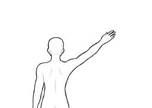
You are testing your patient’s right shoulder abduction strength. Your patient is standing in front of you. You ask your patient to abduct his arm over his head. He is unable to move through full range but is able to move through about 75% of the motion. He has full passive motion, but actively he can only raise his arm about 75% of the motion. What is the manual muscle test grade of his right shoulder abductors:
3-
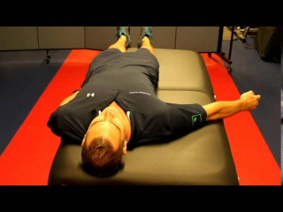
You then test the right shoulder abduction strength of another patient. This patient is unable to raise her arm against gravity. You then have the patient lie down supine. You support the weight of her arm and again ask her to abduct her arm overhead. She is able to move the arm through about 25% of the motion. She has full passive motion, but actively she can only move her arm through 25% of the motion. What is the manual muscle test grade of her right shoulder abductors:
1+
Thinking arthrokinematically, there are 3 basic types of joint surfaces. Which of the following types is the MOST COMMON joint surface:
ovoid
The joint surface influences the type of arthrokinematic motions that can occur at that joint. What type of joint surface do the femoral head and the pelvic acetabulum possess:
ovoid
In the hip joint, which joint surface is the CONVEX member:
femoral head
Osteokinematically, hip abduction and adduction are classified as rotary motions or more specifically swings. What type of arthrokinematic motions occur during hip abduction and adduction:
roll and glide
The concave-convex rule dictates the pattern of rolling and gliding that occurs at a joint. Which of the following statements best describes the arthrokinematics that occur with open chain shoulder abduction, the convex humeral head moving on the concave glenoid fossa:
humeral head rolls superiorly and glides inferiorly
Which of the following statements best describes the arthrokinematics that occur with open chain knee extension, the concave tibial plateaus moving on the convex femoral condyles:
tibia rolls and glides anterior
At the end of open chain knee extension, the tibia laterally rotates to put the knee into the "screw home" position. The lateral rotation is a motion that accompanies knee extension. It occurs because of the geometry of the knee joint and cannot be controlled voluntarily. This type of motion is known as a:
conjunct rotation