A&P- Brain Test
1/36
There's no tags or description
Looks like no tags are added yet.
Name | Mastery | Learn | Test | Matching | Spaced | Call with Kai |
|---|
No analytics yet
Send a link to your students to track their progress
37 Terms
Meninges
membranes that protect the brain and spinal cord; lie between bone and soft tissues of nervous system; 3 layers
3 layers of meninges
dura mater, arachnoid mater, pia mater
dura mater
thick dense connective tissue, outermost layer of the meninges.
-Dural sinuses and epidural space.
arachnoid mater
middle layer; weblike
subarachnoid space contains CSF
pia mater
Innermost layer of the meninges, attached to surface of brain, spinal cord
blood vessels and nerves
nourishes CNS
What is CSF
fluid produced in 4 ventricles of the brain
aids in the selective transfer of substances from blood to CSF
-provides cushion and nutrients to the brain
-maintains stable ionic concentrations in the CNS
-circulates in ventricles, central canal of spinal cord, and subarachnoid space
- takes waste away from brain
-important in immune & metabolic function
What are ventricles?
-ventricles are interconnected cavities within cerebral hemispheres and brain stem
-ventricles are continuous with central canal of spinal cord , filled with CSF
What are the 4 ventricles of the brain?
2 lateral ventricles (1st and 2nd)
3rd ventricle
4th ventricle
What are the major portions of the brain?
cerebrum, diencephalon, cerebellum, brainstem
What is the structure of the cerebrum?
* Cerebral Hemispheres ( 2 Right/Left)
* Corpus Callosum
* Gyri ridges
separated by
* shallow grooves: sulci
*fissures:(deep grooves in surface) logitudinal, transverse
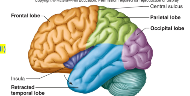
5 lobes of cerebral hemispheres
frontal, parietal, temporal, occipital, insula(island of reil)
corpus callosum
connects cerebral hemispheres, bottom crease of a wrinkle
gyri
ridges of the brain. Raised part of a wrinkle
Sulci
shallow grooves that separate gyri
fissures
deep grooves in the surface of brain
longitudinal- separates cerebral hemispheres
transverse- separates cerebrum from cerebellum
lobes of cerebral hemispheres
1. Frontal
2. Parietal
3. Temporal
4. Occipital
named after the bones that underlie them
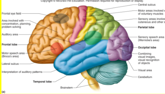
frontal lobe
responsible for concentrating, planning, complex problem solving, and judging consequences of behavior
motor areas control movements of voluntary skeletal muscles
parietal lobe
sensory areas provide sensations of temperature. touch, pressure, and pain involving the skin
association areas function in understanding speech and in using words to express thoughts and feelings
temporal lobe
sensory areas are responsible for hearing
association areas interpret sensory experiences and remember visual scenes, music, and other complex sensory patterns
occipital lobe
sensory area responsible for vision
association areas combine visual images with other sensory experiences
cerebral cortex
thin layer of gray matter, makes up outermost layer of cerebrum
contains 75% of neuron cell bodies in nervous system
white matter of cerebrum
Lies under cerebral cortex
Makes up most of cerebrum
Contains bundles of myelinated axons, that connect neuron cell bodies in cerebral cortex to other portions of nervous system
cutaneous sensory area of cerebral cortex
-parietal lobe
-interprets sensations on skin
(Wernicke's area)
-temporal/parietal lobe
-usually left hemisphere
-understanding and formulating language
visual area
occipital lobe, interprets vision
auditory area
temporal lobe, interprets hearing
sensory area for taste
Near base of the central sulcus
Includes part of insula
Sensory area for smell
arises from centers deep within temporal lobes
Olfactory Bulb
Motor areas of the cortex
primary motor cortex; Broca's area;
frontal eye field
primary motor area
frontal lobe, controls voluntary muscles
most nerve fibers cross over brainstem
Broca's area
-anterior to primary motor cortex
-usually in left hemisphere
-controls muscles needed for speech, actually talking
frontal eye field
Above Broca's area
Controls voluntary movements of eyes and eyelids
parts of the brain stem
midbrain, pons, medulla oblongata
parts of diencephalon
thalamus, hypothalamus, (epithalamus as well)
Thalamus
structure in the center of the brain that acts as a primary relay station for sensory and motor signals traveling to the cerebral cortex.
roles in memory, emotion, and wakefulness/alertness
hypothalamus
located below the thalmus. controlling body temperature, thirst, hunger, SLEEP/WAKE cycles
medulla oblongata
lowest part of brainstem, responsible for regulating INVOLUNTARY body functions such as breathing, heartrate, blood pressure, and digestion. also reflexes and moto pathways