C. 4-6
1/175
There's no tags or description
Looks like no tags are added yet.
Name | Mastery | Learn | Test | Matching | Spaced | Call with Kai |
|---|
No analytics yet
Send a link to your students to track their progress
176 Terms
Metabolism
Sum of all chemical reactions in the body
Cellular Metabolism
Sum of all chemical reactions occurring in a cell; metabolic reactions usually occur in pathways or cycles.
Anabolism requires ATP made during Catabolism
Small molecules are built into larger ones; requires energy. Provides materials for maintenance, cellular growth and repair
Catabolism
Larger molecules are broken down into smaller ones; releases energy
Smaller molecules are bound together to form larger ones. H2O produced in the process. Used to produce polysaccharides, proteins, triglycerides
Dehydration synthesis (Anabolism)
Breaks down larger molecules into smaller ones; A T P is produced. Used to DECOMPOSE carbohydrates, proteins, lipids. H2O to split the substances
Hydrolysis (Catabolism)
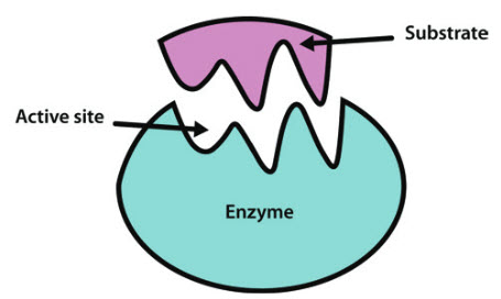
Enzymes
Control rates of both catabolic and anabolic reactions. Greatly increase reaction rates
Enzyme (protein catalysts)/1
Globular proteins that speed up specific chemical reactions. Decreases the energy needed to start a reaction. VERY reusable, because not consumed.
Each enzyme is highly specific. Its unique shape of its must perfectly fit that substrate, which is why individual steps in a metabolic pathway require distinct enzyme
Enzyme (protein catalysts)/2
Regulatory enzyme that catalyzes one step of pathway (chemical reaction)
SET RATE for reaction sequence speed.
NEGATIVE FEEDBACK by inhibiting enzyme to stop pathway for homeostasis and optimal efficiency.
Rate-limiting enzyme (1st in reaction sequence) Number of molecules of this enzyme is limited
Non-protein substance that combines with the enzyme to activate it. Some help fold active site into proper conformation. Some bind enzyme to substrate.
—> A non-protein "helper molecule" that binds to an enzyme to make it functionally active
Cofactor can be ion, element, or small organic molecule (coenzyme)
Coenzyme
Organic molecule that acts as cofactor Most are vitamins, which are essential organic molecules that humans must get from their diet
Denaturation
Inactivation of an enzyme (or any other protein), due to an irreversible change in its conformation. Results in enzyme being unable to bind to substrate
Energy can't created or destroyed, but can be changed from one form to another
Capacity to change something, or the ability to do work. Heat, light, sound, electrical energy, mechanical energy, chemical energy.
Energy is transferred to ATP
40% is released as chemical energy. 60% is released as heat; maintains body temperature
ATP (Adenosine Triphosphate)
Molecule that carries energy in a form the cell can use. Main energy-carrying molecule in the cell; energy from ATP breakdown is used for cellular work
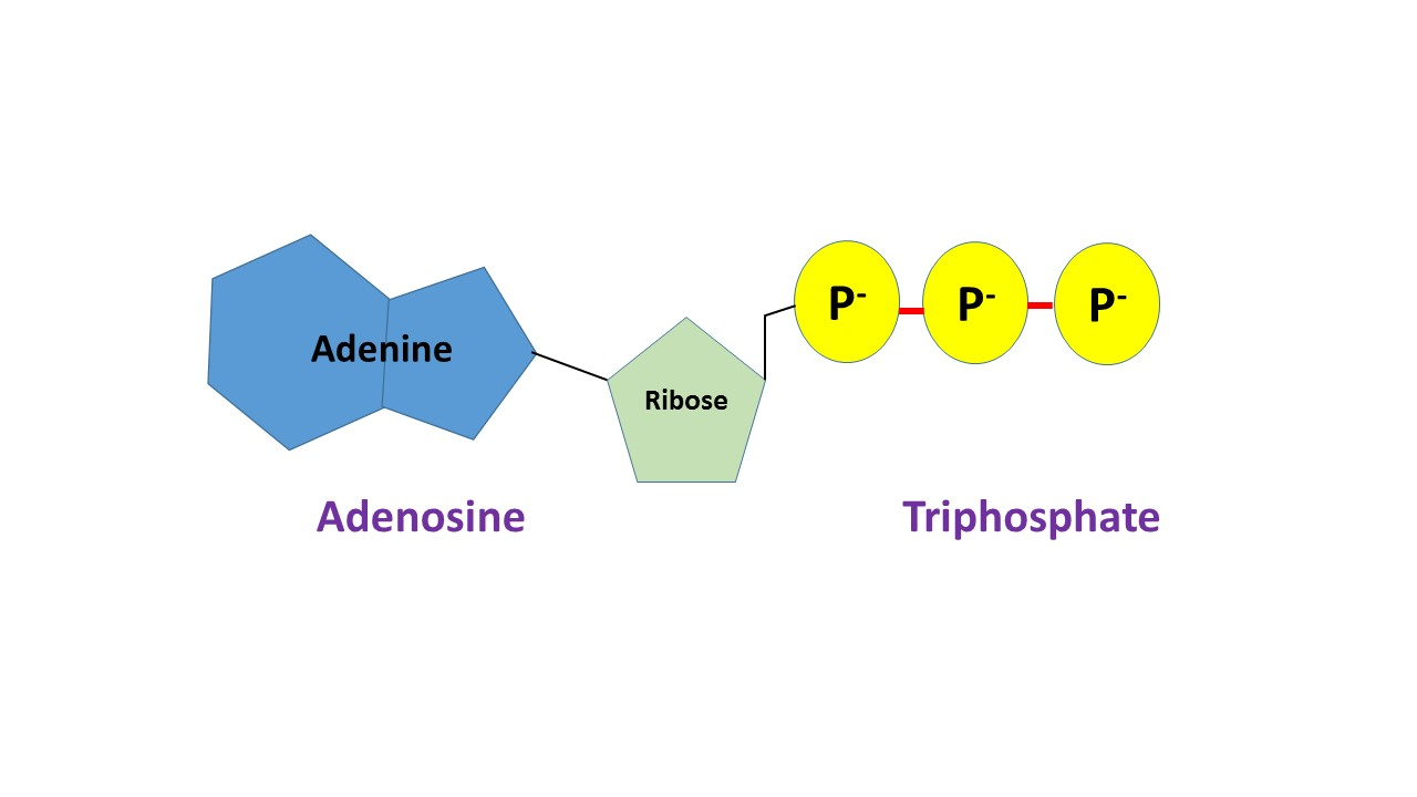
ATP Consists of 3 portions
Adenine, Ribose (a sugar), and 3 phosphates in a chain
When ATP loses terminal phosphate, it become Adenosine Diphosphate (ADP)
ADP can be converted back into ATP by attaching a third phosphate; called phosphorylation, requires energy from cellular respiration.
Glycolysis (anaerobic), Citric acid cycle (aerobic)
Electron transport chain/oxidative phosphorylation (aerobic)
Glycolysis and the Electron Transport Chain are stepwise reaction sequences
Citric Acid Cycle occurs in a metabolic cycle in which the final product reacts to replenish original substrate
Final products of cellular respiration are- Carbon dioxide, Water, ATP (chemical energy, 40%), and Heat (60%)
Cellular respiration need glucose and O2.
Anaerobic reactions (Glycolysis)
do not require O2, and make little ATP
Aerobic reactions (Citric Acid, ETC)
require O2, and make most of A T P
Glycolysis (Cytosal activity) Yields 2 ATP molecules per glucose molecule broken down
Breaks down glucose (6-carbon) into 2 pyruvic acid (3-carbon) molecules. Anaerobic.
3 Phases of Glycolysis
Phosphorylation of glucose, Splitting glucose, and Production of NADH, ATP, and 2 pyruvic acid
In presence of O2, NADH and H+ deliver electrons to the electron transport chain, with oxygen as final electron acceptor
Without O2, they go back to Pyruvic
Aerobic Reactions- In presence of O2, pyruvic acid enters aerobic pathways
Synthesis of Acetyl Coenzyme A, Citric acid cycle, and Electron transport chain
Aerobic reactions begins with pyruvic acid moving from cytosol to mitochondria
Pyruvic acid is used to produce Acetyl CoA
Aerobic cellular respiration end products
CO2, H2O, and up to 36 ATP per molecule of glucose
Begins when acetyl CoA combines with oxaloacetic acid to produce citric acid. Citric acid is changed into oxaloacetic acid through a series of reactions
Citric Acid Cycle repeats as long as pyruvic acid and O2 are available
1 ATP and 8H+ —> FAD
2 CO2 are produced; enters blood and is exhaled
Each citric acid molecule
NADH and FADH2 carry hydrogen and high-energy electrons to the
Electron Transport Chain (ETC)
ETC is a series of enzyme complexes (electron carriers) located
In inner membrane of mitochondria. H2O is formed (oxygen is the final electron "carrier"
All ATP production in complete oxidation of glucose
2 ATP in glycolysis. 2 ATP in citric acid cycle. 28 in electron transport chain
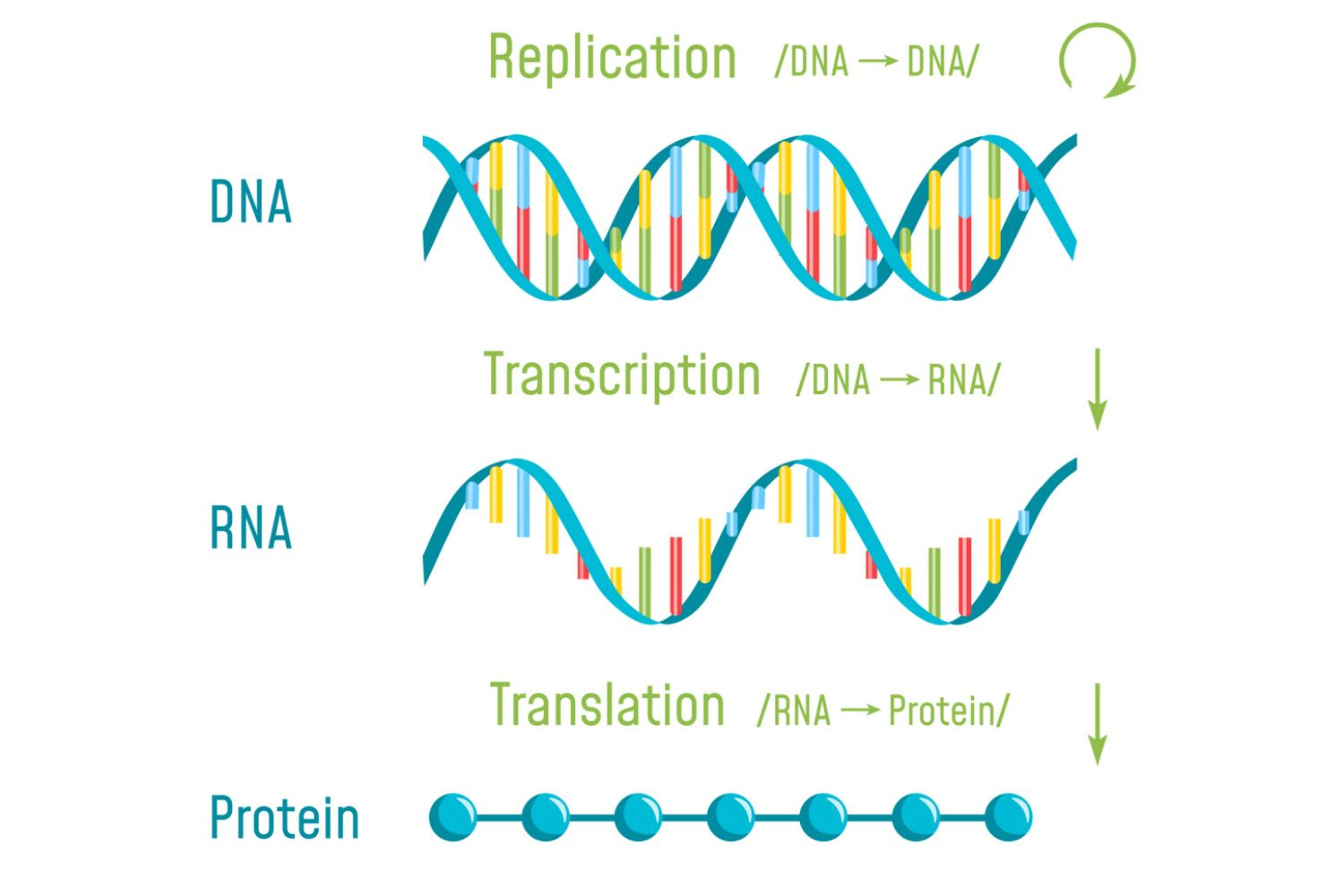
DNA (Deoxyribonucleic Acid)
The genetic material. Molecule that stores information on its sequence of nucleotides, that instructs a cell to how synthesize certain proteins
Enzymes
Blood proteins
tructural proteins of muscle and connective tissue
Antibodies • Cell membrane components
The proteins coded for on DNA function as
Genetic information
Instructions to tell cells how to construct proteins; stored in D N A sequence
Gene
Sequence of D N A that contains information for making 1 protein
Genome
Complete set of genetic information in a cell
Exome
Small portion of the genome that codes for proteins
Gene Expression
Control of which proteins are produced in each cell type, in what amount, and under which circumstances
2 chains of nucleotides of double helix. Resembles ladder twisted into a spiral. Backbone of each strand is a sugar-phosphate chain Bases from the 2 complementary strands are C ̶ G, A ̶ T
Structure of DNA
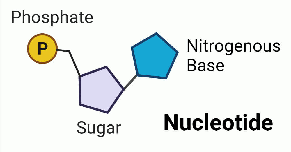
5-carbon sugar (deoxyribose).
A phosphate group.
A nitrogenous base (cytosine, guanine, adenine, or thymine)
Nucleotides. Also building blocks of DNA
Hydrogen bonds break between base pairs. Strands unwind and separate. New nucleotides pair with exposed bases, under direction of DNA polymerase. Other enzymes connect new sugar-phosphate backbone
Steps in DNA replication (interphase)
RNA differs from DNA in following ways
RNA contains Uracil (instead of Thymine), but the other 3 bases are the same (Adenine, Guanine, and Cytosine).Much shorter than DNA
Transcription - process of copying a gene's DNA sequence into messenger RNA (mRNA) inside the nucleus
***Transcription means to copy something via audio
DNA stores master copy of genetic code, and remains in the nucleus
Protein synthesis occurs in cytoplasm. mRNA leaves nucleus and binds to ribosome for protein synthesis
At ribosome, the DNA genetic code carried by mRNA, is used to synthesize a protein. Ribosome and tRNA works together to create a protein.
Translation is the process of converting the genetic code (carried by mRNA) into a sequence of amino acids that becomes a protein

Transcription (In nucleus)
Copying DNA sequence onto an RNA sequence.
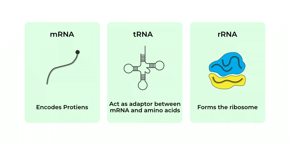
Messenger RNA (mRNA)
The type of RNA that carries genetic code from DNA → ribosome in cytoplasm
RNA Polymerase
Enzyme that catalyzes the formation of mRNA from the proper strand of DNA
Each amino acid is specified by a sequence of 3 bases in DNA
Codons
Aligns amino acids during translation along the mRNA strand on the ribosome. Each ___ contains 3 nucleotide bases, the anticodon, which binds to the complementary codon on the mR N A strand
Transfer RNA (tRNA)
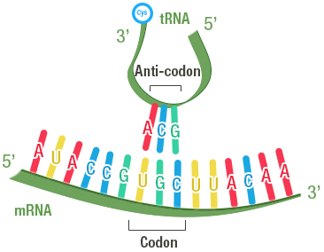
The Initiation codon, AUG, codes for Methionine and signals the start of a protein
3 codons are Stop codons, signaling the end of a protein; these do not have corresponding tRNAs. For each mRNA codon coding for an amino acid
There is corresponding tRNA anticodon
Steps of Transcription (In the Nucleus).
1) RNA polymerase recognizes correct strand of DNA to copy
Then RNA polymerase binds to a gene, unwinds the DNA helix, and synthesize a complementary mRNA molecule matching DNA. Termination signals end of gene. Newly created mRNA strand is released. DNA rewinds BACK into double helix
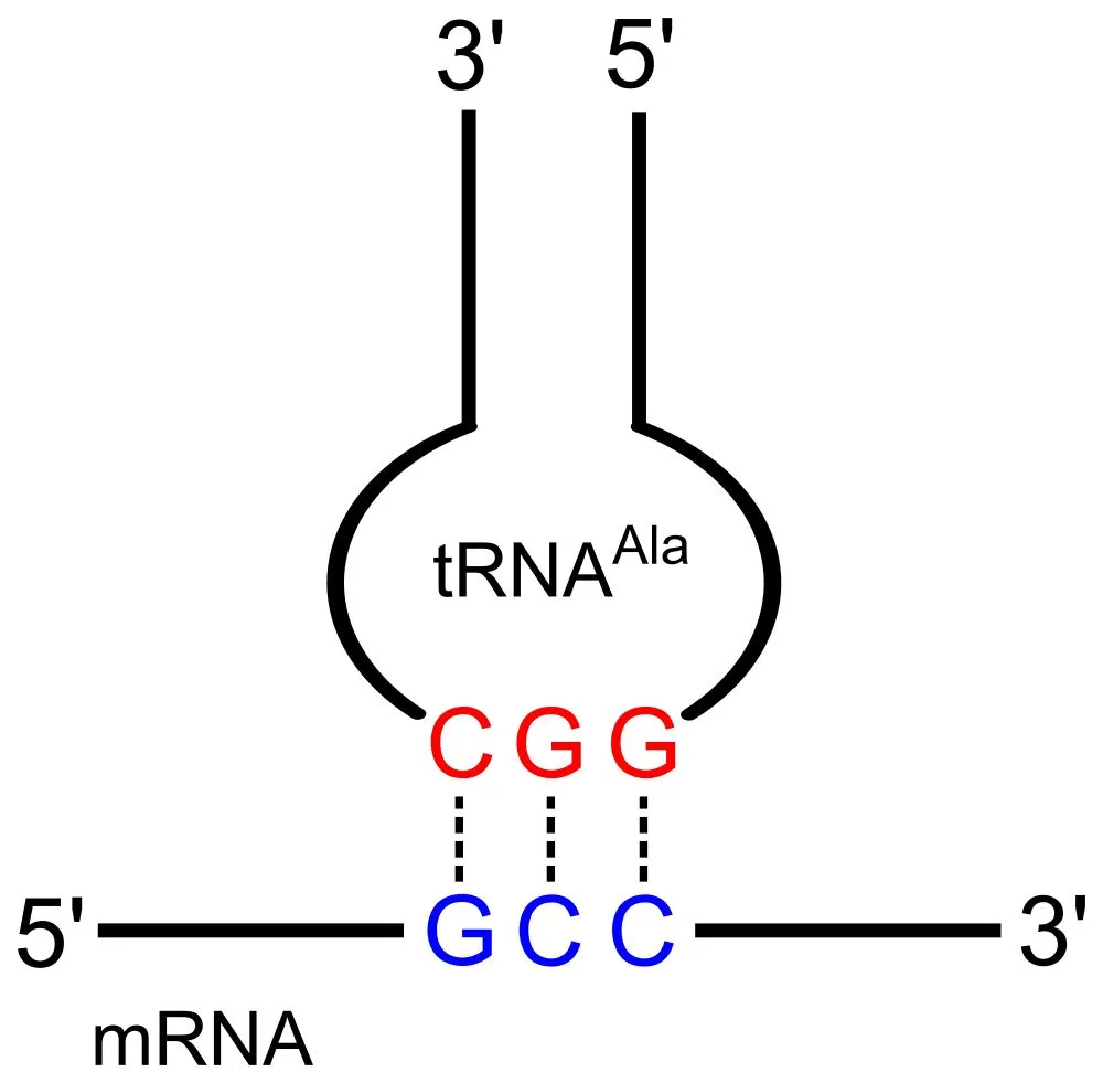
Ribosome binds to the start codon on mRNA. tRNA matches anticodon to codon, bringing specific AMINO ACIDS TO RIBOSOMES. Growing amino acid chain folds into unique functional protein shape
Translation (In the Cytoplasm)
Mutations occur when bases are changed, added, or deleted
Changes in the DNA sequence
Spontaneous Mutations
due to insertion of unstable base into DNA sequence
Induced Mutations
due to exposure to mutagens, chemicals, or radiation that cause mutation
Many mutations affect health, by changing the amino acid sequence, resulting in a nonfunctional
Sickle cell disease is caused by a single nucleotide substitution; this causes production of abnormal hemoglobin, which causes change in shape of red blood cells, in low-oxygen conditions, and extreme pain
Since often 2 to 4 codons specify the same amino acid, some mutations changing the third base of a codon would result in…nothing happening to the protein.
If one copy is mutated, the other copy may provide enough of gene’s normal function to maintain health.
DNA Repair correction of a mismatched nucleotide by a Repair Enzyme.
Epithelial (Protection, secretion, absorption, excretion)
Cover body surface, cover and line inernal organs, compose glands. Lack blood vessels, cells readily divide, cells are tightly packed
Simple Squamous (Diffusion, Filtration)
Alveoli, capillaries, blood vessels. Lines blood and lymphatic vessels.
Simple Cuboidal (Secretion, Absorption)
Kidney tubules, ovary surface, thyroid follicles, and secretory portions of small glands
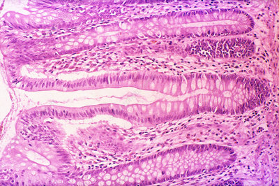
Simple Columnar (Absorption, Secretion, Excretion)
Small intestine, stomach, uterine tubes (ciliated), bronchioles (ciliated)
Pseudostratified Columnar (Mucus Secretion, Mucus Propulsion)
Nasal cavity, trachea, bronchi. Nuclei at two or more levels. Cell reach basement membrane. Cilia & goblin cell.
Stratified Squamous (Protection) - Nuclei at two or more levels.
Skin, oral/esophagus, anal, vagina. Outermost cells are squamous, deeper cells are cuboidal.
Stratified Cuboidal (Protection)
Sweat gland ducts, mammary gland ducts, salivary gland ducts, and pancreas.
Stratified Columnar (Protection, Secretion)
Parts of male urethra, vas deferens, salivary glands. Cube-shaped cells in deeper layers
Transitional (Stretching, Distension)
Urinary bladder, ureters, parts of urethra
Connective (Bind, support, protect, fill spaces, store fat, produce blood cells)
Widely distributed throughout the body. Mostly have good blood supply, cells are farther apart than epithelial cells, with ECM in between
Areolar (Wraps, Cushions, Binds Tissues)
Beneath epithelia, around organs, papillary layer of dermis. Collagen, elastic, and reticular fibers.
Adipose (Energy Storage, Insulation, Protection)
In hypodermis, around kidneys and eyes. Fat-storing cells (adipocytes)
Specialized CT
Hyaline cartilage CT
Ends of bones, nose, and rings in walls of respiratory passages
Elastic cartilage CT
External Ears and Larynx.
Fibrocartilage
Between bony parts of spinal column, parts of pelvic girdle, and knee
Bone
Cells in solid matrix. Supports, protects, provides framework. Bones of skeleton, middle ear
Blood
Cells and platelets in fluid matrix. Transports gases, defends against disease, clotting. Throughout the body in a closed system of blood vessels and heart chambers
Dense Regular CT (Strong Attachment)
Tendons, ligaments. Parallel collagen fibers; resists pulling in one direction.
Dense Irregular CT (Strength, Structural Support)
Dermis of skin, organ capsules. Collagen fibers in every direction for PULL resistance.
Elastic CT (Stretch and Recoil)
Walls of large arteries, vertebral ligaments. Abundant elastic fibers
Muscle (Movement)
Attached to bones, in the walls of hollow internal organs, heart. Able to contract in response to specific stimuli
Skeletal Muscle (Attached to bones)
Striated, voluntary, multinucleated; responsible for body movement, posture, and heat production.
Cardiac Muscle (Heart wall)
Striated, involuntary, branching cells with intercalated discs; pumps blood throughout the body.
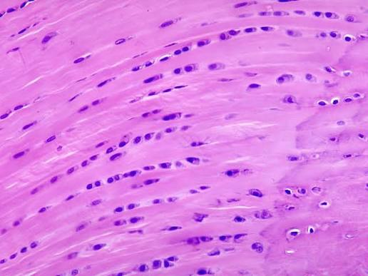
Smooth Muscle (Walls of stomach, intestines, bladder, blood vessels)
1) Central nucleus and short spindle-shaped cells
Non-striated, moves substances through hollow organs and regulates vessel diameter.
Unicellular glands
A single secretory cell. Mucous-secreting cell
Multicellular glands
Glands that consist of many cells
Simple glands
Glands that communicate with the surface by means of ducts that DONT BRANCH before reaching the secretory portion
Simple tubular gland
Straight tube-like gland that opens directly onto surface. Intestinal glands of small intestine
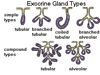
Simple branched tubular gland
Branched, tube-like gland; duct short or absent. Gastric glands

Simple coiled tubular gland (Merocrine)
Long, coiled, tube-like gland; long duct.

Simple branched alveolar gland
Secretory portions of gland expand into saclike compartments along duct. Sebaceous gland of skin
Nervous (Conduct impulses for coordination, regulation, integration, and sensory reception)
Brain, spinal cord, nerves. Cells communicate with each other and other body parts
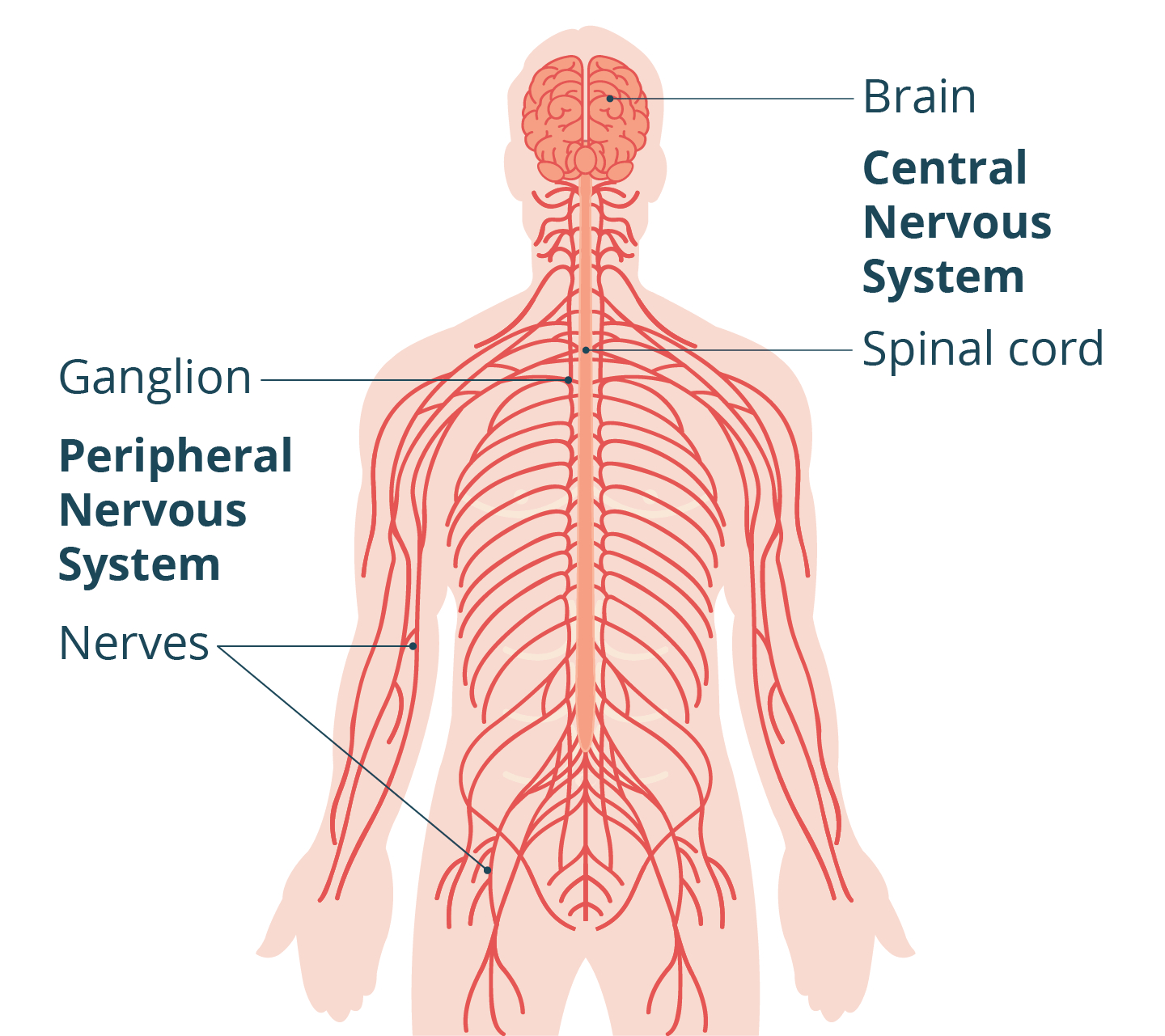
Neurons (Brain, spinal cord, peripheral nerves)
Specialized cells that conduct nerve impulses; responsible for communication, sensory reception, and motor control.
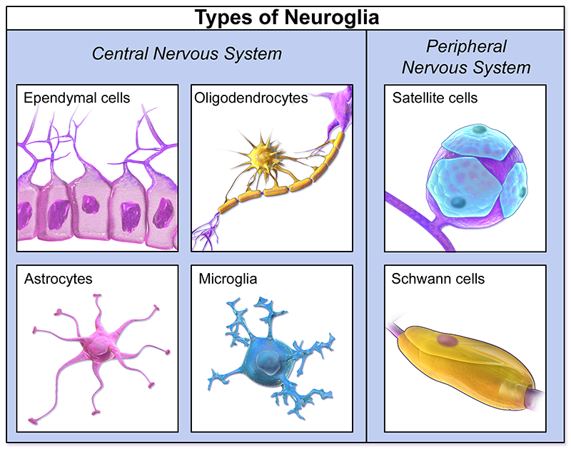
Neuroglia/Glial Cells (Brain, Spinal Cord, Peripheral Nerves)
Support, protect, and nourish neurons, maintaining the nervous system.
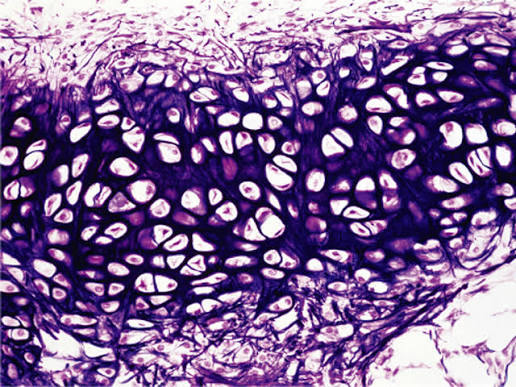
Hyaline Cartilage (Support, Reduce Friction) VERY SMOOTH matrix
Nose, trachea, larynx, ends of long bones. More flexible bones. Most common cartilage
Elastic Cartilage (Flexible Support)
External ear, epiglottis. Contains many elastic fibers
Fibrocartilage (Shock Absorption) STRONGEST cartilage
Intervertebral discs, menisci of knee, pubic symphysis. Thick collagen fiber
Reticular CT (Supportive Framework)
Liver, spleen, lymph nodes. Network of reticular fibers supporting other cells
Tight junctions
Close space between cells by FUSING cell membranes. Cells that line the small intestine