CNS Exam 2 - Tracts and Hindbrain
1/114
There's no tags or description
Looks like no tags are added yet.
Name | Mastery | Learn | Test | Matching | Spaced | Call with Kai |
|---|
No analytics yet
Send a link to your students to track their progress
115 Terms
Where is the cervical enlargement of the spinal cord?
Cord level C5-6
What are the boundaries of the spinal cord?
Superior:
General - foramen magnum
Specific - most sup. ant. rootlet of C1
Inferior:
General - conus medullaris
Specific - L1-2 disc level
How many spinal nerves are there?
31 pairs
SAME DAVE
Sensory Afferent, Motor Efferent
Dorsal Afferent, Ventral Efferent
Which spinal nerve does NOT have a vertebra associated with it?
C8 nerve root
Where doe the S1-Co nerve roots exit the spinal column?
S1-Co correlate with the sacral foramina
S5 and Co exit via the sacral hiatus
Where is the cauda equina?
Begins at the L1-2 vertebral level
What are the two parts of the filum terminale?
Internum: pial part, L1-2 to S1-2
Externum: dural part, S1-2 to coccyx
What is quadriplegia? What are its characteristic symptoms?
Spinal cord injury to C8 or higher
Upper and lower extremity problems
What is paraplegia? What are its characteristic symptoms?
Lower extremity problems
What are the horns of the spinal cord?
Gray matter areas present at most levels of the cord
Anterior/ventral (motor)
Posterior/dorsal (sensory)
Lateral (autonomic, only present at T1-L2 and S2-4)
What are the Rexed laminae?
Specific areas of the gray horns which are not distinguishable through staining
Rexed lamina 1
Most dorsal tip of posterior horn (marginal zone)
Rexed lamina 2
Posterior horn, pain and temperature information (substantia gelatinosa)
Rexed lamina 3-6
Posterior horn, sensory information (touch & pressure)
Rexed lamina 7
Lateral horn, includes:
Nucleus dorsalis (Clarke’s) at C8-L3
Interomediolateral nucleus (sacral paranucleus) at T1-L2 and S2-4
Preganglionic ANS neurons via reticulospinal tracts
Descending axons synapse here
Rexed lamina 8
Medial side of anterior horn
Rexed lamina 9
Centers of anterior and lateral horns, skeletal muscle movement, class A motor neurons (fastest and largest MNs)
Rexed lamina 10
Bridge in center of gray surrounding the central canal (which is continuous with the 4th ventricle), contains unmyelinated gray commissures
What are the 3 funiculi of the spinal cord (bilateral)?
Anterior, posterior, lateral
What is found in the gray matter?
Cell bodies, dendrites, synapses, rich blood supply, unmyelinated fibers
What is found in the white matter?
Axons, less dense blood supply, myelinated fibers (if >1 micron diameter)
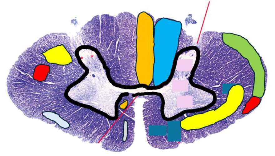
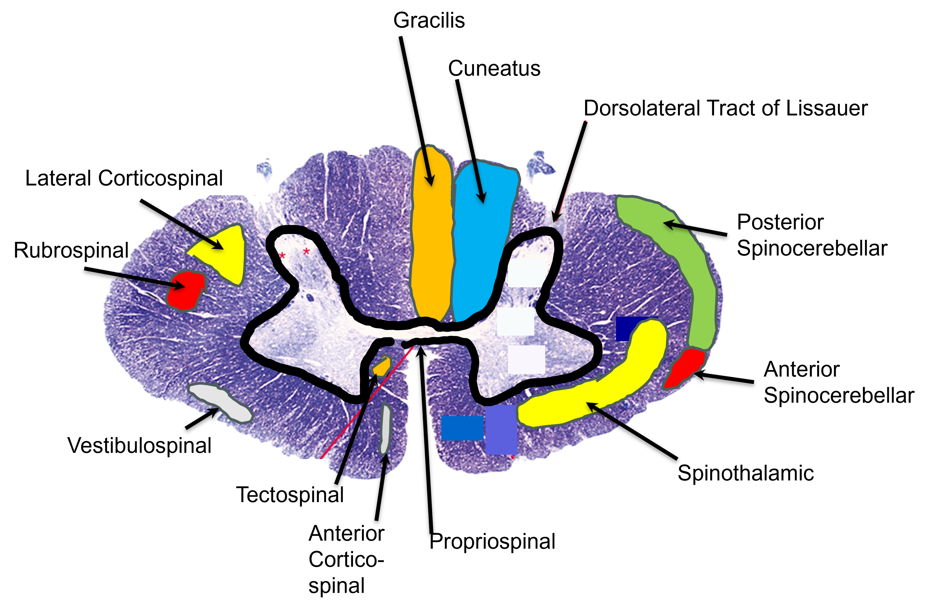
Tracts schematic (UNLABELED)
Tracts schematic (LABELED)
What is characteristic of ascending tracts?
3 neurons in the pathway, “spino___”
What is characteristic of descending tracts?
2 neurons in the pathway, “___spinal”
Gracilis (tract)
Posterior funiculus (med. aspect)
All cord levels (serves T6 and below)
Cell body in dorsal root ganglion
1st neuron synapses in nucleus gracilis in M.O.
NO cross over in cord
2 point touch discrim., vibratory and kinesthetic info
A$$ to Grass (med., lower extremity)
Serves the great toe
Cuneatus (tract)
Posterior funiculus (lat. aspect)
Cord levels T6 and up
Cell body in dorsal root ganglion
1st neuron synapses in nucleus cuneatus in M.O.
NO cross over in cord
2 point touch discrim., vibratory and kinesthetic info
Cuddle with cuneatus (lat., upper extremity)
Serves the carpals
Medial lemniscus (tract)
Cell body in nucleus gracilis/cuneatus of M.O.
2nd neuron of pathway, synapses to VPL of thalamus
Crosses over in M.O.
2 point touch discrim., vibratory and kinesthetic info
If there is a lesion in the gracilis or cuneatus tract, where will you lose sensation for vibration, kinesthesia, and 2 point touch discrimination?
On the SAME side as the lesion, BELOW it
Cuneatus: in the upper extremity
Gracilis: in the lower extremity
Anterior spinothalamic (tract)
Anterior funiculus
All cord levels
Cell body in rexed lamina 3-6
1st neuron synapses in VPL of thalamus
Cross over gradually in cord
Light touch and pressure info (A-LTP)
Lateral spinothalamic (tract)
Lateral funiculus
All cord levels
Cell body in rexed lamina 2
1st neuron synapses in VPL of thalamus
Cross over immediately in cord
Pain and temperature info (L-PT)
If there is a lesion in the ant. or lat. spinothalamic tract, where will you lose sensation for light touch/pressure or pain/temperature?
On the OPPOSITE side as the lesion, BELOW it
Anterior spinocerebellar (tract)
Lateral funiculus (periphery)
All cord levels
Cell body in lumbosacral gray horn
1st neuron synapses in cerebellum via the superior cerebellar peduncle (AS)
Cross over twice (1st in cord, 2nd as entering M.O.)
Gross lower-body anticipatory and proprioceptive movement
Posterior spinocerebellar (tract)
Lateral funiculus (periphery)
Cord levels L3 and up
Cell body in multiple places
Above C8 - lateral cuneate nucleus via cuneocerebellar tract
C8-L3 - in rexed lamina 7 (Clarke’s nucleus)
Below L3 - in gracilis tract (to go up to Clarke’s nucleus)
1st neuron synapses in cerebellum via inferior cerebellar peduncle
NO cross over
Fine proprioceptive movement info:
Lower extremity = below L3
Upper extremity = above C8
If there is a lesion in the anterior spinocerebellar tract, where will you lose sensation for gross lower-body anticipatory proprioceptive movement?
OPPOSITE side of lesion, BELOW it
(Remember, this tract crosses twice: once in the cord, once in the M.O.)
If there is a lesion in the posterior spinocerebellar tract, where will you lose sensation for fine movement proprioception?
SAME side as the lesion, BELOW it
Anterior corticospinal (tract)
10% of info!
Anterior funiculus
Cord levels T6 and up
Cell body in precentral gyrus of cerebral cortex
1st neuron synapses in the anterior horn (lamina 7 to lamina 9)
Cross over in cord
Control of axial muscles of neck and shoulder
pyramidal aka voluntary
Lateral corticospinal (tract)
90% of info!
Lateral funiculus (post. half)
All cord levels
Cell body in precentral gyrus of cerebral cortex
1st neuron synapses in the anterior horn (lamina 7 to lamina 9)
Giant Betz cells go directly to lamina 9
Cross over in pyramids of M.O.
Control of skilled voluntary movements of distal extremities (hands/feet)
pyramidal aka voluntary
Tectospinal (tract)
Anterior funiculus
Cord level C4-5 and up
Cell body in tectum of midbrain (superior colliculus)
1st neuron synapses to C.N. XI to control traps/SCM
Crosses over
Postural reflexes related to visual/auditory acuity (in traps/SCM)
EXTRApyramidal aka INvoluntary
Rubrospinal (tract)
Lateral funiculus
All cord levels
Cell body in tegmentum of midbrain (nucleus Ruber/red nucleus)
1st neuron synapses to anterior horn
Crosses over in midbrain
Contralateral flexors and inhibits extensors
EXTRApyramidal aka INvoluntary
Backup to corticospinal tracts
Bros flex!
Vestibulospinal (tract)
Anterior and lateral funiculi
All cord levels
Cell body in M.O. (lateral vestibular nucleus (Dieter’s))
1st neuron synapses to anterior horn (lamina 7 to lamina 9)
NO cross over
Ipsilateral extensors and inhibits flexors, proper orientation when falling
EXTRApyramidal aka INvoluntary
Extend your arms to keep your balance!
Medial reticulospinal (tract)
Anterior funiculus
All cord levels
Cell body in pons tegmentum
1st neuron synapses to the interomediolateral nucleus (in lamina 7)
NO cross over
ANS control (HR, BP, resp.), alternative to pyramidal tracts
Lateral reticulospinal (tract)
Lateral funiculus
All cord levels
Cell body in M.O.
1st neuron synapses to the interomediolateral nucleus (in lamina 7)
NO cross over
ANS control (HR, BP, resp.), alternative to pyramidal tracts
Propriospinal (not a true tract, spinospinal)
All cord levels
Cell body and synapse in gray matter of cord
1st neurons to be myelinated
Conduct the spinal reflexes
Dorsolateral tract of Lissauer (not a true tract, spinospinal)
All cord levels
Cell body between lamina 1 and post. sulcus of cord
Synapse in posterior cord (lamina 2)
Many contralateral branches
What are the characteristics of upper motor neurons (UMNs)? What happens when there are UMN lesions?
Originate in higher brain center (cortex, brainstem) and influence LMNs
Lesions lead to hyperreflexia, increased muscle tone, clonus, the sign of Babinski
Ex.: cortico___ tracts
What are the characteristics of lower motor neurons (LMNs)? What happens when there are LMN lesions?
Originate in spinal cord, innervate skeletal muscle
Lesions lead to hyporeflexia, hypotonia, atrophy, muscle fibrillations/fasciculations, lack of movement
Ex.: cranial nerves
What are the traits of pyramidal UMNs?
Cell body in cerebral cortex, initiate skilled voluntary movements, conscious control (cortico___ fibers)
What are the traits of extrapyramidal UMNs:
Cell body in brainstem, reflexes and involuntary movements, below consciousness (e.g. tectospinal)
Do EXTRA to smoothen movements!
What is multiple sclerosis?
A CNS demyelinating disease, leads to asymmetrical splotches in the white matter
What is a cord hemisection (Brown-Sequard syndrome)?
Total loss of one side of the cord, loss of function below lesion
What is Guillen-Barre syndrome?
A PNS demyelinating disease, damages one peripheral nerve
What are the parts of the rhombencephalon (midbrain)?
Metencephalon (pons, cerebellum), myelencephalon (M.O.)
What are the parts of the brain stem?
Pons, M.O., midbrain
Where is the medulla oblongata (M.O.)?
Anterior/ventral to cerebellum, inferior to pons, superior to brainstem
Inferior border is the most superior anterior rootlet of C1
What are the functions of the medulla oblongata (M.O.)?
Corticospinal fibers run through, relay via nuclei gracilis and cuneatus, location of cranial nuclei V and VII-XII
What is the decussation of the M.O.?
Where the pyramids meet and pyramidal axons cross
What is contained in the pyramids of the M.O.?
Pyramidal (voluntary) axons, from the corticospinal tracts
What are the olives of the M.O.? The inferior olivary nuclei?
Ventral structures lateral to the pyramids, overlaying the inferior olivary nuclei (internal)
Inferior olivary nuclei relay information to the cerebellum
What is the obex?
The inferior tip of the 4th ventricle, continuous with the central canal; seen at the bottom of the rhomboid fossa of the M.O.
What are the nuclei gracilis and cuneatus?
Gray structures in the M.O. which hold the cell bodies of 2nd order sensory neurons (in gracilis and cuneatus tracts), connecting to the arcuate fibers of the medial lemniscus
What are arcuate fibers?
Found in the M.O., originate in nuclei gracilis/cuneatus, cross the midline and rise through the medial lemniscus (tract) to the VPL of the thalamus
What are the fissures and sulci of the M.O.?
Anterior median fissure
Anterolateral sulcus (between pyramids and olives)
Medullopontine sulcus
Posterior median sulcus
Posterolateral sulcus
What is an apparent origin?
Where a cranial nerve attaches to the CNS
Where are the apparent origins of C.N. IV-VIII and XII?
IV-VI: Posterolateral sulcus of M.O.
VI-VIII: Medullopontine sulcus, moving lateral from the midline
XII: Anterolateral sulcus
What is the apparent origin of CN III?
In the midline at the superior border of the pons
What is a nucleus of origin?
Where lower motor neurons (LMNs) of cranial nerves have their cell bodies
What is a nucleus of termination?
Where 2nd order sensory neurons synapse into the CNS
What are the nuclei for CN III (oculomotor)?
Origin: Oculomotor nucleus (Oc)
Origin: Accessory oculomotor (EW) - parasympathetic
What is the nucleus for CN IV (trochlear)?
Origin: Trochlear nucleus (Tr)
What are the nuclei for CN V (trigeminal)?
Origin: Trigeminal motor nucleus (mT)
Termination: Spinal nucleus of the trigeminal nerve (spT)
Termination: Mesencephalic nucleus (mes)
Termination: Main sensory nucleus (msT)
What is the nucleus for CN VI (abducens)?
Origin: Abducens nucleus
What are the nuclei for CN VII (facial)?
Origin: Facial motor nucleus (Fa)
Origin: Superior salivary nucleus (Ss) - parasympathetic
Termination: Solitary nucleus (Sol)
Termination: Spinal trigeminal nucleus
What are the nuclei for CN VIII (vestibulocochlear)?
Termination: Vestibular nucleus (Ves)
Termination: Cochlear nucleus (C)
What are the nuclei for CN IX (glossopharyngeal)?
Origin: Nucleus ambiguus (Am)
Origin: Inferior salivary nucleus (Is) - parasympathetic
Termination: Solitary nucleus (Sol)
Termination: Spinal nucleus of the trigeminal
What are the nuclei for CN X (vagus)?
Origin: Dorsal nucleus of vagus (dV) - parasympathetic
Origin: Nucleus ambiguus (Am)
Termination: Solitary nucleus (Sol)
Termination: Spinal nucleus of the trigeminal
What is the nucleus for CN XI (spinal accessory)?
Origin: Spinal nucleus of accessory nerve (Ac)
What is the nucleus for CN XII (hypoglossal)?
Origin: Hypoglossal nucleus (Hy)
Oculomotor nucleus
Origin, CN III, serves ciliary and sphincter pupae muscles
Accessory oculomotor nucleus (Edinger-Westphal’s)
Where cell bodies of preganglionic parasympathetic nerves live, which synapse in the ciliary ganglion
Trochlear nucleus
Origin, CN IV, serves the superior oblique
Trigeminal motor nucleus
Origin, CN V, serves muscles of mastication
Spinal nucleus of trigeminal nerve
CN V, VII, IX, X, nucleus of termination for pain & temperature, extends into cervical cord (part of spinotrigeminal pathway)
Solitary nucleus
CN VII, IX, X, nucleus of termination for taste
Nucleus ambiguus
CN IX and X, nucleus of origin
Which CNs have parasympathetic functions?
(Nuclei have preganglionic parasympathetic fibers)
III - Oculomotor
VII - Facial
IX - Glossopharyngeal
X - Vagus
Which CNs are involved in eye musculature?
III - Oculomotor
IV - Trochlear
VI - Abducens
Spinotrigeminal (tract)
1st order cell body = CN V ganglion (spinal nucleus of trigeminal)
2nd order = from spinal nucleus to VPM of thalamus
3rd order = from VPM to post. central gyrus of cerebrum
What happens in a lateral medullary stroke?
Affects the posterior inferior cerebellar artery
Lose pain & temperature sensation on:
Same side of face (spinal nucleus of V)
Opposite side of body (spinothalamic tract)
What are corticobulbar fibers?
UMNs from the cerebral cortex to the M.O., pyramidal (cell body in cortex)
What is the relative location of the pons?
Part of the brainstem; ventral to cerebellum, inferior to midbrain, superior to M.O.
What are the functions of the pons?
Fiber conduction (spinothalamic, corticospinal), home of CN nuclei V-VIII, relay information to cerebellum
Pontine reticular formation: med. reticulospinal tract
What are the external features of the pons?
Anterior bulge (where the basilar artery lies on the ventral surface)
Superior part of rhomboid fossa (4th ventricle)
Middle cerebellar peduncle (pons → cerebellum)
What are the internal structures of the basilar/anterior division of the pons?
Corticospinal fibers (descending pyramidal axons)
Medial lemniscus (post., somatic sensory)
Pontine nuclei (take info from cortex and send to cerebellum via middle cerebellar peduncle)
What are the internal structures of the tegmentum of the pons?
CN nuclei V-VIII (pons = 4 letters = 4 nuclei)
Other extrapyramidal tracts:
Spinothalamic
Ant. spinocerebellar
Tectospinal, rubrospinal
Lateral lemniscus (auditory, most ant. in tegmentum)
What are the 3 pairs of cerebellar peduncles? How do they move information?
Superior: midbrain to cerebellum
Mostly efferent (from cerebellum), some afferent (to cerebellum)
Middle: pons to cerebellum
ONLY afferent
Inferior: M.O. to cerebellum
Mostly afferent, some efferent
SAME DAVE
What are the functions of the cerebellum?
Integrate sensory input, output influences motor neurons
How does the ipsilateral orientation of the cerebellum dictate how it influences the tracts?
Red nucleus (of Ruber): crosses, so R cerebellum affects L nucleus
Lateral vestibular nucleus: NO cross, so R cerebellum affects R nucleus
Cerebral cortex: crosses, so R cerebellum influences L cerebral cortex
What are the 3 lobes of the cerebellum? What is between them?
Anterior and posterior lobes (separated by primary fissure; post. is largest), flocculonodular lobe
Separated by the vermis