Surface Anatomy Of The Forebrain - The Temporal Lobe
1/16
There's no tags or description
Looks like no tags are added yet.
Name | Mastery | Learn | Test | Matching | Spaced | Call with Kai | Chat |
|---|
No analytics yet
Send a link to your students to track their progress
17 Terms
Name the three major gyri of the lateral surface of the temporal lobe in order from superior to inferior.
From superior to inferior, the three major gyri on the lateral temporal lobe are:
Superior temporal gyrus
Middle temporal gyrus
Inferior temporal gyrus
Describe the anatomical boundaries of the superior temporal gyrus.
The superior temporal gyrus is the topmost of the lateral temporal gyri
Superior boundary: Lateral sulcus (Sylvian fissure)
Inferior boundary: Superior temporal sulcus
Describe the anatomical boundaries of the middle temporal gyrus.
The middle temporal gyrus (MTG) sits between two sulci:
Above: Superior temporal sulcus
Below: Inferior temporal sulcus
Describe the anatomical position of the inferior temporal gyrus on both the lateral and inferior surfaces of the temporal lobe.
Lateral surface:
The inferior temporal gyrus (ITG) lies below the inferior temporal sulcus
Inferior surface:
The ITG wraps onto the underside of the temporal lobe
There, it is separated from the occipitotemporal (fusiform) gyrus by the occipitotemporal sulcus
The superior temporal gyrus is bounded superiorly by which sulcus?
Lateral sulcus (Sylvian fissure)
Which sulcus separates the inferior temporal gyrus from the fusiform gyrus on the inferior temporal surface?
Occipitotemporal sulcus
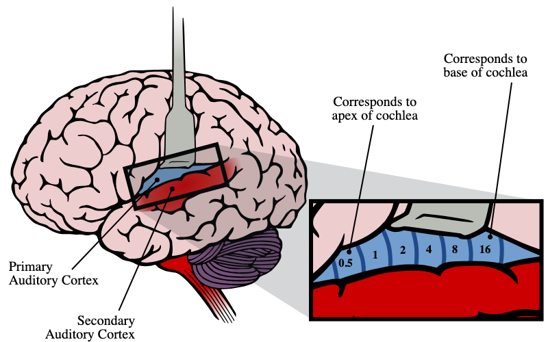
Where is the primary auditory cortex located within the temporal lobe?
The primary auditory cortex sits on the superior surface of the superior temporal gyrus
A patient suffers bilateral damage to the superior surface of the superior temporal gyri. What is the most likely clinical deficit?
Cortical deafness
Bilateral destruction of the primary auditory cortex on the superior temporal gyrus results in cortical deafness — an inability to consciously perceive sounds despite intact peripheral hearing
Which part of the temporal lobe contributes to Wernicke's area, and on which hemisphere is this most commonly located?
Region: Posterior superior temporal gyrus (STG)
Hemisphere: Dominant hemisphere — usually the left
Function: Critical for language comprehension
Wernicke's area is primarily important for which cognitive function?
Language comprehension.
Wernicke's area mediates understanding of spoken and written language; language production (fluency) is the domain of Broca's area in the frontal lobe
A patient produces fluent but unintelligible speech with severely impaired language comprehension following a left MCA territory stroke. Which area is most likely infarcted?
Left posterior superior temporal gyrus (Wernicke's area).
Damage to Wernicke's area produces fluent paraphasic speech with impaired comprehension (Wernicke's aphasia)
Which region of the temporal lobe is primarily involved in higher-order visual processing, and what is the significance of this function?
Region: Mostly the inferior temporal gyrus (ITG) — the key higher‑order visual area of the temporal lobe
Role: Enables conscious visual perception and complex recognition of objects, faces, and scenes
What is the clinical consequence of damage to the inferior temporal gyrus with respect to visual function?
Damage to the inferior temporal gyrus (ITG) causes visual agnosia
The patient can see (acuity and primary visual cortex are intact)
but cannot recognise or interpret what they’re seeing
Where in the temporal lobe is the function of learning and memory localised?
Location: The medial temporal lobe
Key structures: Hippocampus + amygdala (core limbic system components)
Role: Essential for forming new memories and emotional learning
Summarise all four major functions of the temporal lobe and their respective anatomical locations within the lobe.
Auditory processing
→ Primary auditory cortex on the superior surface of the superior temporal gyrusLanguage comprehension
→ Posterior superior temporal gyrus (Wernicke’s area, dominant/left hemisphere)Higher‑order visual processing / conscious visual perception
→ Inferior temporal gyrusLearning & memory
→ Medial temporal lobe, especially the hippocampus and amygdala
A patient presents with inability to consciously perceive visual stimuli despite normal visual acuity. Which temporal region is likely damaged?
This presentation points to damage of the inferior temporal gyrus (ITG)
The ITG is essential for higher‑order visual processing and conscious recognition of what is seen
Damage here causes visual agnosia: the patient can see, but cannot recognise or make sense of visual stimuli
A patient can hear sounds but is unable to understand speech or recognise environmental sounds despite intact basic auditory function on testing. Which specific temporal region is most likely involved?
This pattern points to damage of the posterior superior temporal gyrus — Wernicke’s area in the dominant (usually left) hemisphere
Primary hearing is intact but comprehension is lost, affecting both speech and environmental sound recognition