SAM3.8: Vitreous, Retina, and optic n.
1/50
There's no tags or description
Looks like no tags are added yet.
Name | Mastery | Learn | Test | Matching | Spaced | Call with Kai |
|---|
No analytics yet
Send a link to your students to track their progress
51 Terms
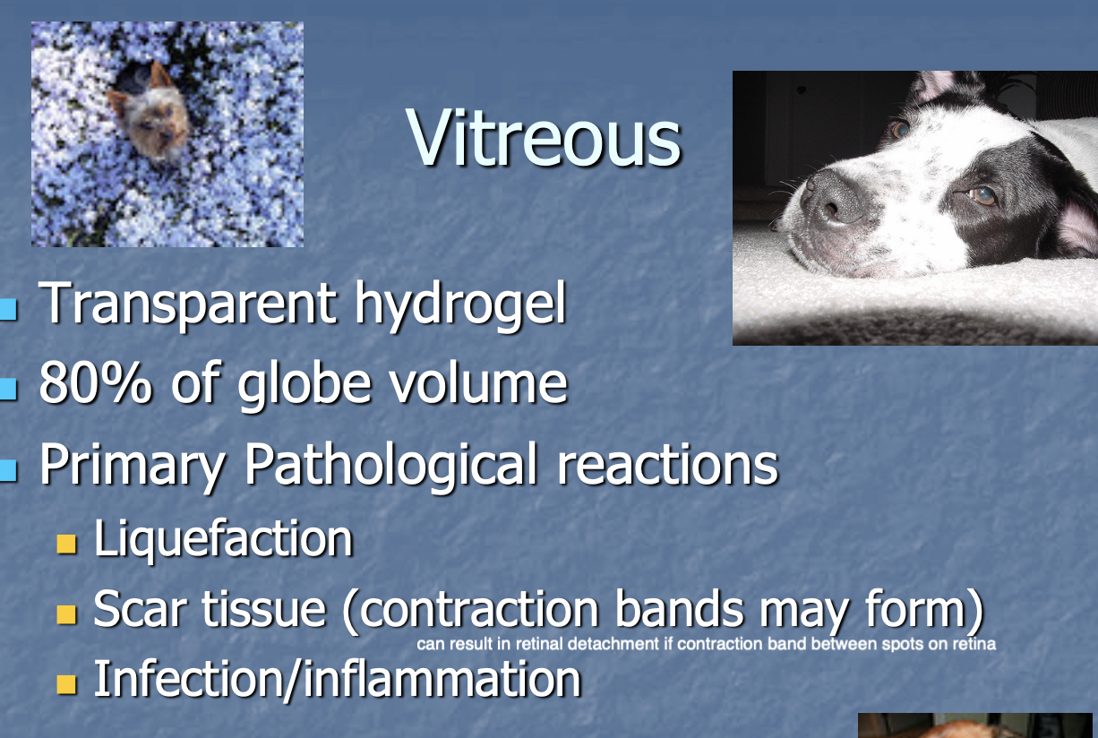
Vitreous is
a transparent hydrogel that takes up 80% of globe volume.

Primary Pathological rxns of the vitreous (3)
Liquefaction.
Scar tissue.
Infection/inflammation.
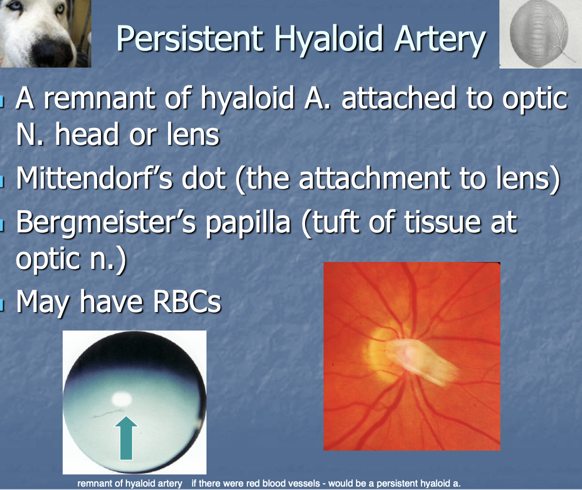
Persistent hyaloid a. is
a remnant of hyaloid a. attached to optic n. head or lens.
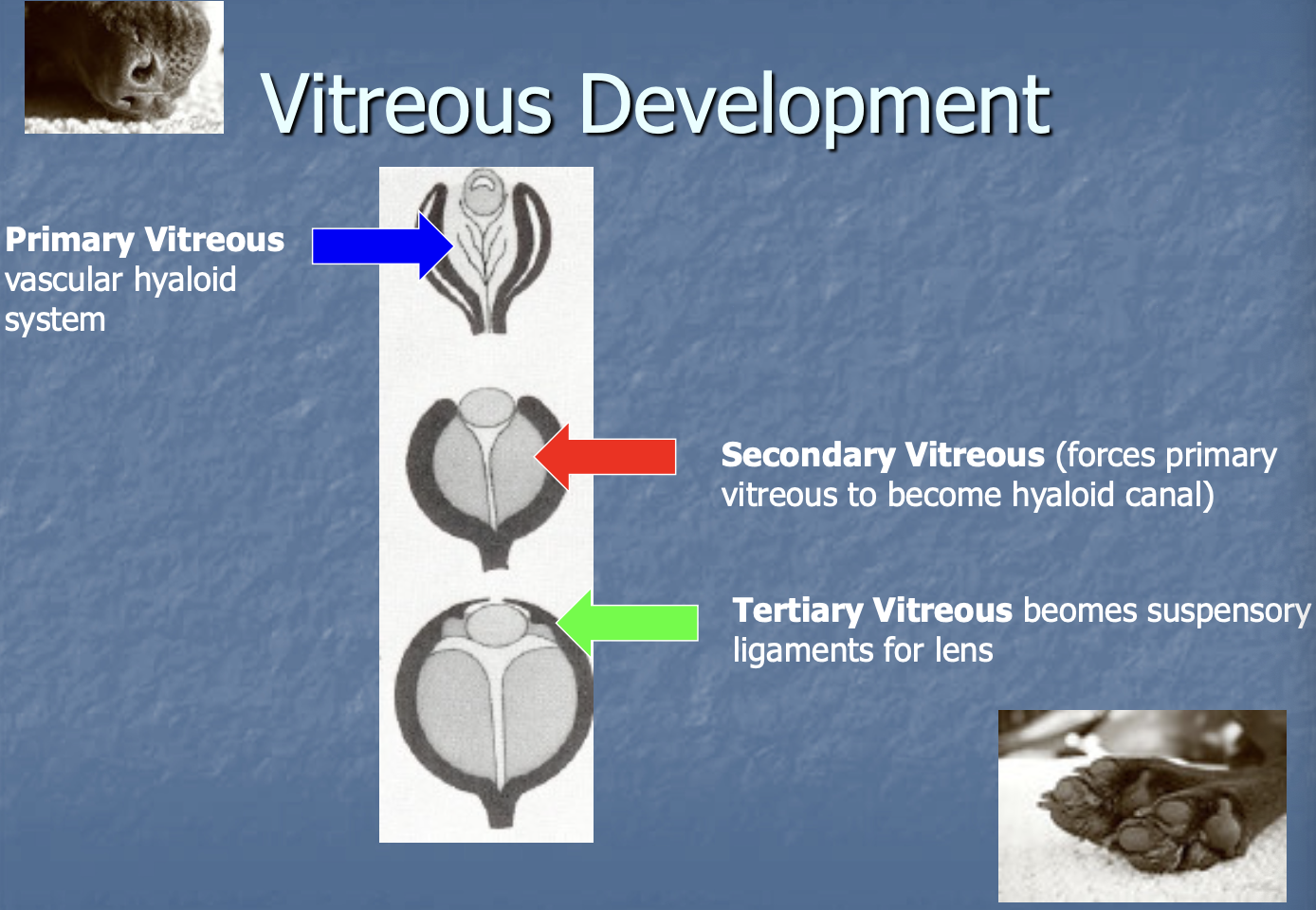
Vitreous development (3)
Primary - vascular hyaloid system.
secondary - forces primary to become hyaloid canal.
tertiary - becomes suspensory lgmt for lens.
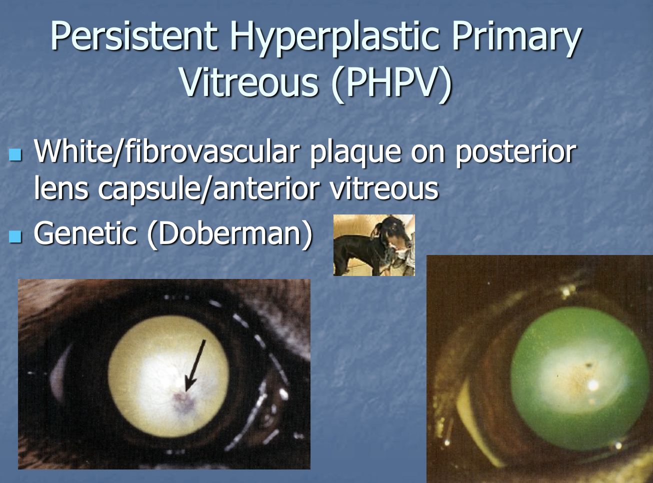
Persistent hyperplastic primary vitreous (PHPV) (2)
White/fibrovascular plaque on posterior lens capsule/anterior vitreous.
Genetic in Dobies.
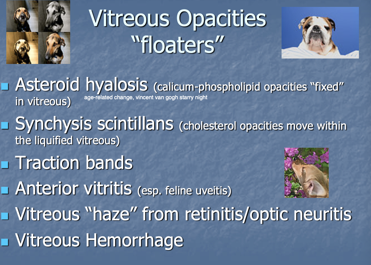
Vitreous Opacities (floaters) include (6)
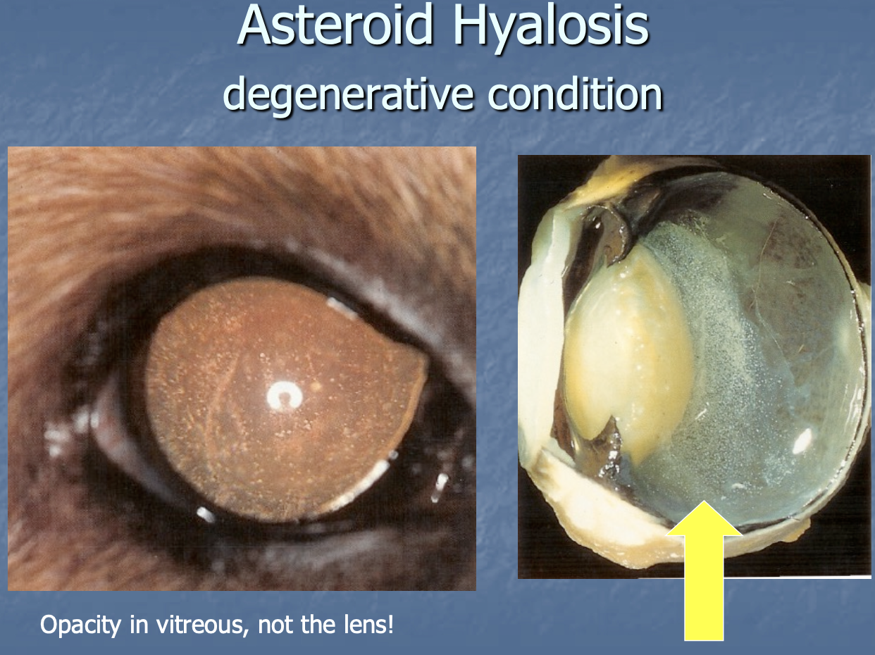
Asteroid hyalosis (calcium opacities fixed in vitreous).
Synchysis scintillans (cholesterol moving w/in liquified vitreous).
Traction bands.
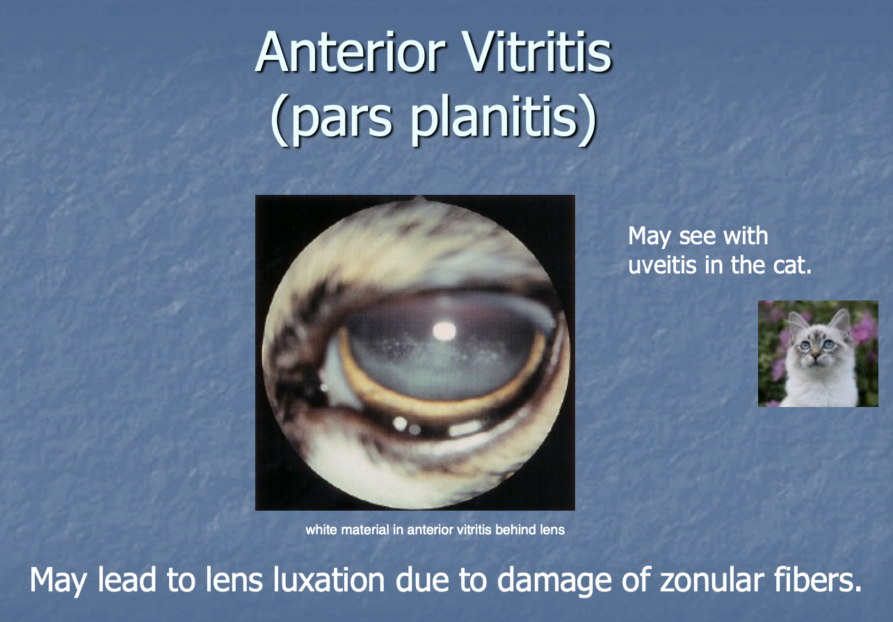
Anterior vitritis.
Vitreous haze from retinitis/optic neuritis.
Vitreous hemorrhage.
Anterior hyalosis is a
degenerative condition where opacity is seen in the vitreous due to calcium phospholipid fixed in vitreous.
Anterior vitritis (pars planitis) may be seen w/
uveitis in cats - may lead to lens luxation due to damage of zonular fbs.
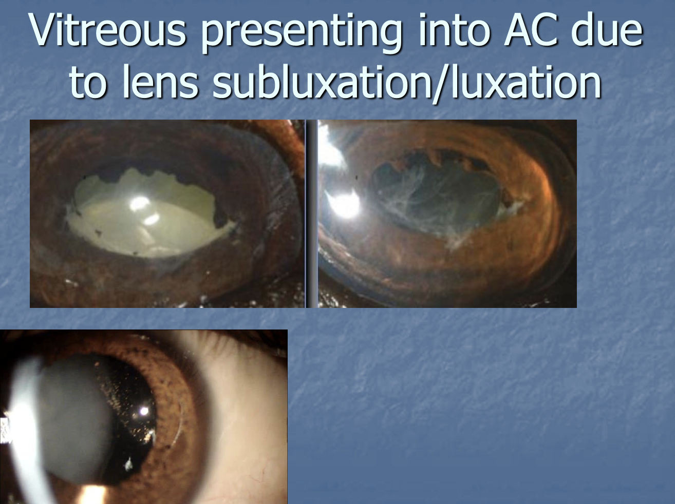
Seeing vitreous in the anterior chamber is concerning for
lens luxation.
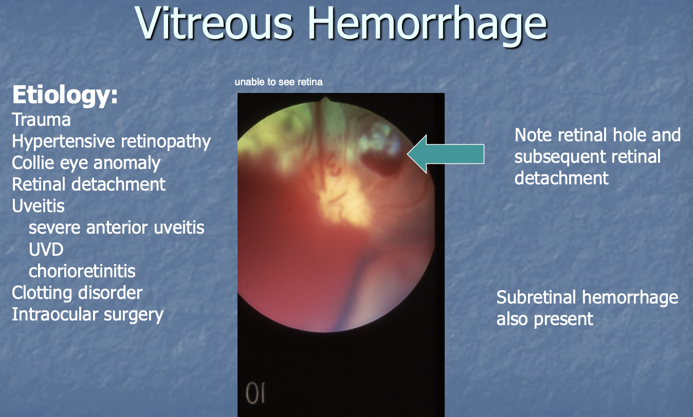
Vitreous hemorrhage etiology (7)
Trauma.
Hypertensive retinopathy.
Collie eye anomalies.
Detached retina.
Uveitis.
Clotting disorder.
Intraocular Sx.
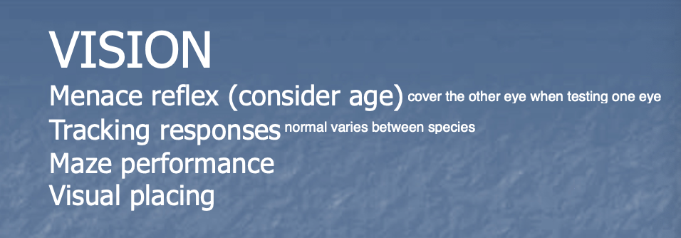
Vision is tested by (4)
menace reflex (may need to wait until 4m of age).
Tracking responses.
Maze performance.
Visual tracking.
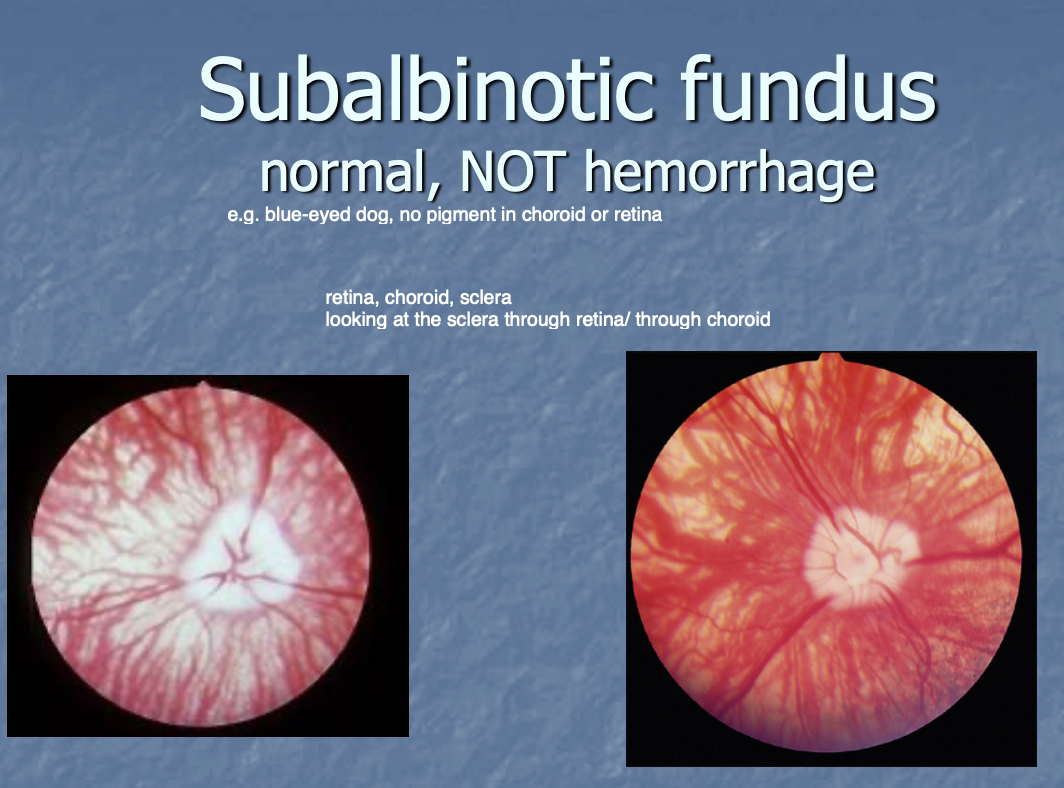
Subalbinotic funduse
normal - seen in dogs that lack pigment in the back of the eye.
Looks like hemorrhage (is not!), but you are just seeing choroid vessels and sclera .
Retinal dysplasia can present w/
folds or geographical detachment.
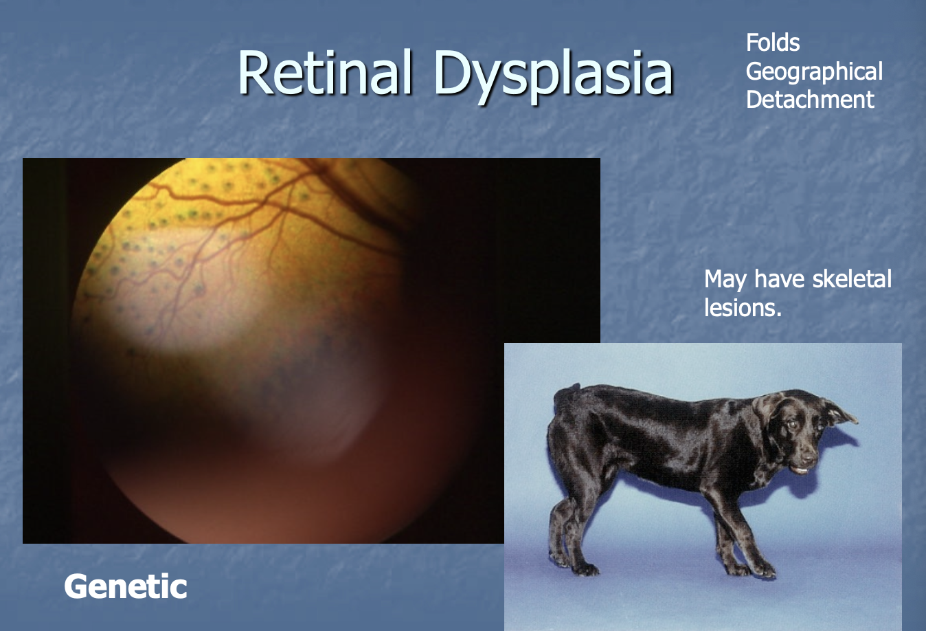
Retinal dysplasia genetic prediposition
Labradors - will also see forelimb/skeletal abnormalities.

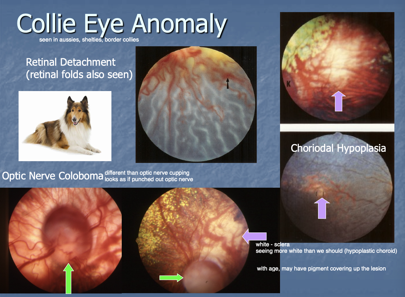
Collie eye anomaly - in order of severity less to most(3)
Choroidal hypoplasia.
Retinal detachment - can see retinal folds.
Optic n. coloboma.

Collie eye anomaly - the dogs can have
one or multiple of the anomalies associated w/ dz.

Choroidal hypoplasia can only be
seen when they are puppies - are masked when they get older and can only Dx w/ genetic test.
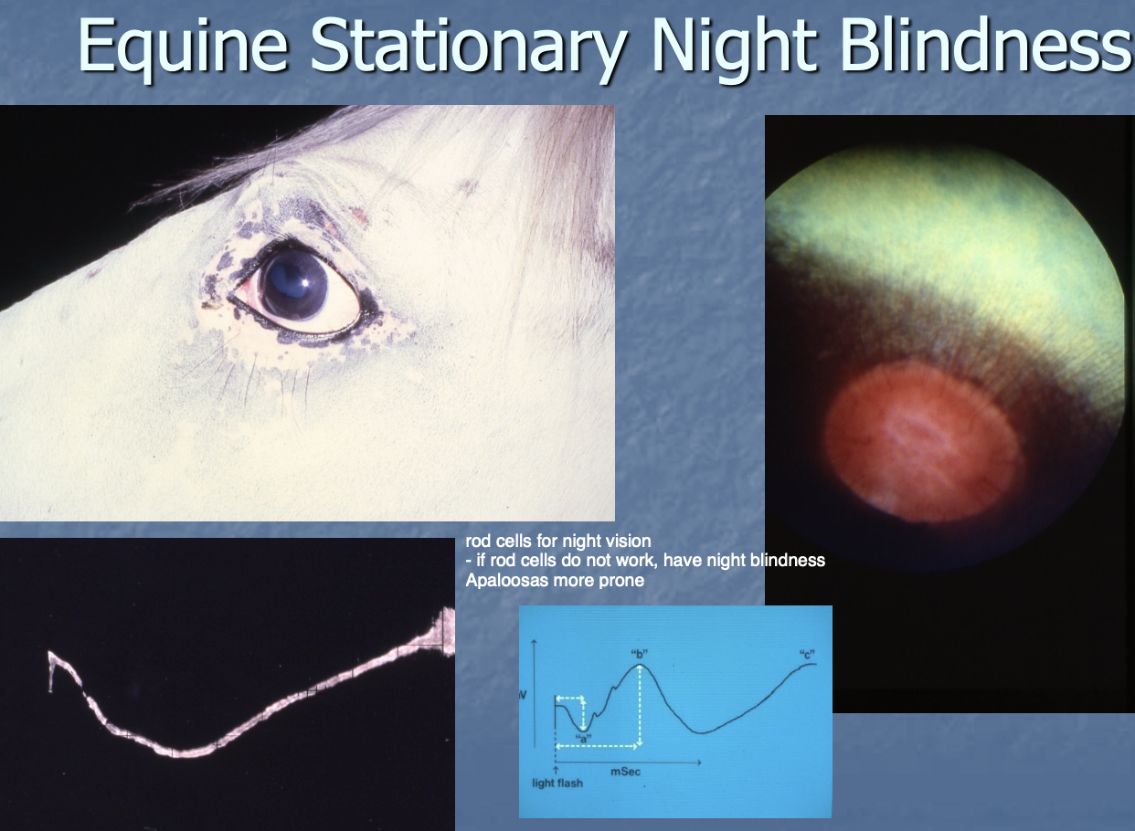
Equine Stationary night blindness (2)
rods of the retina are abnormal which affects night vision.
Cones are unaffected so daytime vision is unaffected.
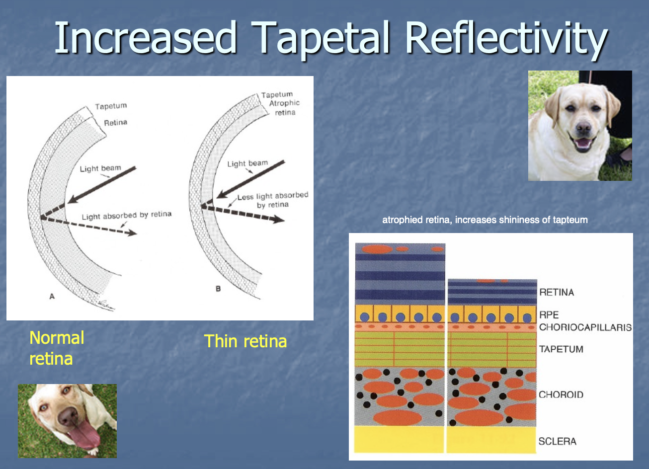
Progressive retinal atrophy (PRA) is seen in
canines (and occasionally cats).

PRA appearance of the eye (2)
gradual thinning of the retina and tapetum is more reflective (b/s of loss of retinal tissue).
Decreased size and diameter of vessels.
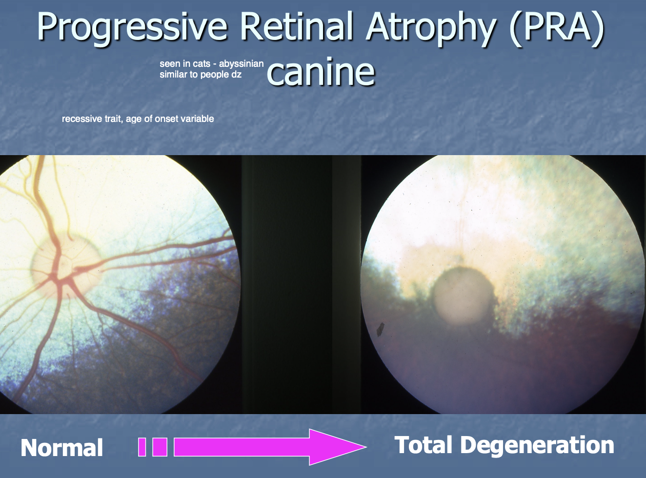
PRA: retina thins resulting in
increased tapetal reflectivity
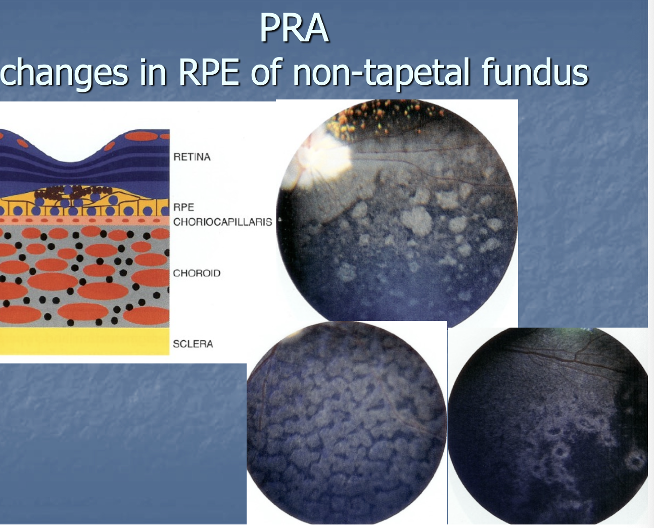
PRA: Non-tapetal retina
changes in RPE of non-tapetal retina - may see pigment build-up.
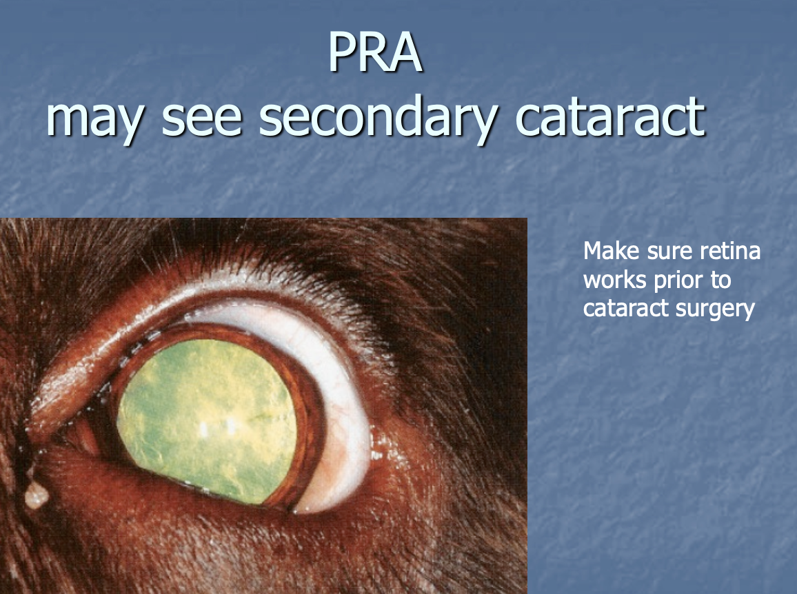
PRA may have
secondary cataract - make sure that the retina works prior to catracts Sx.
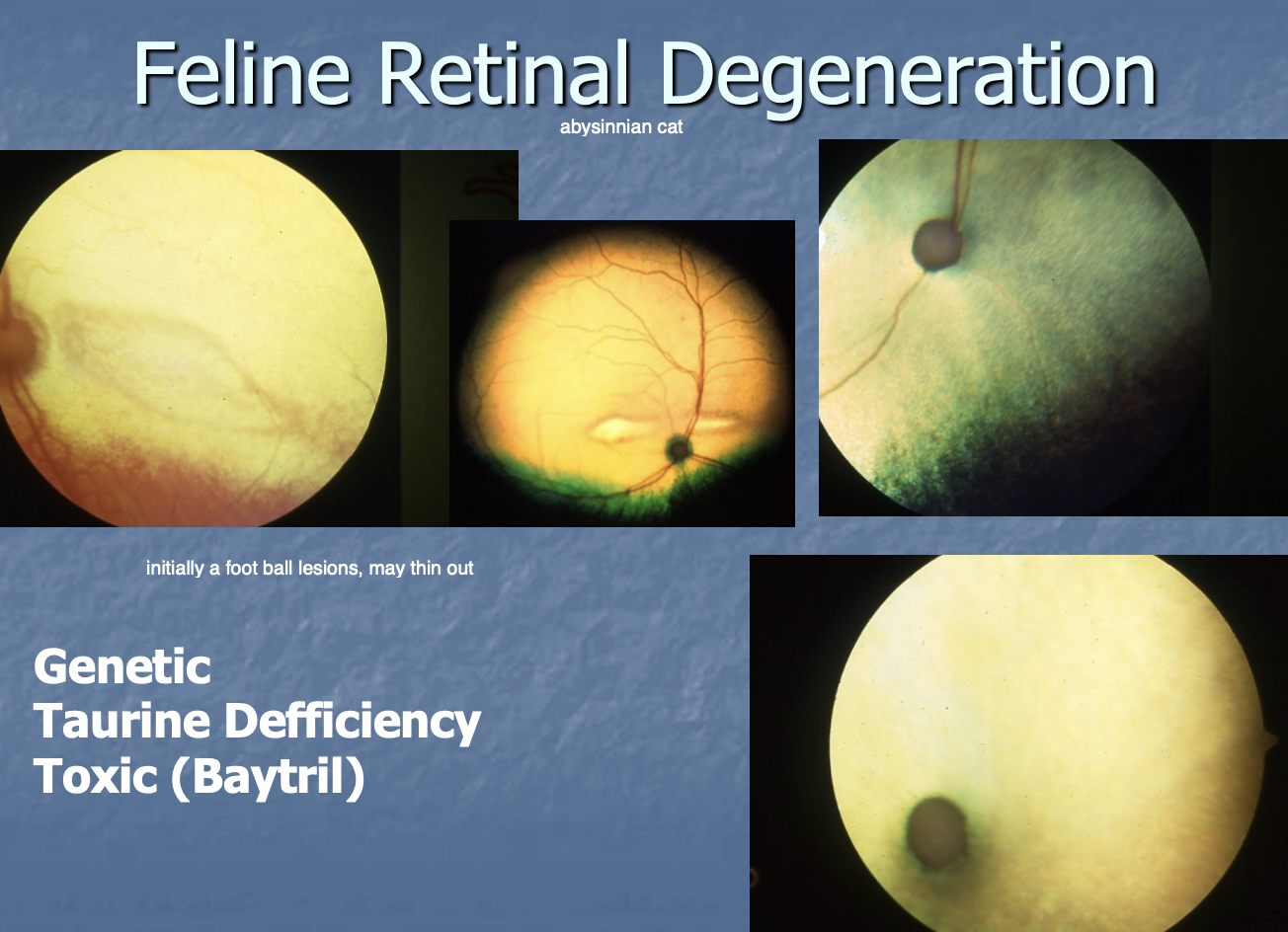
Feline Retinal Degeneration is seen w/ (3)
Genetics.
Taurine deficiency.
Toxic - Enro (baytril).
Sudden Acquired Retinal Degeneration (SARD) is seen in
canines w/ an acute onset

CS/CP of SARD - may have (3)
PU/PD or polyphagic.
Increased ALP.
Decreased sense of smell.

Etiology of SARD
unknown - potentially autoimmune.
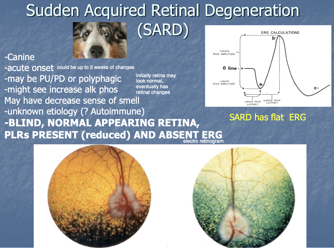
SARD - eye appearance (4)
Blind.
Normal appearing retina.
PLRs present but reduced.
Absent ERG (flat electroretnogram).

red light, blue light in a normal dog
will constrict w/ both
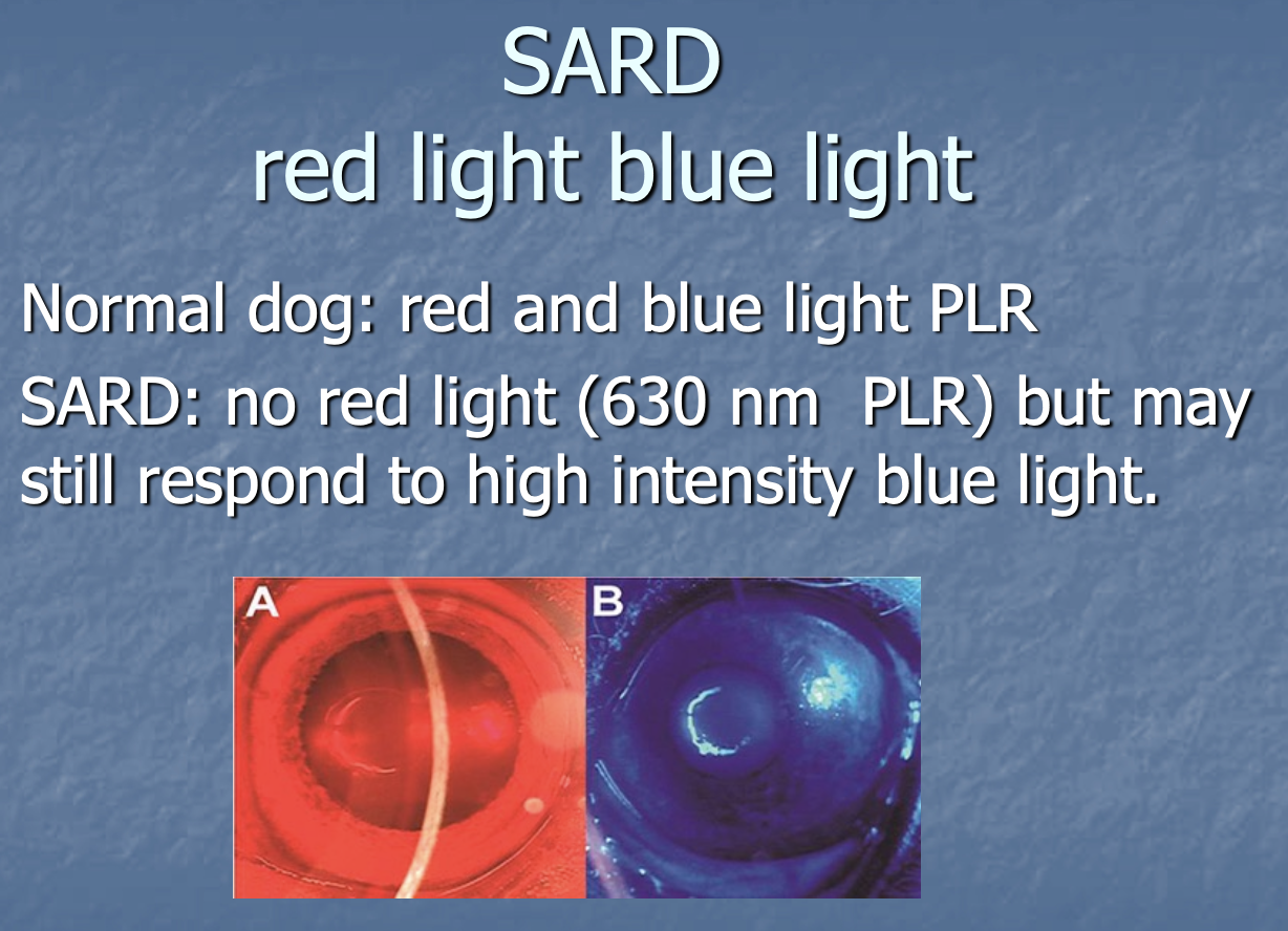
red light, blue light in a SARD dog
no constriction w/ red light.
may still respond to high intensity blue light.

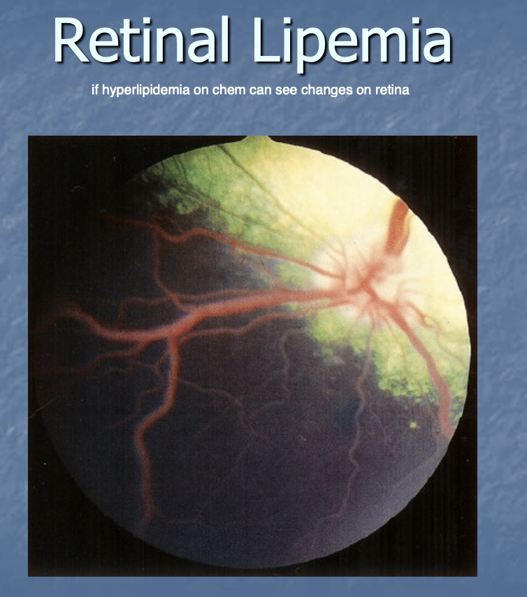
Retinal lipemia may be a sign of
hyperlipidemia
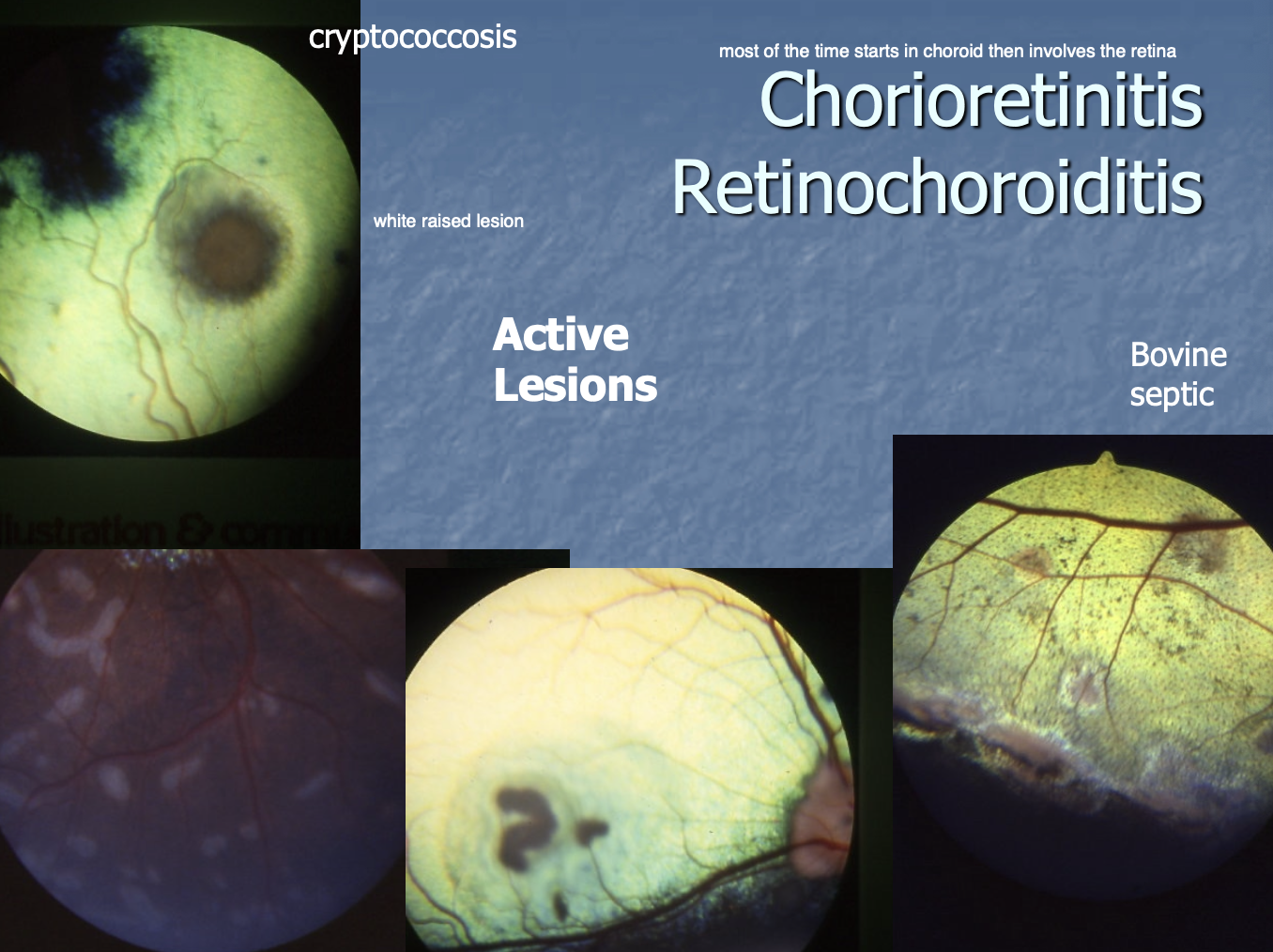
Active chorioretinitis is when there is
there is active inflammation occurring
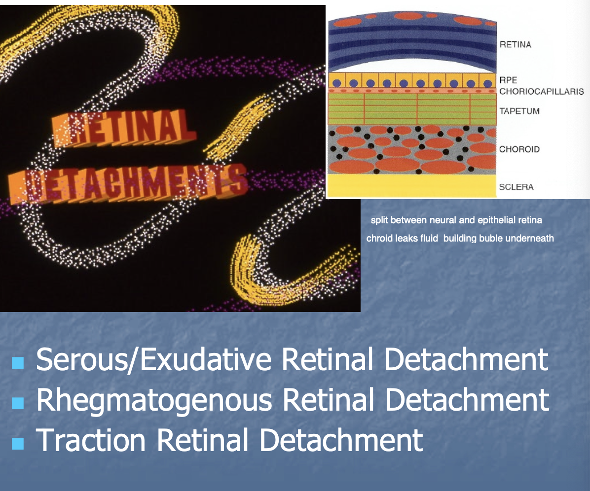
Retinal detachment def
split between neural and epithelial retina

Types of Retinal detachment (3)
Serous/Exudative.
Rhegmatogenous.
Traction.
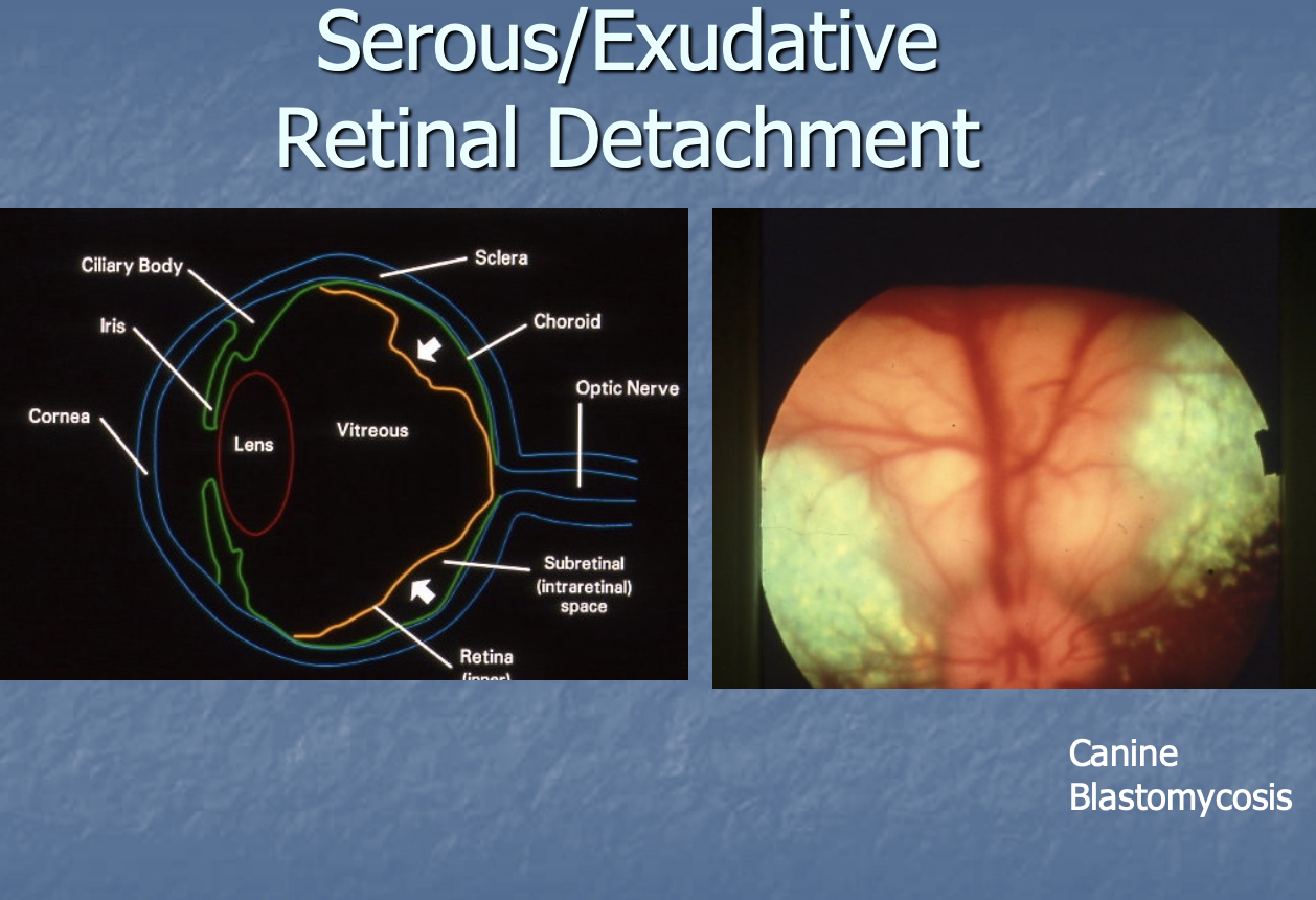
A cause of serous/exudative retinal detachment
canine blastomycosis
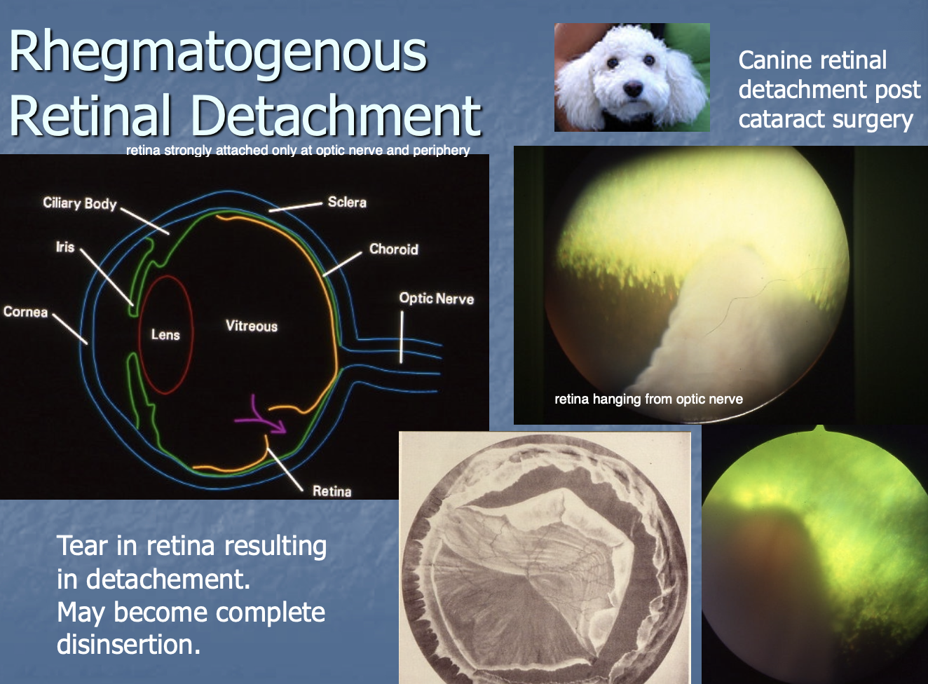
Rhegmatogenous Retinal Detachment
tear in retina resulting in detachment, may become disinsertion.
Traction retinal detachment
retina pulled in different directions

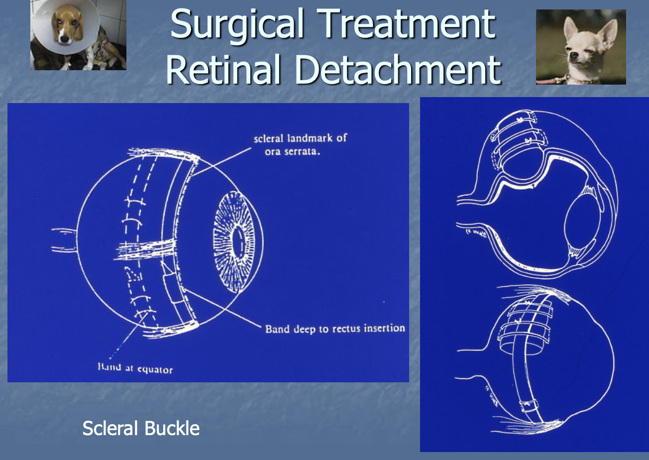
Sx tx of retinal detachment
scleral buckle
laser or cryo retinopexy
Hypertensive retinopathy is seen in
older cats
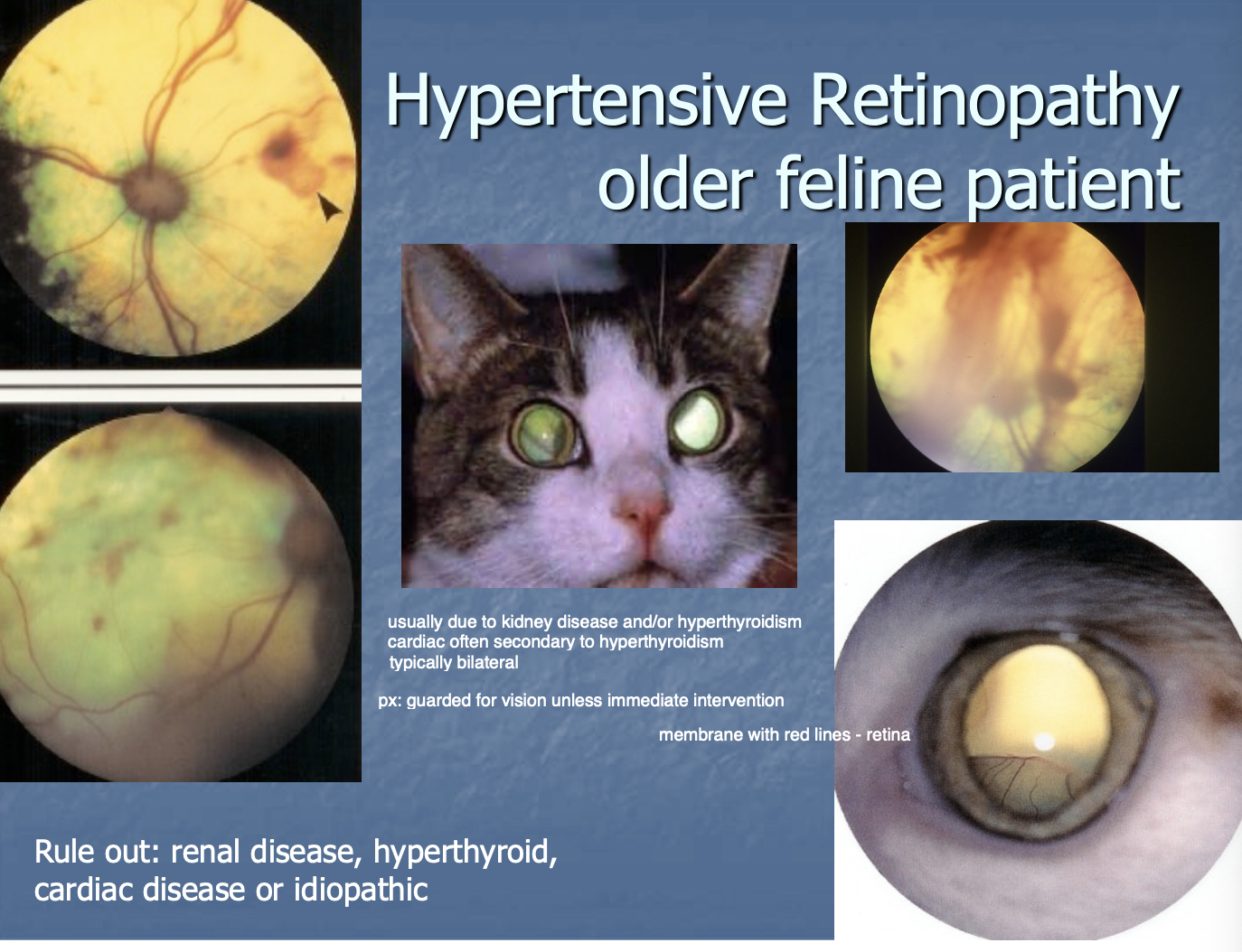
Hypertensive retinopathy r/o's (4)
renal dz.
hyperthyroid.
cardiac dz.
idiopathic.

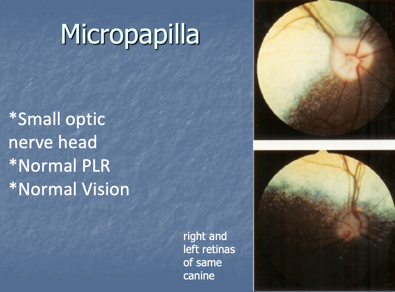
Micropapilla is
a small optic n. head, but has normal PLR and normal vision.
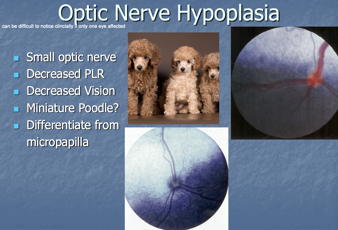
Optic n. hypoplasia def
small optic n. w/ decreased PLR, vision.

Optic n. hypoplasia is seen
in mini poodles.

Optic n. hypoplasia has to be differentiated from
micropapilla.
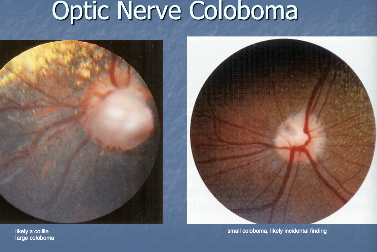
Optic. n. coloboma def
congenital absence of optic disc,
usually presnets as gap, hole, figure, notch-shaped defect
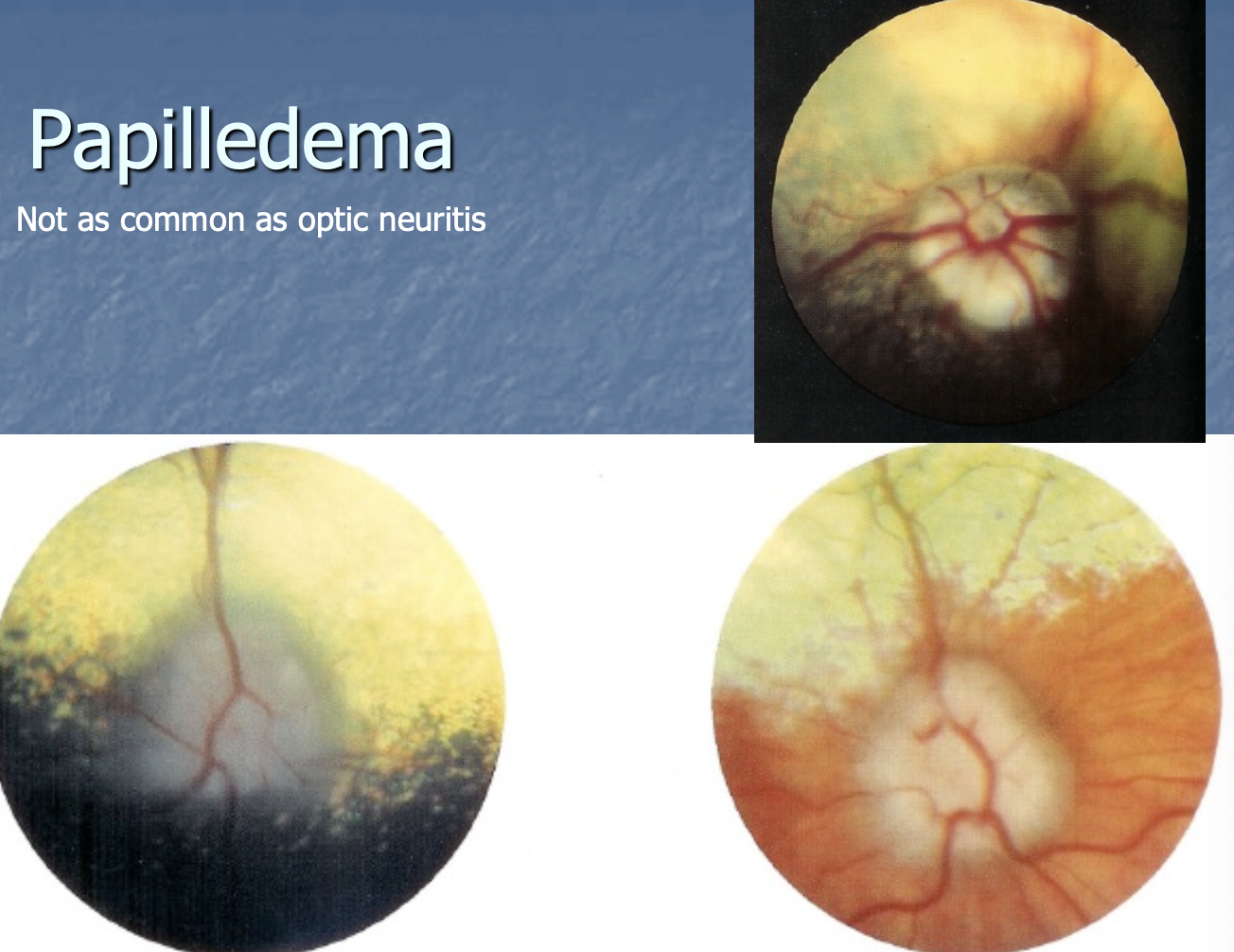
Papilledema def
edema of optic disc with normal vision
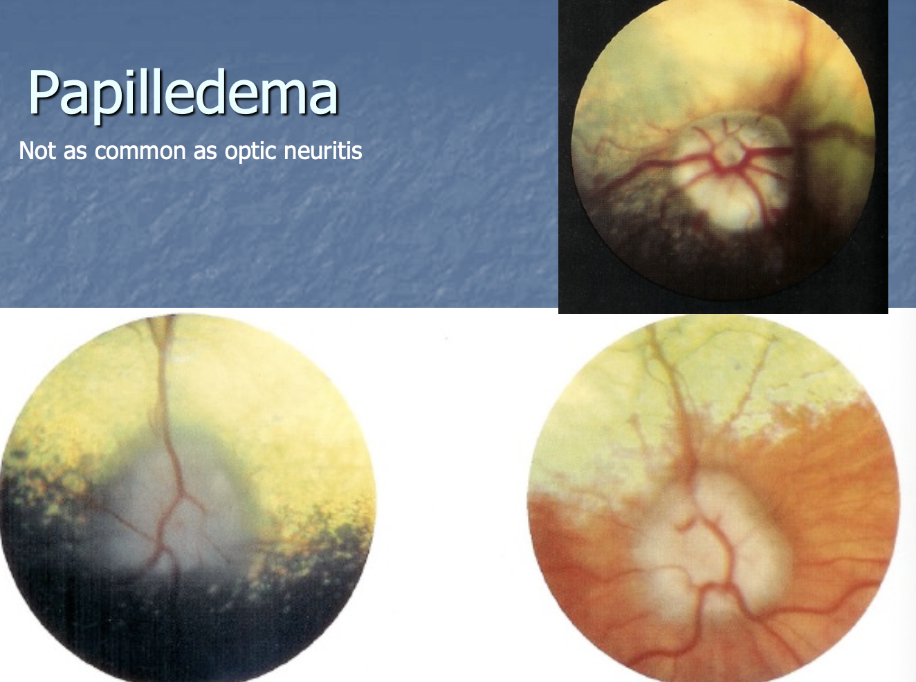
Papilledema is not
as common as optic neuritis.
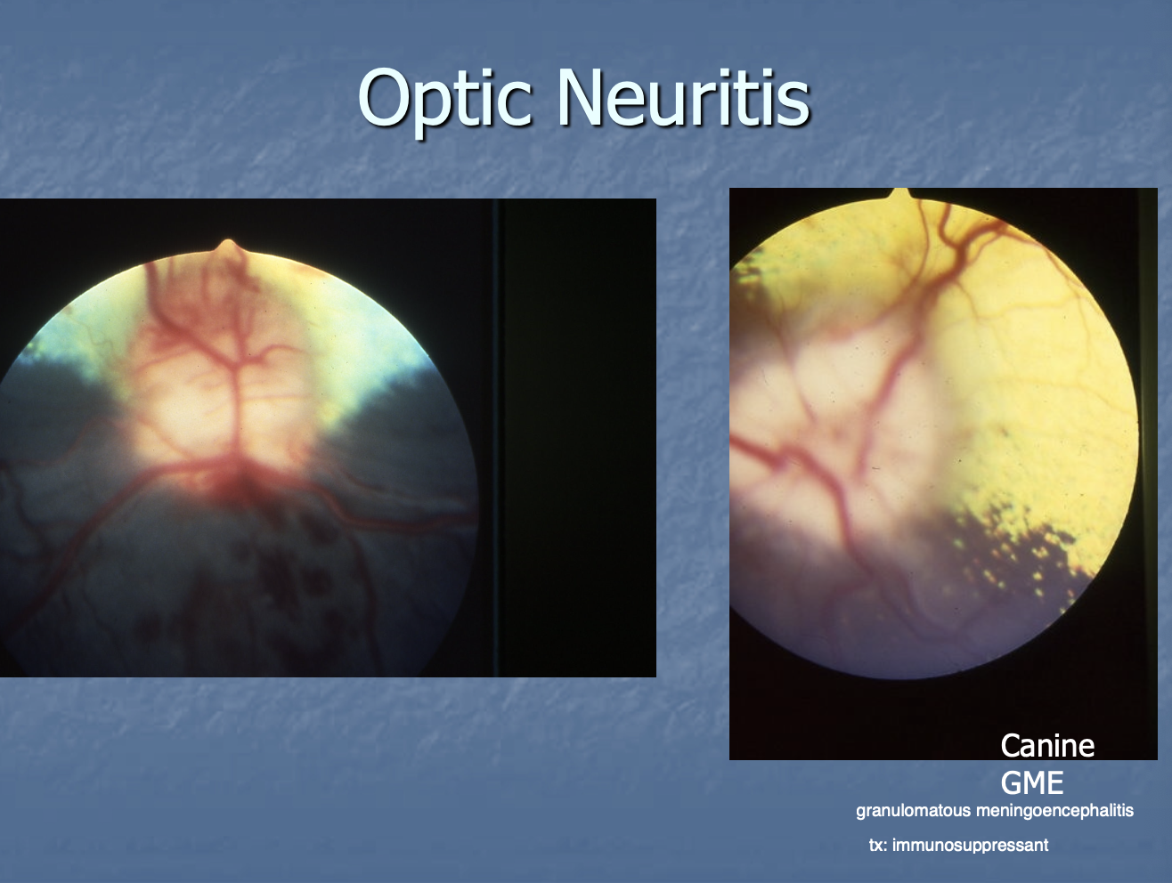
Optic neuritis def
inflammation of optic disc with decreased vision.
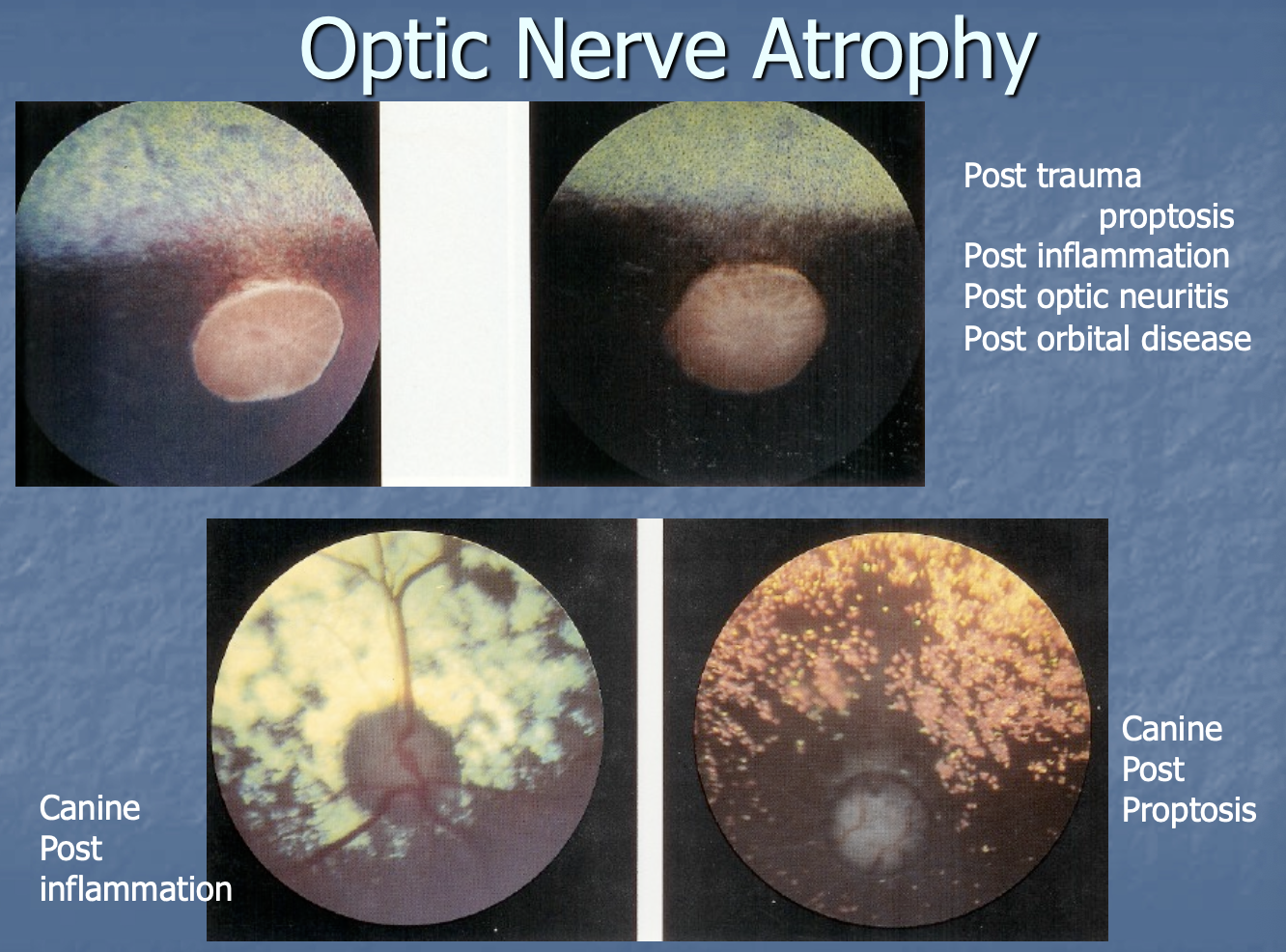
Optic n. atrophy can be seen post (4)
trauma/proptosis.
inflammation.
optic neuritis.
orbital dz.
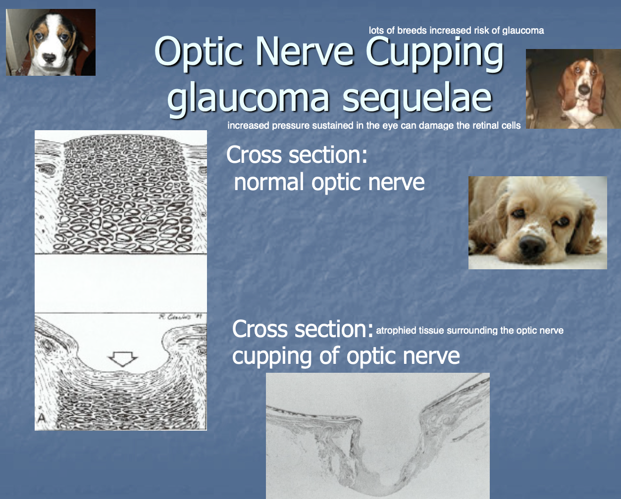
Optic disc cupping def
abnormal depression in optic disc (assoc w/ glaucoma)
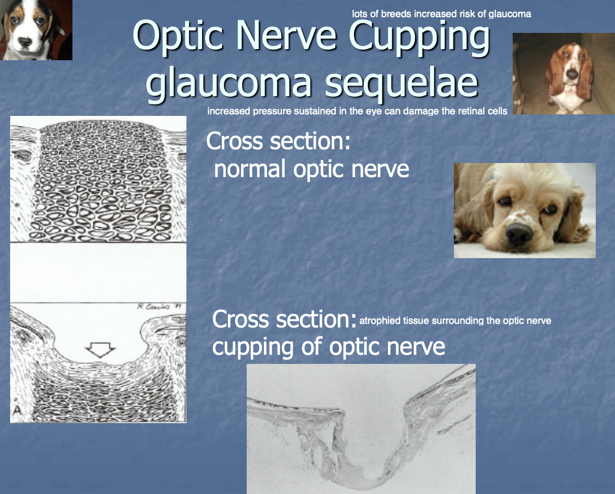
Optic n. cupping is seen
as a sequellae to glaucoma (inc IOP can damage retinal cells)