biopsychology
1/140
There's no tags or description
Looks like no tags are added yet.
Name | Mastery | Learn | Test | Matching | Spaced | Call with Kai |
|---|
No analytics yet
Send a link to your students to track their progress
141 Terms
nervous system is based on ___ +___ signals
electrical
chemical
functions of the nervous system
collect, process and respond to information from the environment
co-ordinate working of organs and cells in the body
subsystems of the nervous system
central nervous system
peripheral nervous system
central nervous system function
origin of all complex commands and decisions
central nervous system components
brain
spinal chord
CNS - brain
centre of conscious awareness
outer layer = cerebral cortex only found in mammals
highly developed in humans + distinguishes our higher mental functions form other animals
divided into 2 hemispheres
only a few living creatures have no brain eg. jellyfish
cerebral cortex is ___mm thick
3
cerebral cortex function
most sophisticated part of our brain
carries out essential functions
eg. memory, thinking, learning, problem-solving, consciousness, sensory functions
CNS - spinal chord
extension of the brain
passes messages to and form the brain
connects nerve to the PNS
responsible for reflex actions eg. pulling hand away from something hot
peripheral nervous system function
sends information (via millions of neurons) to the CNS from outside
transmits messages from the CNS to muscles + glands in the body
peripheral nervous system components
autonomic nervous system
somatic nervous system
PNS - autonomic nervous system
involuntary
governs vital functions in the body
transmits information to and from internal body organs
eg. breathing, HR, digestion, sexual arousal and stress responses
PNS - somatic nervous system
voluntary
governs muscle movement
transmits information from receptors cells in sense organs to the CNS
receives information from the CNS that directs muscles to act
transmits + receives information from all sense apart from sight
reflex arc
an automatic and rapid response to a stimulus, which minimises any damage to the body from potentially harmful conditions eg. touching something hot
autonomic nervous system subdivisions
sympathetic nervous system
parasympathetic nervous system
sympathetic nervous system function
associated with the fight or flight’ response with the endocrine system
SPNS prepares body for physical activity when the hypothalamus detects a stimulus which requires attention/action (eg. running away from or fighting the threat)
SPNS triggered when the body is in an ‘alert’ state (eg. crossing the road)
adrenaline realised from adrenal glands to fuel physical activity required along with physiological changes (eg. increased HR, wider bronchial passages, decreased large intestine activity, pupil dilation, sweating)
SPNS enables fast automatic response to possible threat or dangerous situation
can also occur when someone is highly elated or excited
fight or flight response limitation - beta bias
biological research generally favours male animals because female behaviour is affected by regular hormonal changes due to ovulation - therefore ignores any possible differences
early research into fight or slight response - assumed that both males and female would respond to threatening situations with fight or flight
more recent research - found that any approach is the tend and befriend
oxytocin is more plentiful in women, as a stress response women have increased oxytocin production
this reduces the fight or flight response + enhances the tend and befriend
therefore the original research minimised gender differences which resulted in a misrepresentation of women’s behaviour
parasympathetic nervous system function
‘rest and digest’ system
the body at rest to preserve energy
helps conserve activity levels by decreasing activity which may be needed later
regulates bodily functions eg. digestion and urination
slows HR and breathing + lowers blood pressure
body enters state of relaxation, enables it to go into recovery mode
endocrine system is based on secreting ___ into the ____
hormones
bloodstream
hormone definition
biochemical substance that circulates in the blood but only affects target organs
produced in large quantities but disappear quickly
very powerful effects
endocrine system hormone action
most hormones affect cells in 1+ body organs → leads to many diverse and powerful responses
act slowly but has widespread + powerful effects
eg. puberty = slow release of testosterone/oestrogen
major gland of the endocrine system
pituitary gland - located in the hypothalamus
controls the release of hormone from all other endocrine glands in the body
‘master gland’
endocrine system - thyroid gland
produces thyroxine - affects cells in the heart (increases heart rate) and cells throughout the body (increases metabolic rates)
affects growth rates
endocrine system - pancreas
produces insulin
produces hormones to regulate blood glucose levels + digestive enzymes such as pancreatic amylase + lipase
endocrine system - adrenal glands
produces adrenaline
endocrine system - testes
male sex hormone
produces testosterone
endocrine system - ovaries
female sex hormone
produces oestrogen
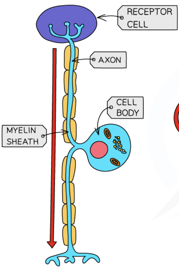
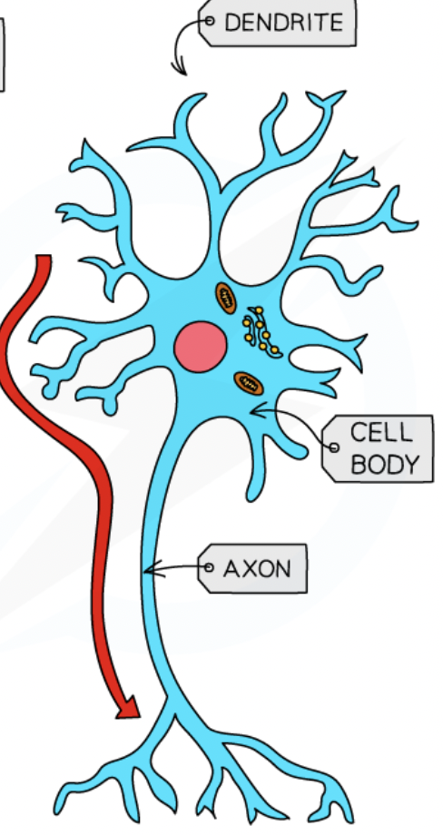
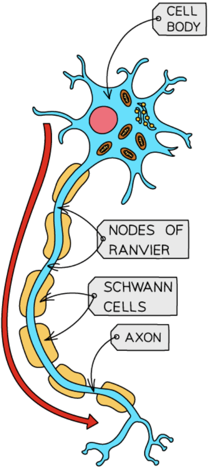
structure of a neuron (nucleus, dendrites, axon, myelin sheath, nodes of ranvier, axon terminal)
cell body containing nucleus - where DNA is stored
dendrites - receive/transmit signals from other neurons
axon - carries electrical charge from the cell body down the length of the neuron, covered in protective fatty myelin sheath
gaps in the myelin sheath = nodes of ranvier (speed up electrical transmissions)
axon terminal - end of the axon, communicate with the next neuron across the synapse
neuron + function + location
sensory neuron - carry messages from the PNS to the CNS
located in the PNS in clusters = ganglia
sensory neurons have __axons and __dendrites
short
long
neuron + function + location
relay neuron - connect sensory neurons to the motor/other relay neurons
located mostly within the brain and the visual system
___% of all neurons are relay neurons
97
relay neurons have __axons and __dendrites
short
short
neuron + function + location
motor neuron - connect the CNS to effectors eg. muscles + glands
cell bodies located in the CNS but their long axons form part of the PNS
motor neurons have __axons and __dendrites
long
short
neuron action potential
causes when a neuron is activated by a stimulus and the inside of the cell becomes positively charge for a split second
creates an electrical impulse that travels down the axon towards the end of the neuron
signals within neurons are transmitted ___
electrically
signals between neurons are transmitted ___ across the ___
chemically
synapse
process of synaptic transmission
electrical impulse reaches the presynaptic terminal
triggers the release of neurotransmitters in synaptic vesicles
neurotransmitters diffuse across the synapse
taken up b the postsynaptic receptor site on the dendrites of the next neuron
chemical message is converted back into an electrical impulse
process repeats
neurotransmitters
brain chemicals
only travel in one direction (presynaptic → postsynaptic)
each has its own molecular structure that fits perfectly into a postsynaptic receptor site (lock&key)
each have specialist function
have either an excitatory or inhibitory effect on the neighbouring neuron
inhibitory neurotransmitters
prevents an action potential in the post-synaptic neuron
increases the neurons -ve charge
makes it less likely to fire
eg. serotonin
excitatory neurotransmitters
stimulate an action potential in the post-synaptic neuron
increases the neuron’s positive charge
makes it more likely to fire (send electrical signals down its axon to communicate with other cells)
eg. adrenaline
summation
process deciding whether a postsynaptic neuron fires or not
inhibitory/excitatory are summed
if final effect on the post-synaptic neuron is inhibitory → less likely to fire (+vice versa)
action potential is only triggered if the sum of the inhibitory/excitatory signals at any one time reaches the threshold
phineas gage case study
worked on railroad - preparing to blast a section of rock with explosives to create a new railway line
explosion hurled a metre-length pole through his left cheek, behind his left eye and exiting his skull form the top of his head
took a portion of his brain with it - most of his left front lobe
survived but his personality changed
went from calm+reserved to quick-tempered + rude
suggests the frontal lobe may be responsible for regulating mood
localisation theory
Broca + Wernicke
theory that different areas of the brain are responsible for specific behaviours, processes or activities - therefore if a certain area is damaged then the function associated with that area is also affected
previous theories supported the holistic theory - all parts of the brain involved in the processing of thought + action
cerebral cortex function
outer layer of both hemispheres
divides into 4 centres = lobes all associated with different function
frontal, parietal, occipital, temporal
location + function of the motor area
back of the frontal lobe
controls voluntary movement in the opposite side of the body
location + function of the somatosensory area
front of both parietal lobes
processes sensory information eg. touch, heat,pressure
location + function of the visual cortex
in the occipital lobe
receives and process visual information
left visual cortex receive info from the right visual field + vice versa
location + function of the auditory area
temporal lobes
analyses speech-based information - damage may produce partial hearing loss
function of the cerebellum
monitors + regulates motor behaviour
learning centres of the brain
restricted to the left side of the brain (in most people)
Wernicke and Broca
Wernicke’s areas + aphasia
in the temporal lobe
responsible for language comprehension
damage causes:
Wernicke’s aphasia - produce language fine, but difficulty understanding, words produced were nonsense words
Broca’s areas + aphasia
in the frontal lobe
responsible for speech production
damage causes:
Broca’s aphasia - slow + laborious speech that lacks fluency, often struggle with prepositions and conjunctions, can understand language
localisation of brain function strength - evidence from neurosurgery
neurosurgery sometime last resort method for treating some mental disorders by targeting specific areas of the brain which are involved
eg. cingulate gyrus implicated in OCD
44 people with OCD underwent the surgery
after 32 weeks - 30% met criteria for successful response to the surgery, 14% for partial response
success of the procedures suggests that behaviours associated with some mental disorders may be localised - supports the theory
localisation of brain function strength - supporting evidence
Phineas Gage case study - supports localisation
Petersen - brain scans demonstrated how Wernicke’s area was active during a listening task + Broca’s area was active during a reading task
review of LTM studies - semantic/episodic memories reside in different parts of the PFC
confirm localised areas for everyday behaviours
therefore objective methods for measuring brain activity have provided scientific evidence that many brain functions are localised
localisation of brain function limitation - challenging evidence
Lashley - removed 10-50% of rat’s cortex
rats were learning the route through a maze
no area found more important than others in terms of their ability to learn the route
process of learning seemed to require every part of the cortex rather than one particular area
suggests that higher cognitive process (eg. learning) may not be localised but instead distributed holistically in the brain
hemispheric lateralisation
theory that the 2 halves of the brain function entirely separate
some localised areas appear in both hemispheres (eg. vision, motor, somatosensory)
left hemisphere function
language centres (analyser)
Broca and Wernicke’s areas
right hemisphere function
emotion linked to language (synthesiser)
only produce basic words + phrases
lateralisation - motor function
contralateral wiring
right hemisphere controls movement on the left side of the body
left hemisphere controls movement on the right side of the body
lateralisation - vision function
contralateral and ipsilateral wiring
each eye receives light from the left and right visual fields
left visual field of birth eyes is connected to the right hemisphere
right visual field of birth eyes is connected to the left hemisphere
enables the visual areas to compare slightly different perspectives + aids depth perception
contralateral meaning
cross wiring of brain functions
ipsilateral meaning
same sided wiring of brain functions
hemispheric lateralisation strength - research support
PET scans used to identify which brain areas were active during a visual processing task
participants asked to look at the global elements of an image - regions of the RH were more active
asked to focus on the finer detail - specific areas of the LH were more active
suggests that in terms of visual processing, hemispheric lateralisation is a feature of the connected brain as well as the split-brain
hemispheric lateralisation limitation - LH and RH roles may be wrong
general idea that LH is the analyser and RH is the synthesiser
research suggests that people don’t have a dominant side of their brain that creates a different personality
analysis of brain scans from 1000+ people aged 7-29 - found that people used certain hemispheres for certain tasks (supports lateralisation)
but there was no evidence of a dominant side of the brain
therefore suggests that the notion of right/left brained people in incorrect
reason for split-brain procedure
corpus callosum connects the right and left hemisphere
epileptic seizure - brain experiences excessive electrical activity that travels between the hemispheres
when the corpus callosum is cut it prevents these connections and splits the brain in 2 halves that can’t communicate with each other
Sperry’s split-brain research procedure
11 people who had already had a split brain operation
set up - image could be projected to a participants RVF and the same/different image could be projected to the LVF
normal brain - corpus callosum would share information between hemispheres + give a complete picture of the visual world
split brain - information can’t be conveyed between hemispheres
Sperry’s split-brain research findings
object shown to RVF, processed by LH, could describe what was seen
object shown to LVF, processed by RH, said nothing was there
messages from the RH weren't relayed to the language centres in the LH, preventing them from being spoken aloud
asked to select the matching object without seeing their hands - could do this correctly using their left hand, connected to the RH
image shown to LVF, could correctly pick up the matching object using their left hand
funny picture shown to LVF, emotional response happened eg. giggle, but responded saying they saw nothing
show how certain functions are lateralised in the brain + supports the view that LH is verbal and the RH is silent, but emotional
split-brain research strength - support from recent research
recent findings show that split-brain participants performed better than control group with connected brains on certain tasks
were faster at identifying the odd one out in an array of similar objects
this is because in the normal brain - the LH’s better cognitive strategies are weakened by the influence of the inferior RH
therefore supports Sperry’s earlier findings that the left and right brain are distinct
split-brain research limitation - generalisability
difficult to establish causal relationships from Sperry’s research
behaviour of the split-brain participants was compared to a neurotypical control group
however none of the participants in the control group had epilepsy = major confounding variable
any observed differences between the groups may have been the result of epilepsy rather than the split brain
therefore some of the unique cognitive abilities of the split-brain participants may have been due to their epilepsy, limited support for the lateralisation theory
split-brain research limitation - ethics
the split-brain procedures were already performed, not for the research
procedures were explained to participants + informed consent was obtained
however, trauma from the operation may have meant they didn’t fully understand the implication of what they’d agreed to
they were tested repeatedly over a length period which may have been stressful over time
therefore the ethics behind the research are questioned
brain plasticity
the brain is able to change throughout life
during infancy - brain experiences a rapid growth in the number of synaptic connections, peaking at 2-3 years
at 2-3 years the brain has around ____synaptic connections per neuron which is ___than an adult brain
15,000
x2 more
synaptic pruning
as we age, rarely-used connections are deleted
frequently used connections are strengthened
enables lifelong plasticity - new neural connections are formed in response to new demands on the brain
research into plasticity - taxi drivers
Maguire
studies brains of London taxi drivers
found more grey matter in the hippocampus than a matched control group
associated with spatial and navigational skills in humans + animals
London taxi drivers take a test in their training - assess recall of the city street + possible routes
finings show that this learning experience alters the structure of the taxi drivers brains
longer they’d been on the job = more structural difference (+ve correlation)
research into plasticity - medical students
imaged brains of medical students 3 months before and after their final exams
learning-induced changes were observed in the hippocampus + parietal cortex
presumed to be a result of their learning
brain plasticity limitation - -ve consequences
evidence shows the brains adaptation to prolonged drug use leads to reduced cognitive functioning in later life and increases chance of dementia
also 60-80% of amputees develop phantom limb syndrome, due to reorganisation in somatosensory cortex after limb loss - unpleasant and painful experience
therefore the brain’s ability to adapt is not always beneficial
brain plasticity strength - life-long ability
study - participants aged 40-60
found that 40 hours of golf training produced changes in the neural representations of movement
using fMRI - observed increased motor cortex activity in the noise golfers compared to a control group → suggests more efficient neural representations after training
therefore suggests that neural plasticity can continue throughout the lifespan
brain plasticity limitation - research on seasonal changes
research suggests that there may be seasonal plasticity in the brain in response to environmental changes
eg. suprachiasmatic nucleus regulates the sleep/wake cycle - evidence suggests that it shrinks in all animals during spring and expands throughout autumn
however - most research on seasonal plasticity has been done on animals (mostly songbirds) which has limited application to human behaviour
functional recovery
occurs in the brain after trauma = neural plasticity
healthy brain areas may take over functions of damaged areas
can occur quickly (spontaneous recovery) then slow down after weeks/months
brain can rewire/reorganise itself by forming new synaptic connections
functional recovery - axonal sprouting
when axons with a similar job become aroused to a higher level to compensate for lost ones
functional recovery - denervation super sensitivity
when a nerve cell axon is damaged, a new one is sprouted
can have the -ve consequence of oversensitivity to messages eg. pain
functional recovery - recruitment of homologous areas
if damage occurs in the LH, RH may pick up the task
eg. damage to Broca’s area → equivalent area in the RH may develop language skills to make up for lost ability
functional recovery strength - real-world application
understanding the processes of plasticity has contributed to the field of neurorehabilitation
understanding of axonal growth encourages new therapies to be tried
eg. constraint-induced movement therapy is used with stroke patients - repeatedly practise using the affected part of their body, while the unaffected arm is restrained
therefore shows that research into functional recovery is useful as it helps medical professionals know when intervention need to be made
functional recovery limitation - cognitive reserve
level of education may influence recovery rates
study revealed that the more time people with a brain injury had spent in education (indication of their cognitive reserve), the greater their chances of a disability-free recovery
40% of those with a DFR had 16+ years of education compared to 10% for those with less than 12 years of education
therefore implies that people with brain damage who have sufficient DFR are less likely to achieve a full recovery
functional recovery limitation - small samples
1 study treated patients with Total Anterior Circulation Stroke (TACS) with stem cells
all patients recovered compared to the typical level of 4% recovery - provides strong support for the use of stem cells helping functional recovery
however this study only had 5 participants and no control group
therefore the research may lack validity and is difficult to draw conclusions from
ways of studying the brain
fMRI - functional magnetic resonance imaging
EEG - electroencephalogram
ERP - event-related potentials
Post-mortem examinations
fMRI
detects changes in the blood oxygenation + flow that occur as a result of neuron activity in specific parts of the brain
more active area consumes more oxygen so more blood is directed there
produces 3 dimensional images showing which parts of the brain are involved in a particular mental processes
important implication for our understanding of localisation of function
fMRI +ve
doesn’t rely on the use of radiation - virtually risk-free + non-invasive + straightforward
produces images with very high spatial resolution - clear detail by mm, clear picture of how brain activity is localised
fMRI -ve
expensive compared to other neuroimaging techniques
poor temporal resolution - 5 second time-lag behind the image on the screen and the initial firing of neuronal activity
therefore may not truly represent moment-to-moment brain activity
EEG
measures electrical activity within the brain via electrodes fixed to an individual’s scalp using a skull cap
scan recording represents the brainwave patterns that are generated from the action of thousands of neurons
provides an overall account of brain acitivty
often used as a diagnostic tool - unusual arrhythmic patterns of acidity activity can indicate neurological abnormalities
eg. epilepsy, tumours, sleep disorders
EEG +ve
useful in studying the stages of sleep + diagnosis of conditions eg. epilepsy
extremely high temporal resolution - (today) can accurately detect brain activity at a resolution of 1 millisecond
real-world usefulness
EEG -ve
generalised nature of the information received - from thousands of neurons
not useful for pinpointing the exact source of neural activity
doesn't allow researchers to distinguish between activities originating in different but adjacent locations
ERPs
EEG has scientific + clinical applications, but the raw data is an overly general measure of brain activity
EEG data contains all neural responses associated with sensory/cognitive/motor events that may be of interest
researchers developed ways of isolating these responses by using a statistical averaging technique
extraneous brain activity is filtered out, leaving only those responses need for the presentation of a specific stimulus or the performance of a specific task
ERPs = remaining data - type of brainwave that are triggered by particular events
ERPs +ve
bring more specificity to the measurement of neural processes that could be achieved from raw EEG data
excellent temporal resolution
frequently used to measure cognitive functions + deficits
eg. allocation of attentional resources + maintenance of working memory
ERPs -ve
lack of standardisation in ERP methodology between different research studies - difficult to confirm findings
in order to establish pure data, background noise + extraneous material must be completely eliminated - not always easy to achieve
Post-mortem examination
technique involving the analysis of a person’s brain following their death
brains tested are likely to be from those who have a rare disorder + have experience unusual deficits in cognitive processes or behaviour during their lifetime
damaged areas of the brain are examined after death to establish the likely cause of the affliction that the person experienced
may also involve comparison to a neurotypical brain to assess the extent of the difference
Post-mortem examination +ve
vital in providing a foundation for early understanding of key processes in the brain
Broca + Wernicke relied on them to establish links between language, brain and behaviour - decades before neuroimaging was possible
used to study HMs brain to identify areas of damage - then associated with his memory deficits
Post-mortem examination -ve
observed damage to the brain may not be linked to the deficits under review, but instead due to some other unrelated trauma or decay
ethical issues of consent from the individual before death - some may not be able to provide informed consent
eg. HM couldn’t provide this consent, but post-mortem research was still conducted on his brain
challenged usefulness of post-mortem studies in psychological research