dental embry, histo, and anatomy ch 5-6 quiz
 Call Kai
 Learn
Learn Practice Test
 Spaced Repetition
 Match
Match Flashcards
 Knowt Play
1/112

There's no tags or description
Looks like no tags are added yet.
Name | Mastery | Learn | Test | Matching | Spaced | Call with Kai |
|---|
No analytics yet
Send a link to your students to track their progress
113 Terms
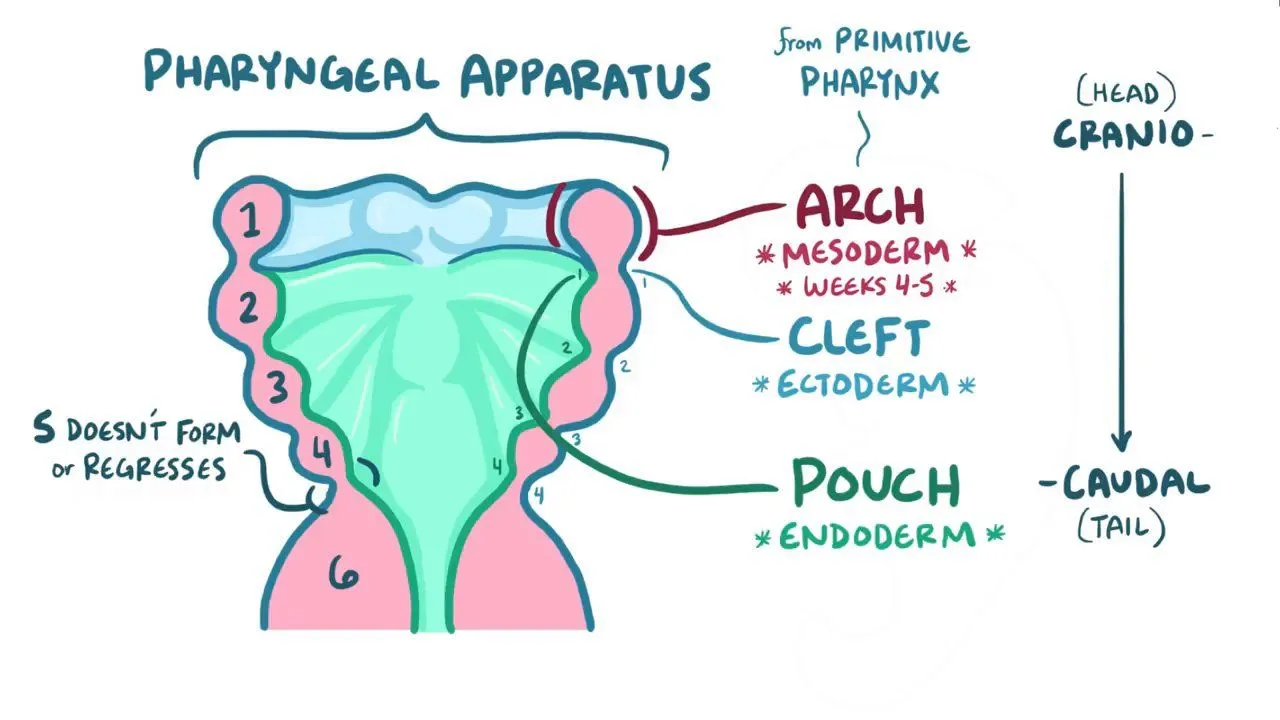
pharyngeal arches/ branchial apparatus
consists of arches 1-6, negating arch 5 as it goes away
begins development in week four due to embryonic folding
consists of arches, grooves, membranes, and pouches
each arch has its own cartilage, muscle, vascular, and cranial nerves
first pharyngeal arch
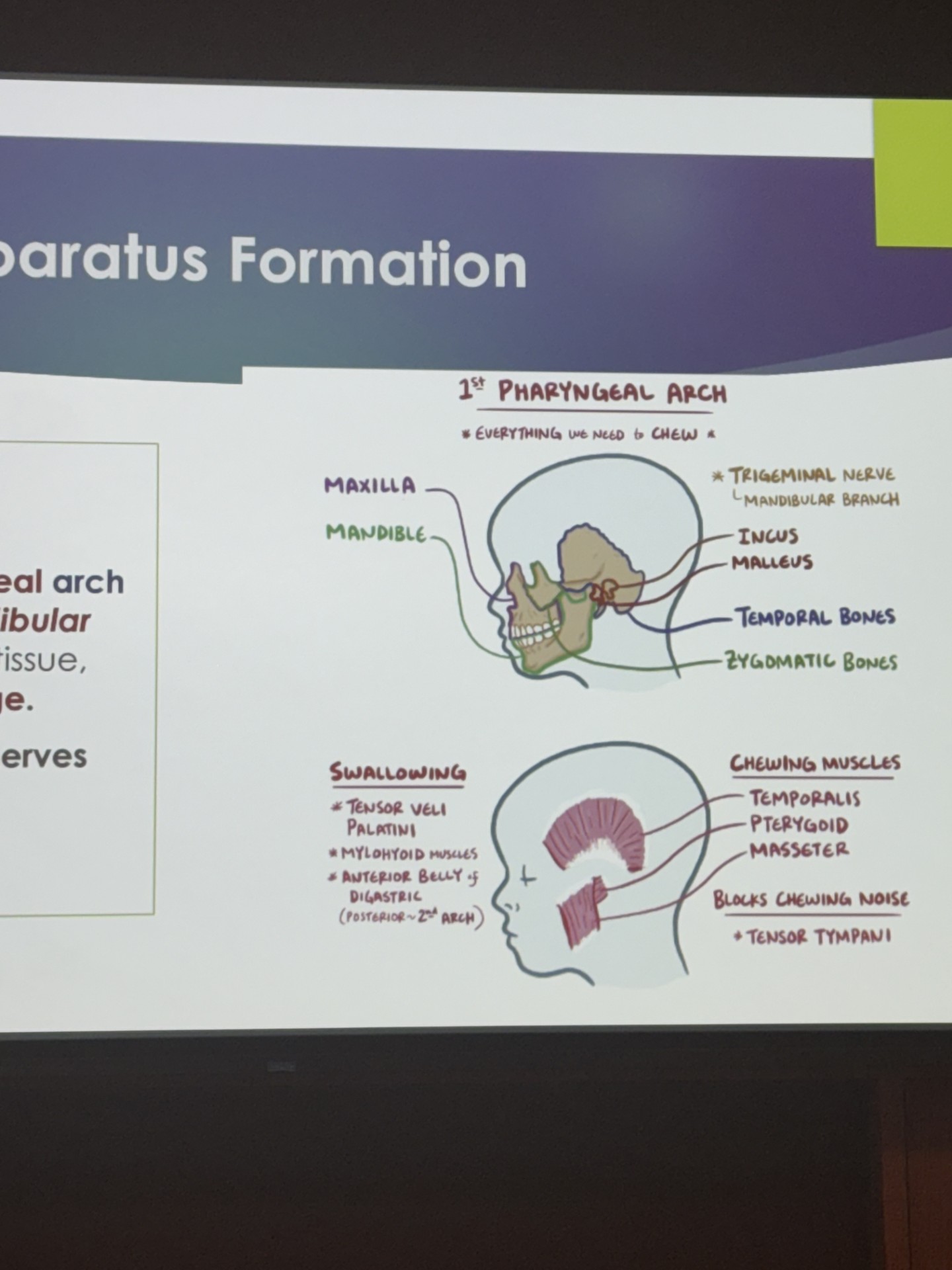
also known as the mandibular arch
both mandibular processes fuse to form mandible, lower lip, and mandibular teeth
contains meckel cartilage
innervated by the trigeminal nerve (mandibular branch)
muscles and structures needed for chewing
second pharyngeal arch
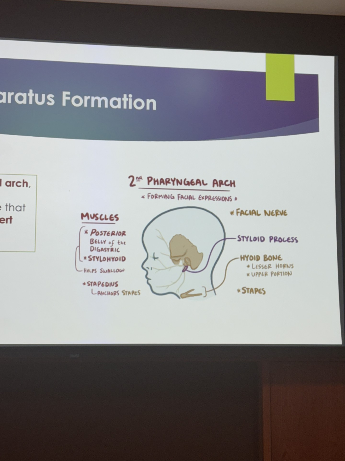
known as hyoid arch
contains reinchert cartilage
innervated by facial nerve
develops horns of hyoid bone and muscles/ facial features designated to creating facial movements and expressions
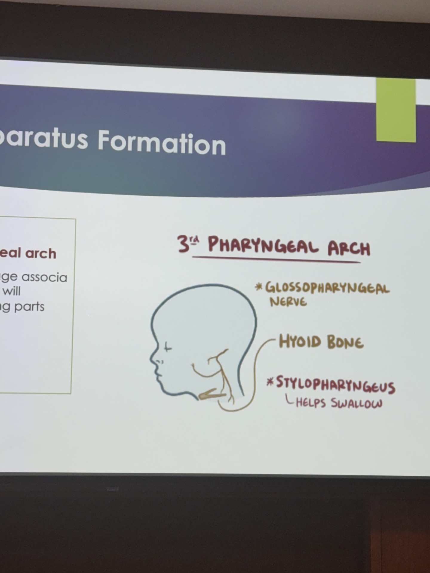
third pharyngeal arch
creates the rest of the hyoid bone
contains unnamed cartilage
innervated by the glossopharyngeal cranial nerve
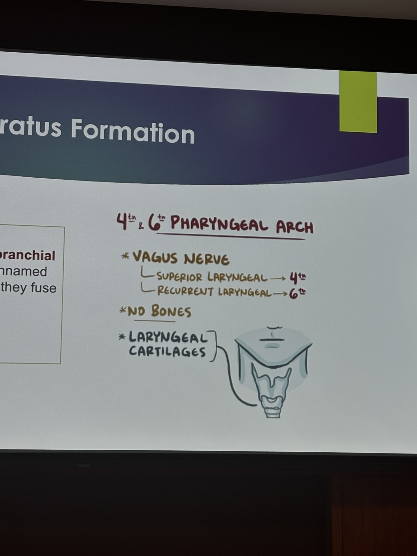
fourth and sixth pharyngeal arch
contain unnamed cartilage
innervated by the vagus nerve
create the laryngeal cartilages
the development of the orofacial structures including the palate, nasal septum, nasal cavity, and tongue, occurs during the _ to _week of prenatal development, spanning the later part of the embryonic period and early part of fetal period
fourth to twelfth week
the formation of the palate begins during the __ week of prenatal development within the embryonic period
fifth week
the palate is then completed during the __ week within the fetal period
twelfth
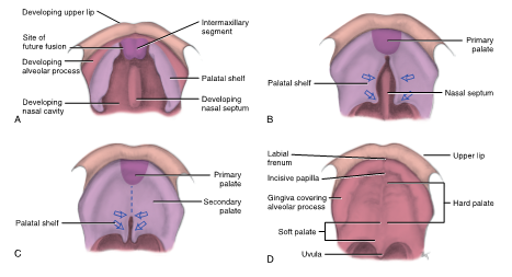
fifth to sixth week of palatal development
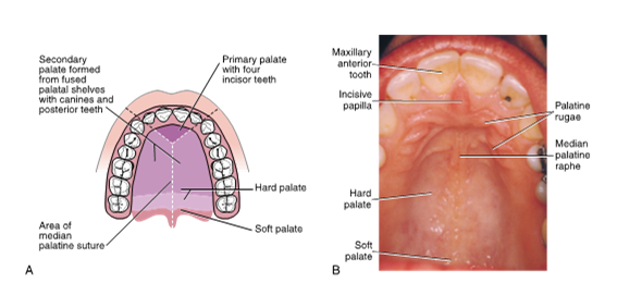
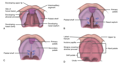
The primary palate is formed from the inferior growth of the intermaxillary segment
triangular shape, anterior 1/3 of the hard palate
gives rise to the 4 anterior maxillary incisors
served only as a partial separation between the developing oral cavity proper and the nasal cavity
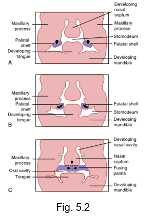
sixth to twelfth week of palatal development
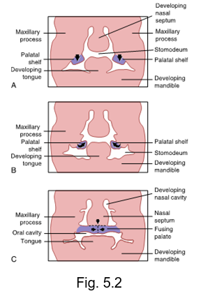
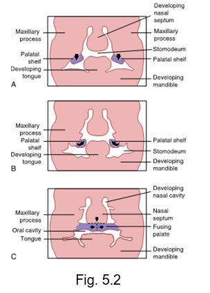
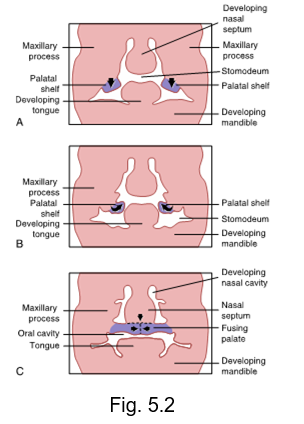
Bilateral maxillary processes will give rise to palatal shelves
secondary palate begins to form and migrate as palatal shelves grow inferiorly and deep on the inside of the stomodeum in a vertical direction towards each other
tongue forming on the floor of the primitive pharynx initially fills the cavity, but will contract and move inferiorly
because of an unknown elevating force, palatal shelves will begin to “flip” up and grow in a superior direction after movement of tongue
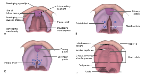
two palatal sheleves elongate and move medially, joining in the middle and fusing to form secondary palate
posterior 2/3rds of hard palate, soft palate, uvula is the last thing to fuse
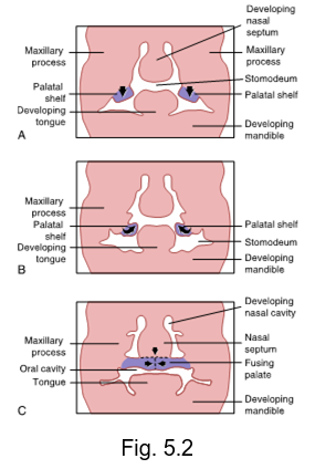
intermaxillary segment arises as a result of the fusion between the two __ nasal processes internally
medial
inferior growth and fusion of the medial nasal processes internally forms the intermaxillary segment that will eventually give rise to the anterior 1/3 of the hard palate and the 4 anterior maxillary incisors
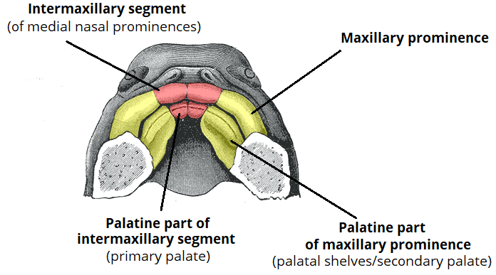
palatal shelves
also known as lateral palatine processes
will grow horizontally towards each other after flipping in superior direction to form secondary palate
will fuse with nasal septum
median palatine raphe
indicate the line of fusion of the palatal shelves
suture located in the middle of the mucosa lining of the hard palate
twelfth week of palatal development
final palate takes form
palate undergoes fusion of all three processes (primary palate and two palatal shelves that have created secondary palate)
posterior part of primary palate meets the secondary palate due to increased growth, structures gradually fuse in an anterior to posterior direction (primary palate to median raphe to uvula)
mature oral cavity becomes completely separated from the nasal cavity
failure of fusion of the palatal shelves with the primary palate and/or with each other results in __ __ with varying degrees of disability
cleft palate
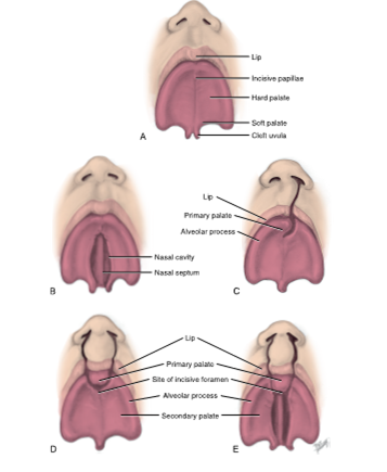
cleft uvula
is the least complicated form of cleft palate
may still cause velopharyngeal insufficiency (VPI)
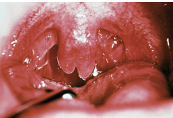
velopharyngeal insufficiency (VPI)
a disorder that prevents the velum or soft palate from closing against the posterior pharyngeal wall during speech in order to close off the nasal cavity, which is needed for the production of most speech sounds
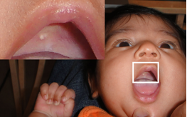
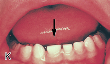
Epstein pearl
small white papule seen in the midline of the palate of an infant
represents epithelial tissue that becomes trapped during palatal fusion
nasal cavity formation
forms in the same time frame as palate from the fifth to twelfth week of prenatal development
later serve as part of respiratory system
nasal septum is a growth from the fused medial nasal processes
vertical nasal septum then fuses with the horizontally oriented final palate after it has formed
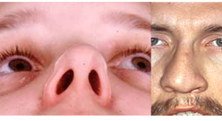
deviated septum
occurs when the thin wall that makes up the nasal septum inside the nose is displaced to one side
nasal septum separates the right and left nasal cavities
the tongue begins to develop during the __ to __week of prenatal development
fourth to eighth
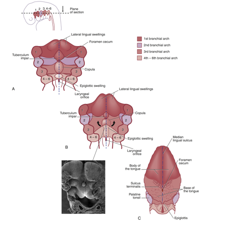
the body of the tongue develops from the __ pharyngeal arch
first pharyngeal arch
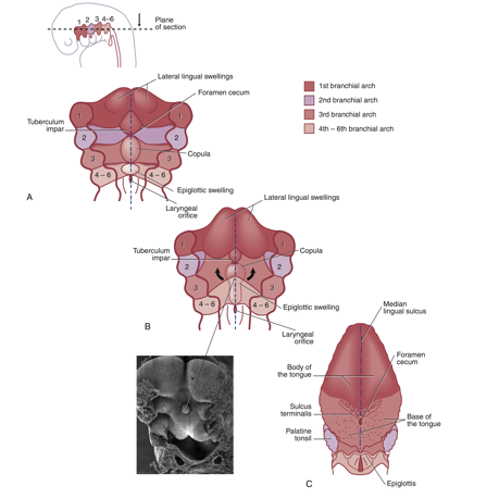
the base of the tongue originates from the _,_, and _ pharyngeal or bracnhial arches
the base of the tongue originates from the
second
third
and fourth pharyngeal arches
fourth week to eighth week of the body of the tongue development
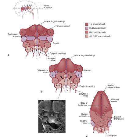
The body begins to form as a triangular median swelling called the tuberculum impar is formed on the mandibular arch, and lateral lingual swellings appear on either side of the tuberculum impar
paired swellings grow and merge, growing over the top of the tuberculum impar
These swellings form the anterior 2/3rds of the body of the tongue, and around swellings, the cells degenerate, forming a sulcus and freeing the body of the tongue from the floor except for the midline lingual frenum
tuberculum impar
median tongue bud
triangular median swelling that appears during the body of the tongues development
appears in the midline and formed on mandibular arch (first pharyngeal arch)
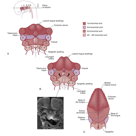
median lingual sulcus
is a superficial demarcation of the line of fusion of the two lateral lingual swellings, as well as of a deeper fibrous structures
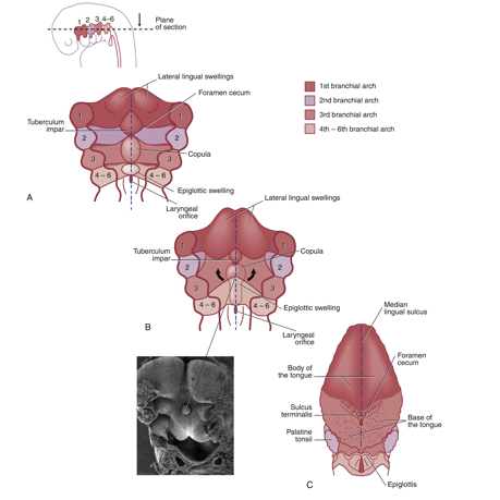
fourth week to eighth week of the base of the tongue development
posterior to the fused anterior swellings, a pair of swellings, the copula, becomes evident
gradually overgrows the hyoid arch (second pharyngeal arch) and this overgrowth will form the posterior one-third of the mature tongue, the base
farther posterior to copula is a projection of a third median swelling, the epiglottic swelling and marks the development of the most posterior region of tongue and site of epiglottis
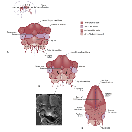
eighth week of tongue development
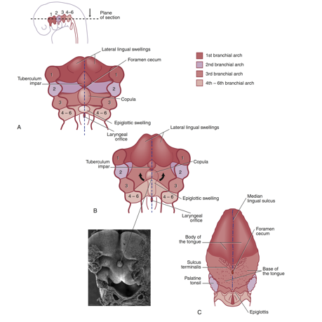
completed tongue: merging of anterior swellings of body and copula of base after copula overgrows hyoid arch (2nd pharyngeal arch) during eighth week of prenatal development
fusion is superficially demarcated by the sulcus terminalis on the mature dorsal surface of tongue
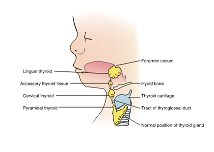
sulcus terminalis points backwards toward a small pit-like depression, the foramen cecum, which is the beginning of the thyroglossal duct
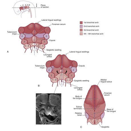
the thyroglossal duct shows the origin of the __ gland and the migration pathway of the __ gland into the neck region
thyroid gland
later this duct will close off and become obliterated before birth
ankyloglossia
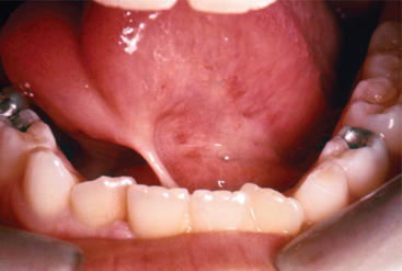
“tongue-tied”
tongue is “anchored down”
results from a short attachment of the lingual frenum that extends to the tongue apex
incisive foramen
important structure in the hard palate that allows nerves to sprout through
formed due to the merging and fusion of primary palate and secondary palatal shelves
when the triangular primary palate and elongated secondary palatal shelves meet, it leaves a hole that we know as the __ __
odontogenesis
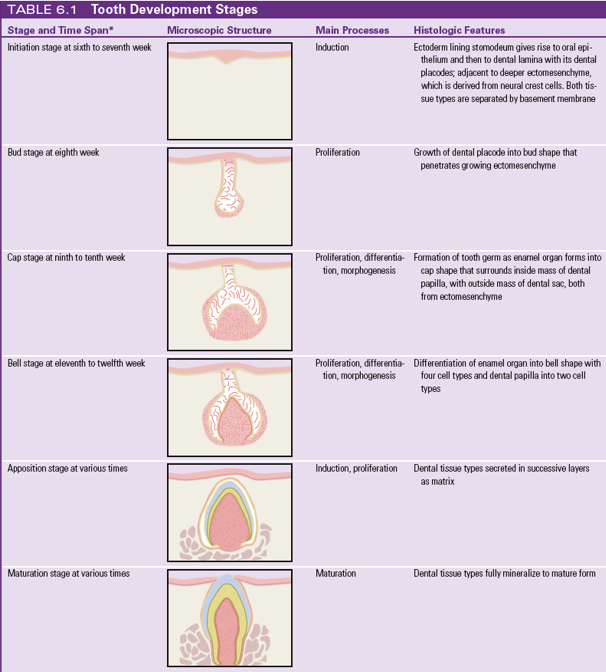
is the process of tooth development
starts between weeks 6-7 of prenatal development
typically includes 6 stages
initiation, and then the initial identifiable stages are:
bud
cap
bell
progresses to the apposition
maturation
dentition
used to describe the natural teeth in the jaws
primary (deciduous) and permanent
initiation stage (weeks 6-7)
the first stage of odontogenesis
at the beginning of the sixth week, the embryo’s stomodeum (primitive mouth) is lined by ectoderm that gives rise to oral epithelium
in the oral epithelium ectomesenchyme interacts with dental lamina but is seperated by an acellular structure known as the basement membrane
during the later part of seventh week oral epithelium thickens to form dental lamina and vestibular lamina
dental placodes form
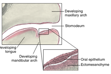
oral epithelium
consists of two horse-shoe shaped bands of tissue at the surface of the stomodeum
one band for each future dental arch
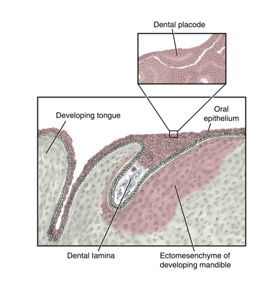
ectomesenchyme
a tissue derived from neural crest cells that have migrated to the region
in this instance it is located deep to the forming oral epithelium
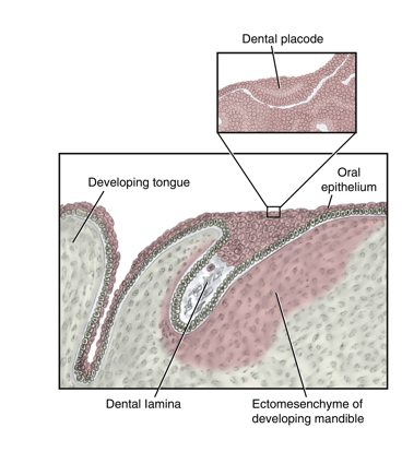
basement membrane
an important acellular structure that separates the oral epithelium and the ectomesenchyme within the stomodeum
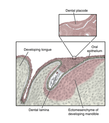
dental lamina
2 u-shaped bands of thickened epithelium formed during induction
occurs in each developing jaw where the two future curved dental arches of the primary dentition will form
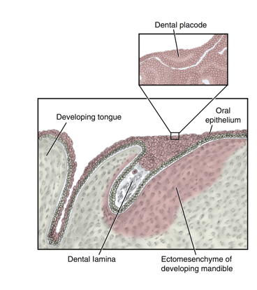
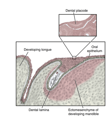
dental placodes
resemble the other placodes developing
each placode consist of plate-like thickened epithelium associated with an underlying neural crest derived mesenchyme (neuroectoderm)
function as first signalling centers of the tooth
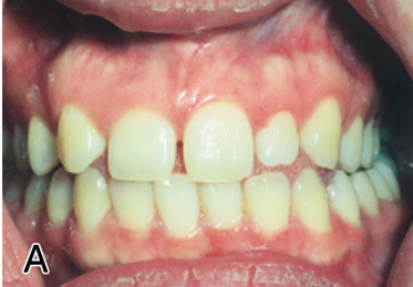
anodontia
lack of initiation within the dental lamina results in the absence of a single tooth, multiple teeth, or even an entire dentition
also known as hypo (less) dontia
hereditary
most common teeth for partial anodontia
permanent third molar
maxillary lateral incisor
mandibular second premolar (because it is the only premolar to have 3 cusps)
ectodermal dysplasia
can be associated with anodontia
many of the components of the tooth germ are indirectly or directly of ectodermal origin
dysplasia- displaced
displaced ectoderm
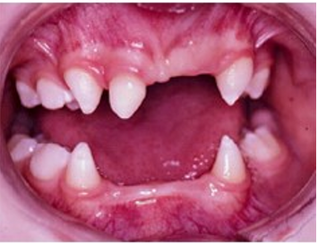
supernumerary teeth
known as hyperdontia
extra teeth initiation from persisting clusters of the dental lamina
have a hereditary etiology
commonly between the maxillary central incisors, distal to the maxillary thid molar, and premolar region of both dental arches
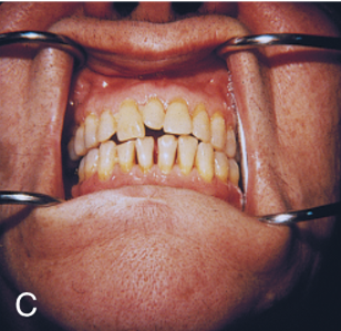
mesiodens
term for a supernumerary tooth located between the maxillary central incisors
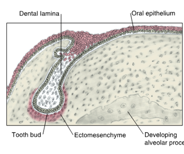
bud stage (week 8)
second stage of odontogenesis
extensive proliferation of the dental lamina into 10 buds per arch that penetrate the ectomesenchyme
basement membrane remains between the dental lamina bud and the surrounding growing and condensing ectomesenchyme
each bud will later form the tooth germ
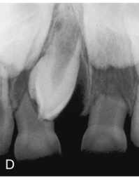
distomolar
supernumerary tooth located distal to the maxillary third molar
perimolar
supernumerary tooth located in the premolar region of both dental arches
macrodontia
abnormally large teeth
result of abnormal proliferation during bud stage
most commonly affects maxillary lateral incisors and third molars
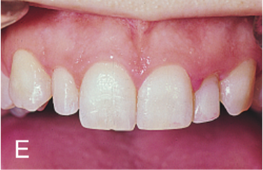
microdontia
abnormally small teeth
result of abnormal proliferation during bud stage
most commonly affects maxillary lateral incisors and third molars
cap stage (weeks 9-10)
third stage in odontogenesis
differentiation and morphogenesis begin
unequal growth in different parts of tooth bud, leads to the formation of cap shape overlying the ectomesenchyme
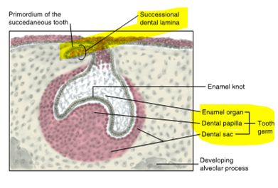
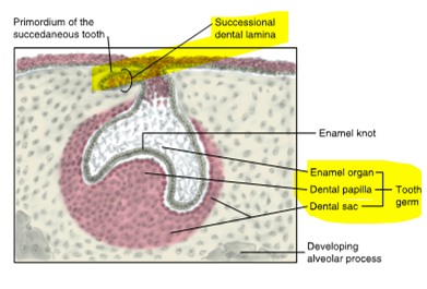
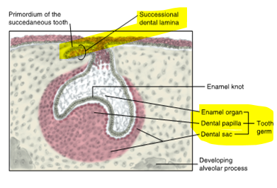
primordium of the tooth (tooth germ) forms containing tissue
enamel organ
dental papilla
dental sac
innermost margin of cap consists of enamel knot
during later part, site of origin called the successional dental lamina begins to form lingual to developing primary tooth germs
enamel organ
forms enamel
tooth germ
formation of tooth bud in cap shape with deep central depression
dental papilla
forms dentin and pulp (DP)
tooth germ
condensed mass of ectomesenchyme within concavity of enamel organ
dental sac (follicle)
forms cementum, periodontal ligament, alveolar process
tooth germ
condensed mass of ectomesenchyme surrounding the outside of enamel organ
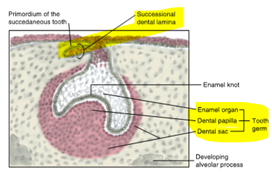
successional dental lamina
appears during the 10th week
each primordiym for these initially formed permamnent teeth appears as an extension of the dental lamina into the ectomesenchyme that grows lingually to developing primary tooth germs
start to form permanent dentition
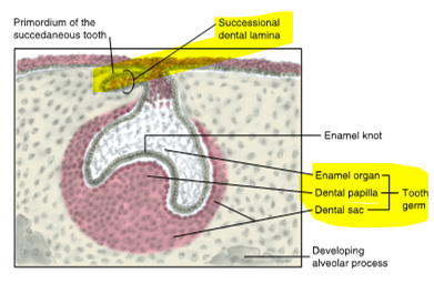
succedaneous teeth
permanent teeth formed with primary predecessors
anterior teeth and premolars
crown of each permanent succedaneous tooth will reupt lingual to roots of its primary predecessor
nonsuccedaneous
have no primary predecessors
the six permanent molars per dental arch
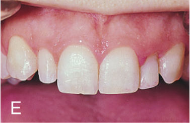
dens in dente
translates to tooth inside a tooth
during cap stage enamel organ may abnormally invaginate by growth into the dental papilla
(enamel folds into crown)
commonly found in permanent maxillary incisors (lateral)
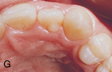
gemination
occurs as the single tooth germ tries unsuccessfully to divide into two tooth germs, resulting in a notably large, single-rooted tooth with common pulp cavity
developmental disturbance during cap stage
gemini (two heads, two germs)
root is one but the head is two
normal number of teeth
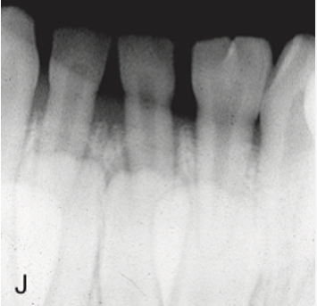
fusion
union of two adjacent tooth germs, possibly resulting from pressure in the area
a different head and root, but are joined together
Two tooth buds merge as a result of pressure
fewer teeth in the arch
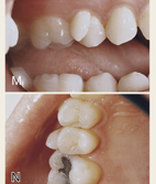
tubercles
small, round enamel extensions forming extra cusps
results from an enamel knot malformation
common on molars
bell stage (weeks 11-12)
fourth stage of odontogenesis
continuation of ongoing process of proliferation, differentiation, and morphogenesis
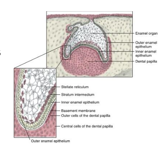
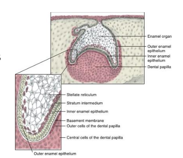
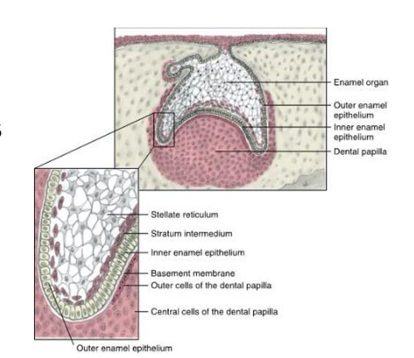
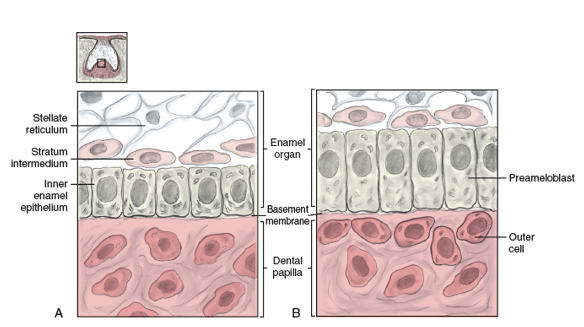
four different types of cells are now formed within the enamel organ
(from outer to inner)
outer enamel epithelium (OEE)
stellate reticulum
stratum intermedium
inner enamel epithelium (IEE)
assumes a three dimensional bell shape
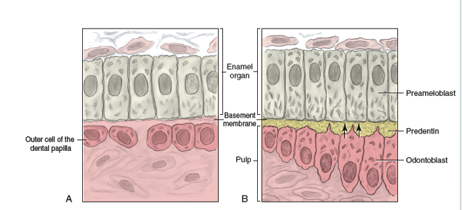
dental papilla cell differentiation
outer cells → become odontoblasts
inner cells → become pulp tissue
dental sac increases amount of collogen fibers forming around enamel organ
dental lamina is disintegrating so tooth continues development divorced from oral epithelium
outer enamel epithelium (OEE)
outer cuboidal cells of the enamel organ
serves as a protective barrier for the rest of the enamel organ during production
inner enamel epithelium (IEE)
innermost tall columnar cells of the enamel organ
will differentiate into enamel-secreting cells known as ameloblasts
basement membrane still remains between the IEE and adjacent dental papilla during bell stage
establishes crown pattern of tooth by ways of folding
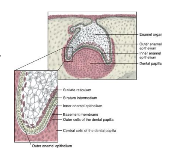
ameloblasts
enamel secreting cells that will form enamel matrix
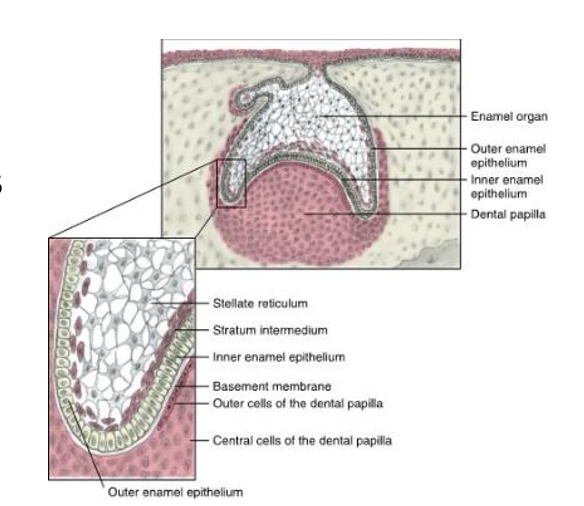
stellate reticulum
consists of star-shaped cells in many layers, primarily located in the center of the enamel organ, and forms a network
synthesize glycosaminoglycans, in which water is drawn in between the cells and stretches them apart
even though they are being stretched, they remain in contact with each other through cell junctions by way of desmosomes
located between the outer and inner enamel epithelium
help support future production of enamel
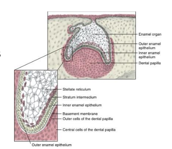
stratum intermedium
made up of a compressed layer of flat to cuboidal cells
help support the future production of enamel
is a layer of two or three cells like a layer cake between IEE and stellate reticulum
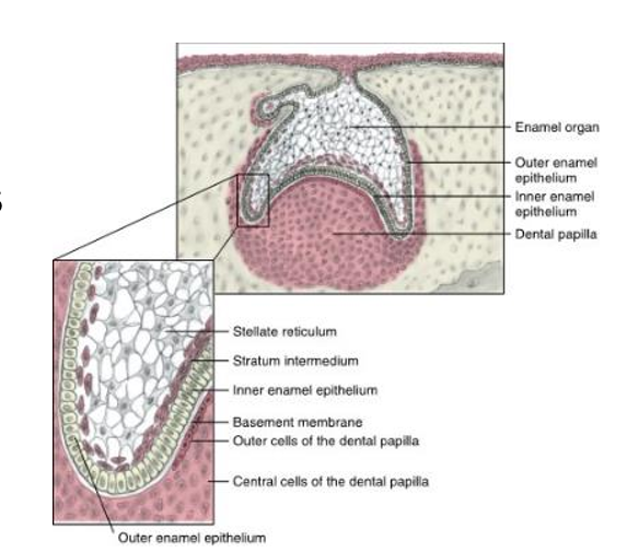
outer cells of dental papilla
become odontoblasts during the bell stage
form dentin
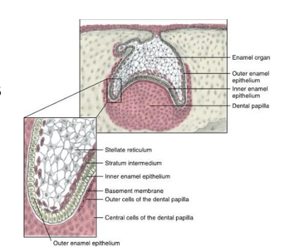
central cells (inner cells) of dental papilla
become primordium of the pulp tissue during the bell stage
apposition stage (secretory stage)
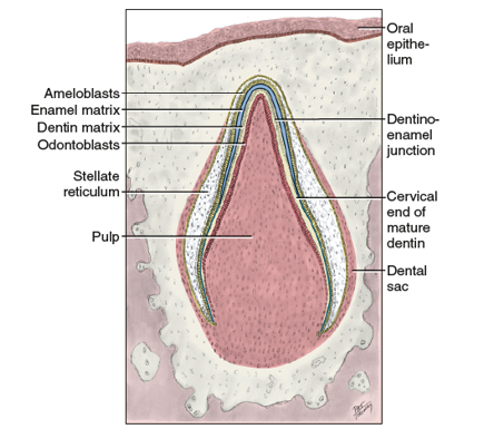
one of the final stages of odontogenesis
enamel, dentin, and cementum are secreted in successive layers upon those already present
originally secreted as a matrix
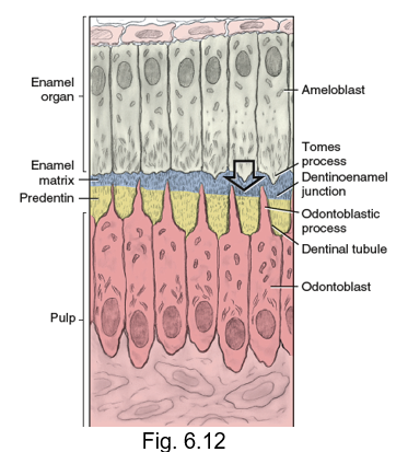
odontoblasts secrete pre-dentin → triggers ameloblasts to form enamel matrix
basement membrane disintegrates, forming the dentinoenamel junction (DEJ)
matrix
an extracellular substance or surrounding medium
serve as frameworks
maturation stage
the final stage of odontogenesis
reached when the matrices of the hard dental tissue types subsequently fully mineralize to their correct levels
enamel, dentin, and cementum fully mineralize
apposition
add
adding layers on top of one another
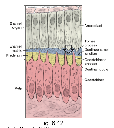
pre-ameloblasts
after the formation of IEE
“baby” or “learning” version of enamel makers
innermost cells grow even more columnar as they elongate and differentiate into __
The nucleus of each cell moves away from the center of the cell to the position furthest away from the basement membrane during cellular repolarization
will induce dental papilla cells to differentiate into dentin-forming cells (odontoblasts) and then will themsleves differentiate into ameloblasts after contacting predentin
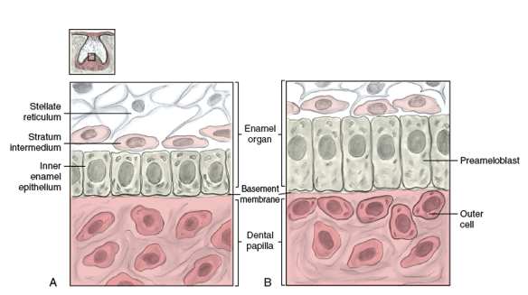
odontoblasts
dentin building cells
located in the outer cells of the dental papilla
induced by the pre-ameloblasts
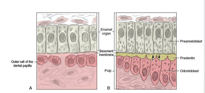
repolarization of cells
nuclei moves from the center of the cell to a position in the cell that is farthest from the basement membrane
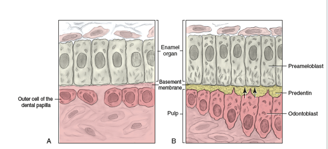
dentinogenesis
The appositional growth of the dentin matrix that will form dentin
amelogenesis
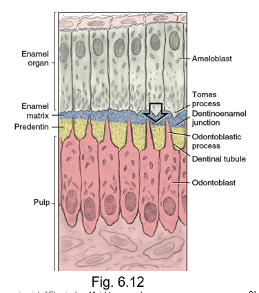
the appositional growth of the enamel matrix
begun by the ameloblasts
laying it down on their side of the now disintegrating basement membrane
tomes process
the enamel matrix is directly secreted from the __ __
an angled distal part of each ameloblast that faces the fully disintegrated basement membrane
dentinoenamel junction
enamel matrix comes in contact with predentin and mineralizes the disintegrating basement membrane
inner junction between the dentin and enamel tissue
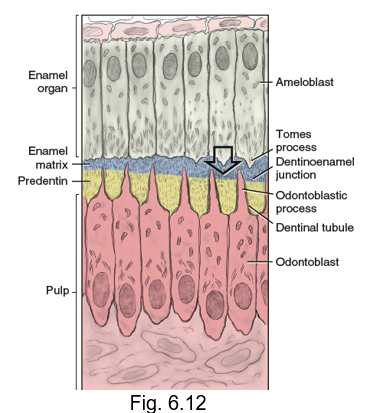
odontoblastic process
odontoblasts leave attached cellular extensions in the length of the predentin called __ __ as they move away from DEJ
each process is contained in a mineralized cylinder, the dentinal tubule
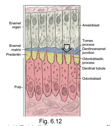
enamel dysplasia
local or systemic
faulty (defective) development of enamel
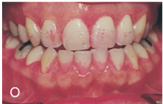
local enamel dysplasia
may result from trauma or infection occurring to a small group of ameloblasts
systemic enamel dysplasia
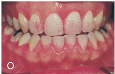
involves larger numbers of ameloblasts that may result from traumatic birth, systemic infections, nutritional deficiencies, or dental flurosis
dental fluorosis
may occur alongside enamel hypoplasia or hypocalcification
hypomineralization occurs due to excessive systemic fluoride level that causes white spots or brown putting
may occur due to oxidative stress to the ameloblast from excess fluroide
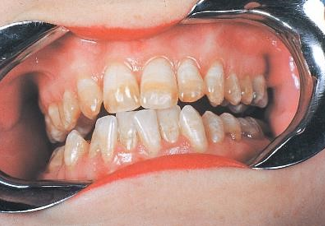
enamel hypoplasia
a type of enamel dysplasia
develops during apposition stage
results from a reduction in the quantity of the enamel matrix
teeth appear with pitting and grooves in the enamel surface or in the development of horizontal lines across the enamel of the crown
noted in the presence of Hutchinson incisors and mulberry molars
enamel hypocalcification
A disturbance developed in the apposition stage that results in a reduction in the quality of the enamel maturation
teeth appear more opaque, yellower, or even browner within because of an intrinsic staining of enamel
could have “turner spots” that are single affected areas, or could affect the entire crown
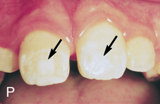
amelogenesis imperfecta
a type of enamel dysplasia
has a hereditary etiology and can affect all teeth of both dentitions
teeth either have no enamel or very thin enamel that chups off, leaving yellow crowns of dentin
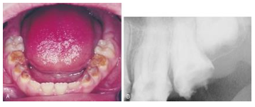
dentin dysplasia
faulty development of dentin
result of interference with the metabolic processes of odontoblasts during dentinogenesis
more rare than enamel dysplasia but can be caused by systemic factors
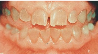
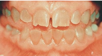
dentinogenesis imperfecta
a type of dentin dysplasia
a hereditary basis
blue-gray or brown teeth with opalescent sheen
root development begins long after __
the crown is completely shaped and the tooth is starting to erupt into the oral cavity
tooth is formed starting from crown then moving to apex of root
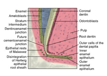
cervical loop
structure responsible for root development
the most cervical part of enamel organ
bilayer rim that consists of only inner enamel epithelium (IEE) and outer enamel epithelium (OEE)
grows deeper into surrounding ectomesenchyme of dental sac and forms the Hertwig epithelial root sheath (HER)
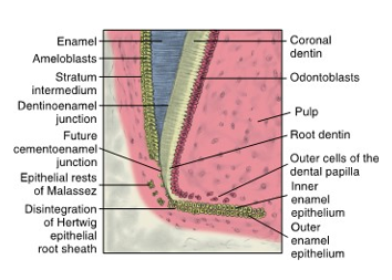
Hertwig epithelial root sheath
__
function is to shape the root(s) by inducing dentin formation in the root area, so that it is continuous with coronal dentin
will determine if the root will be curved or straight, short or long as well as single or multiple
there is differential growth of __ that causes the root trunk of each multirooted tooth to divide into two or three roots
induces odontoblastic differentiation but fails to differentiate into enamel-forming ameloblasts (no enamel in roots)
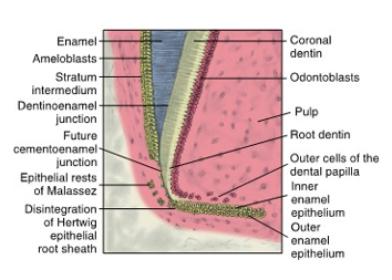
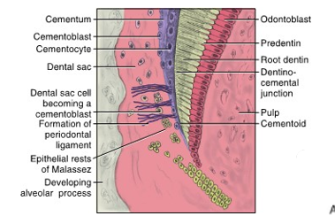
epithelial rests of Malassez (ERM)
After the disintegration of Hertwig epithelial root sheaths, these cells may possibly form
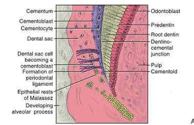
cementogenesis
The process of forming the cementum that surrounds the root area of a tooth
occurs when HERS disintegrates
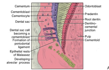
cementoblasts
cells that build the cemetum
form after contact of dental sac cells with dentin surface
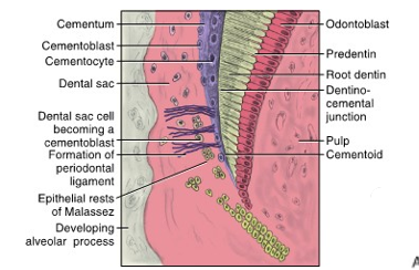
cementocytes
many cementoblasts become entrapped by the cementum they produce and become mature __
surrounded and entrapped by cementoid, and once they mineralize, they make cementum
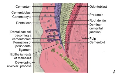
cementoid
cementoblasts move to cover the root dentin area and undergo cementogenesis, laying down cementum matrix, also known as __
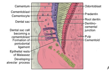
dentinocemental junction (DEJ)
basement membrane disintegrates and _ is formed where the membrane was located
a result of the apposition of cementum over the dentin
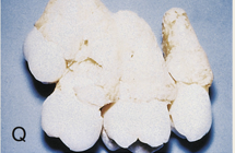
concrescence
excess cementum formation that rarely occurs
con man produced too much cementum
enamel pearl
misplaced ameloblasts migrated to the surface of root by localized failure of HERs to separate from dentin
enamel ectopically forms over cemental root surface
small spherical enamel projection on root especially at CEJ