Cartilage (SDL)

0.0(0)
Studied by 0 people
call kaiCall Kai
learnLearn
examPractice Test
spaced repetitionSpaced Repetition
heart puzzleMatch
flashcardsFlashcards
GameKnowt Play
Card Sorting

1/18

encourage image

There's no tags or description

Looks like no tags are added yet.

Last updated 12:20 AM on 5/29/26
Name
Mastery
Learn
Test
Matching
Spaced
Call with Kai

No analytics yet

Send a link to your students to track their progress

19 Terms
1
New cards
<p>Part 1</p>

Part 1

knowt flashcard image
2
New cards
term image

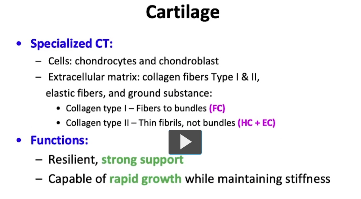
Cartilage is a specialized connective tissue. It has characteristic cells called chondrocytes and chondroblasts. It has a matrix that surrounds the cells known as the extracellular matrix, which has very specific components, such as collagen fibers, type I and type II. As well as elastic fibers, and what is known as ground substance.

Normally collagen type I is found as fibers to bundles in the so called fiber cartilage, and collagen type II, which is the term for collagen fibrils, and contains no ramifications in a branching sense as if they were fasicles. They won’t be seen in collagen type II, and this will be present in hyaline cartilage and elastic cartilage.

Cartilage tissue is superbly strong, very good for resilience. It’s strong and at the same time, within it’s strength, it has a certain degree of flexibility. It is also capable of growth and development and in a moment you will see these mechanisms of reproduction.
<p>Cartilage is a specialized connective tissue. It has characteristic cells called chondrocytes and chondroblasts. It has a matrix that surrounds the cells known as the extracellular matrix, which has very specific components, such as collagen fibers, type I and type II. As well as elastic fibers, and what is known as ground substance. </p><p>Normally collagen type I is found as fibers to bundles in the so called fiber cartilage, and collagen type II, which is the term for collagen fibrils, and contains no ramifications in a branching sense as if they were fasicles. They won’t be seen in collagen type II, and this will be present in hyaline cartilage and elastic cartilage.</p><p>Cartilage tissue is superbly strong, very good for resilience. It’s strong and at the same time, within it’s strength, it has a certain degree of flexibility. It is also capable of growth and development and in a moment you will see these mechanisms of reproduction. </p>
3
New cards
<p>On screen we have an image of a vertebral column, and these blue structures I’m pointing to is where we have famous intervertebral discs. </p><p>It’s a structure made of cartilage. It’s a good tissue for support as well as compression. </p><p>As the column moves, these structures function as cushioning and are resistant to the pressure column is subjected to during movement. In the same manner, the hyaline cartilage is the base model used to form the axial skeleton of our skeletal system. </p>

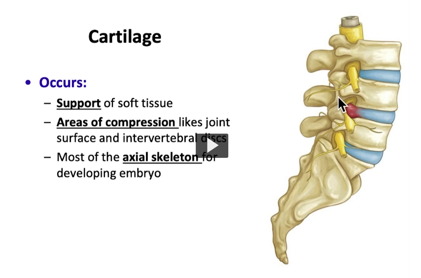
On screen we have an image of a vertebral column, and these blue structures I’m pointing to is where we have famous intervertebral discs.

It’s a structure made of cartilage. It’s a good tissue for support as well as compression.

As the column moves, these structures function as cushioning and are resistant to the pressure column is subjected to during movement. In the same manner, the hyaline cartilage is the base model used to form the axial skeleton of our skeletal system.

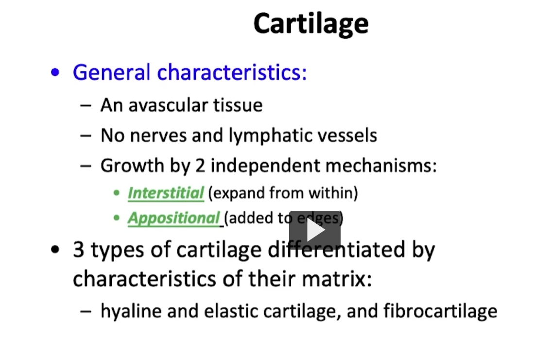
Cartilage is an avascular tissue without nerves or lymphatic vessels. The curious thing about cartilage is that it has two mechanisms it uses to reproduce itself.

What is known as the interstitial mechanism and the appositional mechanism. It’s interesting because cartilage is a specialized connective tissue. Remember that connective tissue is characterized by plenty of vascularity and innervation, and cartilage does not have that.

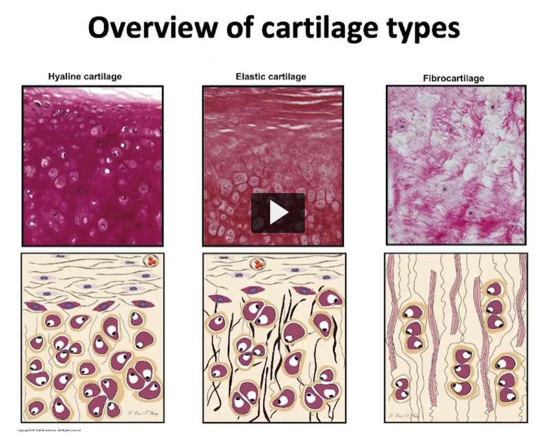
Very similar to the characteristics of epithelial tissue, cartilage can be divided into three modalities.
<p>Cartilage is an avascular tissue without nerves or lymphatic vessels. The curious thing about cartilage is that it has two mechanisms it uses to reproduce itself. </p><p>What is known as the interstitial mechanism and the appositional mechanism. It’s interesting because cartilage is a specialized connective tissue. Remember that connective tissue is characterized by plenty of vascularity and innervation, and cartilage does not have that. </p><p>Very similar to the characteristics of epithelial tissue, cartilage can be divided into three modalities.  </p>
4
New cards
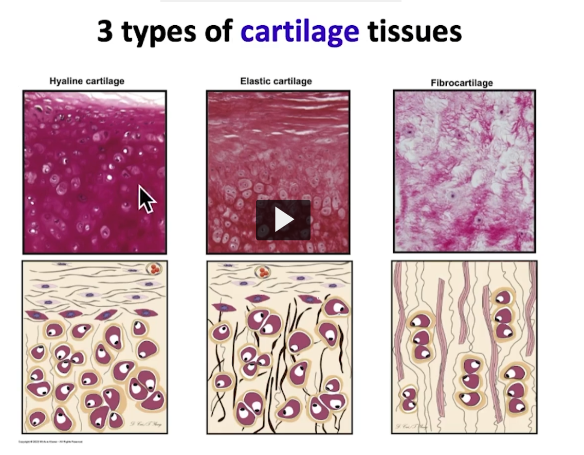
<p>For example, hyaline cartilage, elastic cartilage, and fibro cartilage </p><p>Where can I see hyaline cartilage? In the anterior portion of your nose, there is a mass of hyaline cartilage. The costal cartilage of your rib cage, where you join the rib to the sternum in the thoracic cage </p><p>The elastic cartilage contains elastic fibers in its extracellular matrix, it can be seen in the auricle of your external ear. </p><p>Finally, fibrocartilage that can be found in intervertebral discs and in the meniscus. </p><p>She is going to explain the characteristics of cartilage. We will start with hyaline and eventually we will discuss the other two, I will show you what makes each one of them distinctive. which I think is the easiest way to visualize these three types of cartilage. </p>

For example, hyaline cartilage, elastic cartilage, and fibro cartilage

Where can I see hyaline cartilage? In the anterior portion of your nose, there is a mass of hyaline cartilage. The costal cartilage of your rib cage, where you join the rib to the sternum in the thoracic cage

The elastic cartilage contains elastic fibers in its extracellular matrix, it can be seen in the auricle of your external ear.

Finally, fibrocartilage that can be found in intervertebral discs and in the meniscus.

She is going to explain the characteristics of cartilage. We will start with hyaline and eventually we will discuss the other two, I will show you what makes each one of them distinctive. which I think is the easiest way to visualize these three types of cartilage.

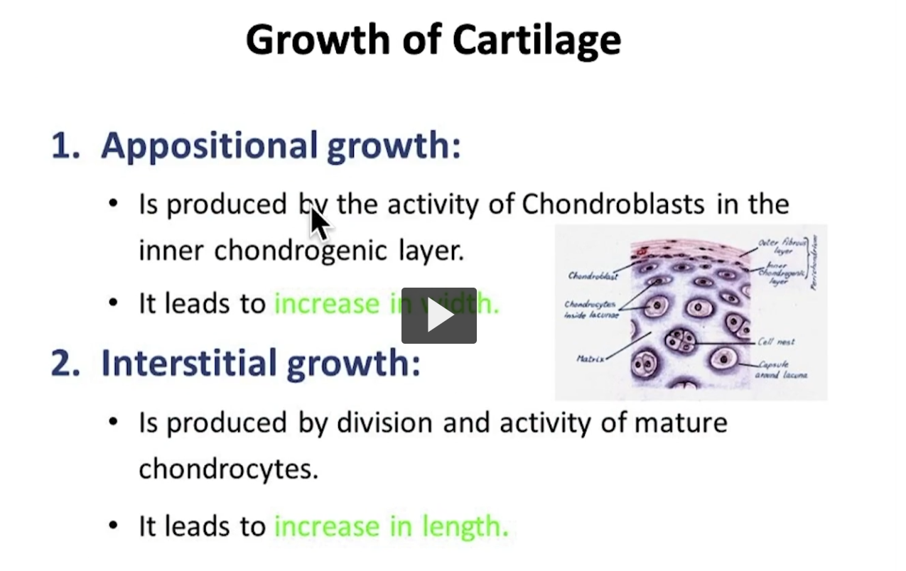
it’s interesting to see how cartilage can develop and grow. Cartilage has two types of mechanism of growth. Interstitial growth and appositional growth.

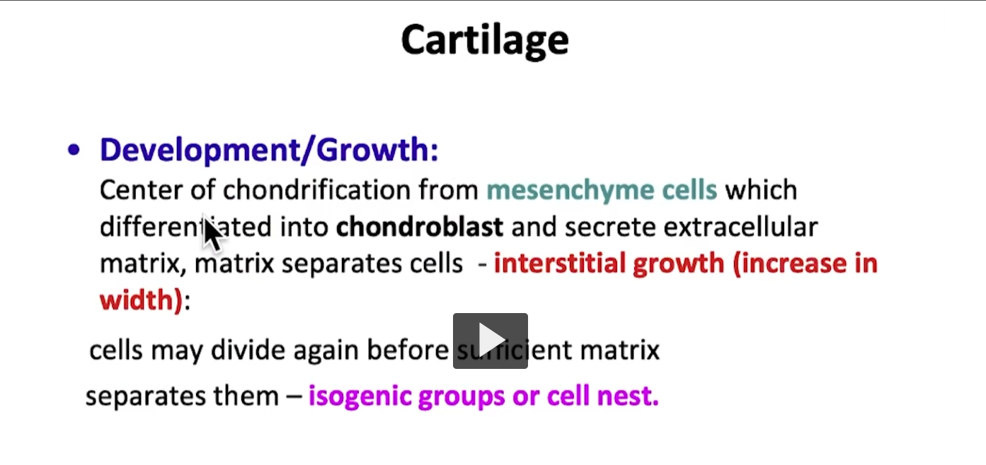
Let’s begin with interstitial growth. Remember, we are going to focus on hyaline cartilage but in the same manner it can be used for elastic cartilage and fibrocartilage. There is no doubt that cartilage comes from embryonic tissue, from mesenchymal cells that later differentiate into chondroblasts.

These chondroblasts will then secrete the extracelluar matrix of cartilage, and once submerged and surrounded within the extracellular matrix, we begin forming chondrocytes to obtain mature cartilage tissue.

Once the chondrocyte is established, the chondrocytes begin to divide by mitosis to maintain a population of cartilage within that mature cartilage tissue, and consequently maintain cartilage.

Basically, it’s been found that the interstitial growth which is where chondrocytes reproduce themselves, increases the width of the cartilage tissue.

One of the most peculiar characteristics you will see in cartilage tissue is isogenic groups. These groups arise and originate from a common origin and are a product of the mitotic cell division process.

The second process of division or reproduction is called appositional growth, which favors longitudinal growth of cartilage. Basically it’s an avascular tissue, it contains in its periphery dense connective tissue. That dense connective tissue, because it is a type of connective tissue, is highly vascularized and can supply the nutrients needed to cartilage by way of a diffusion mechanism. That connective tissue that is found in the periphery is known as perichondrium. You will notice that normally, in the periphery of cartilage, you will see the presence of this type of dense connective tissue known as perichondrium. The perichondrium helps with appositional growth. How does it help? It’s been found that the perichondrium is subdivided in an external layer, known as the fibrous layer, which is a classic type of dense connective tissue, and an internal layer, that we call the chondrogenic layer. Why chondrogenic? It has been found that this layer, which is the most proximal to the cartilage contains fibroblasts, which is a typical cell found in connective tissue.

The fibroblast differentiates into chondroblasts, when then differentiate into chondrocytes that we have established the chondrocyte and we have established the growth of cartilage.

The perichondrium is well associated with hyaline cartilage, it is associated with elastic cartilage, but we must emphasize there is no perichondrium in fibrocartilage. Those are areas with a lot of compression is not ideal place vascularized structures in those types of areas with high compression. For example, the intervertebral discs, basically, in the same manner, it has been found that the articular cartilage that covers the ends of long bones in the articulation surface is made up of hyaline cartilage. Similarly it is also an area that does not contain perchondrium. Where can you have perchondrium? In hyaline cartilage, except for the articular surfaces of long bones. There is perichondrium and elastic cartilage and there is no perichondrium in fibrocartilage.
<p>it’s interesting to see how cartilage can develop and grow. Cartilage has two types of mechanism of growth. Interstitial growth and appositional growth.</p><p>Let’s begin with interstitial growth. Remember, we are going to focus on hyaline cartilage but in the same manner it can be used for elastic cartilage and fibrocartilage. There is no doubt that cartilage comes from <strong>embryonic tissue</strong>, from <strong>mesenchymal cells </strong>that later differentiate into chondroblasts.</p><p>These chondroblasts will then secrete the extracelluar matrix of cartilage, and once submerged and surrounded within the extracellular matrix, we begin forming chondrocytes to obtain mature cartilage tissue.</p><p>Once the chondrocyte is established, the chondrocytes begin to divide by mitosis to maintain a population of cartilage within that mature cartilage tissue, and consequently maintain cartilage.</p><p>Basically, it’s been found that the interstitial growth which is where chondrocytes reproduce themselves, increases the width of the cartilage tissue.</p><p>One of the most peculiar characteristics you will see in cartilage tissue is isogenic groups. These groups arise and originate from a common origin and are a product of the mitotic cell division process.</p><p>The second process of division or reproduction is called appositional growth, which favors longitudinal growth of cartilage. Basically it’s an avascular tissue, it contains in its periphery dense connective tissue. That dense connective tissue, because it is a type of connective tissue, is highly vascularized and can supply the nutrients needed to cartilage by way of a diffusion mechanism. That connective tissue that is found in the periphery is known as perichondrium. You will notice that normally, in the periphery of cartilage, you will see the presence of this type of dense connective tissue known as perichondrium. The perichondrium helps with appositional growth. How does it help? It’s been found that the perichondrium is subdivided in an external layer, known as the fibrous layer, which is a classic type of dense connective tissue, and an internal layer, that we call the chondrogenic layer. Why chondrogenic? It has been found that this layer, which is the most proximal to the cartilage contains fibroblasts, which is a typical cell found in connective tissue.</p><p>The fibroblast differentiates into chondroblasts, when then differentiate into chondrocytes that we have established the chondrocyte and we have established the growth of cartilage. </p><p>The perichondrium is well associated with hyaline cartilage, it is associated with elastic cartilage, <strong>but we must emphasize there is no perichondrium in fibrocartilage. </strong>Those are areas with a lot of compression is not ideal place vascularized structures in those types of areas with high compression. For example, the intervertebral discs, basically, in the same manner, it has been found that the articular cartilage that covers the ends of long bones in the articulation surface is made up of hyaline cartilage. Similarly it is also an area that does not contain perchondrium. Where can you have perchondrium? In hyaline cartilage, except for the articular surfaces of long bones. There is perichondrium and elastic cartilage and there is no perichondrium in fibrocartilage. </p><p></p>
5
New cards
<p>This slide is a summary so that you can review the interstitial and appositional growth. </p>

This slide is a summary so that you can review the interstitial and appositional growth.

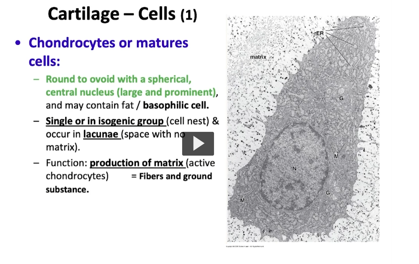
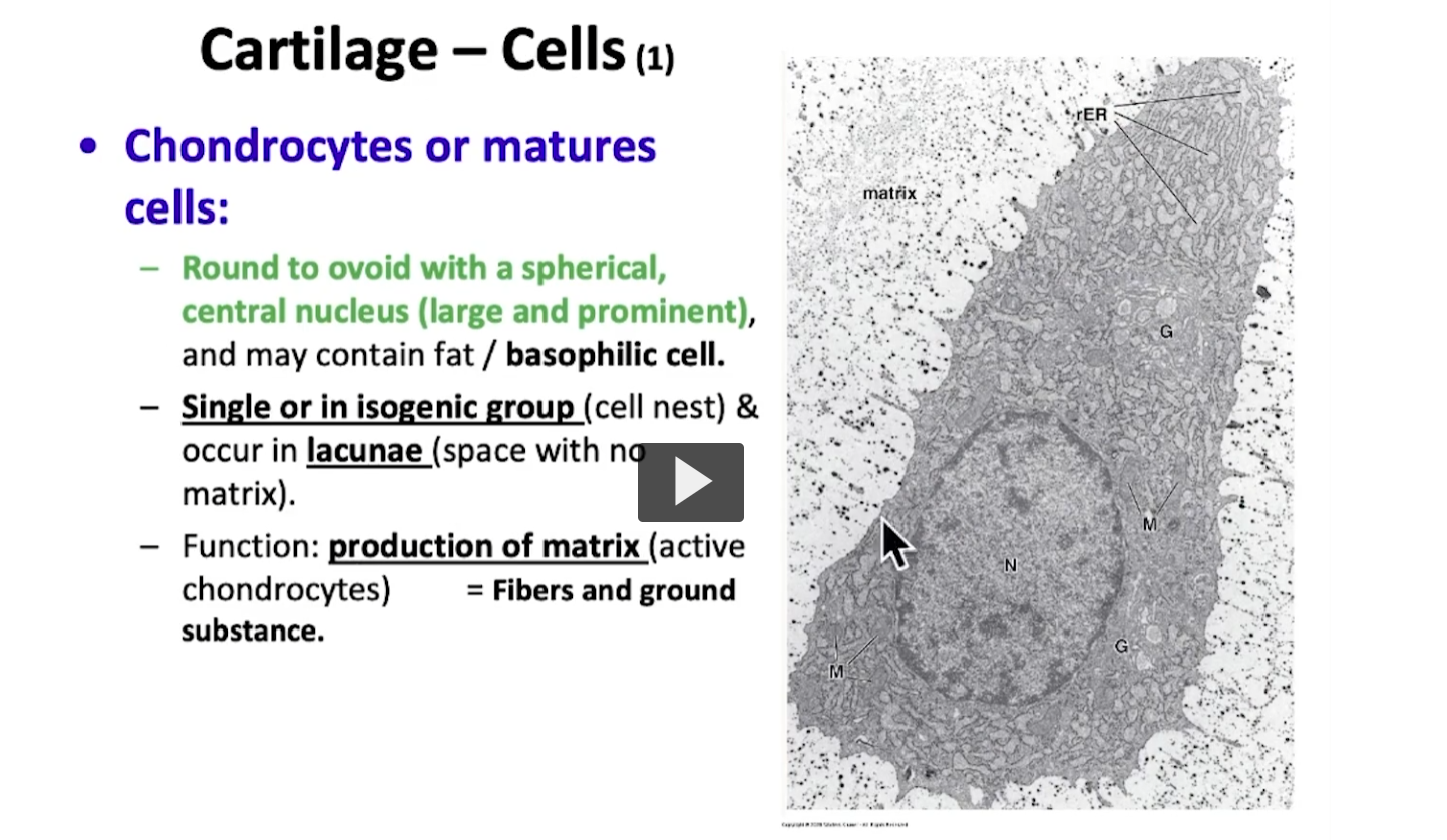
In this slide, we will begin talking about the cells. In this electron micrograph, we have a chondrocyte. A typical cell of mature cartilage tissue. It is a big cell, a round or oval shaped central spherical nucleus and contains all of the typical organelles of a cell. It also has a basophilic nucleus within its cytoplasm, containing a rough endoplasmic reticulum and Golgi Apparatus because the chondrocyte is responsible for synthesizing the matrix of cartilage tissue. The chondrocytes are well associated with isogenous groups and you will also see them in what is known as lacunae.

You will be able to observe that every chondrocyte creates a space within the matrix, and that space where the chondrocyte is placed is named a lacunae.
<p>In this slide, we will begin talking about the cells. In this electron micrograph, we have a chondrocyte. A typical cell of mature cartilage tissue. It is a big cell, a round or oval shaped central spherical nucleus and contains all of the typical organelles of a cell. It also has a basophilic nucleus within its cytoplasm, containing a rough endoplasmic reticulum and Golgi Apparatus because the chondrocyte is responsible for synthesizing the matrix of cartilage tissue. The chondrocytes are well associated with isogenous groups and you will also see them in what is known as lacunae. </p><p>You will be able to observe that every chondrocyte creates a space within the matrix, and that space where the chondrocyte is placed is named a lacunae.  </p>
6
New cards
<p>you will see in this other image a chondrocyte. This chondrocyte as you can see is oval shaped, a prominent nucleus that is centric and spherical and contains a numerous amount of organelles and scaffolding needed for it to succeed in its function of synthesizing  the extracellular matrix. </p>

you will see in this other image a chondrocyte. This chondrocyte as you can see is oval shaped, a prominent nucleus that is centric and spherical and contains a numerous amount of organelles and scaffolding needed for it to succeed in its function of synthesizing the extracellular matrix.

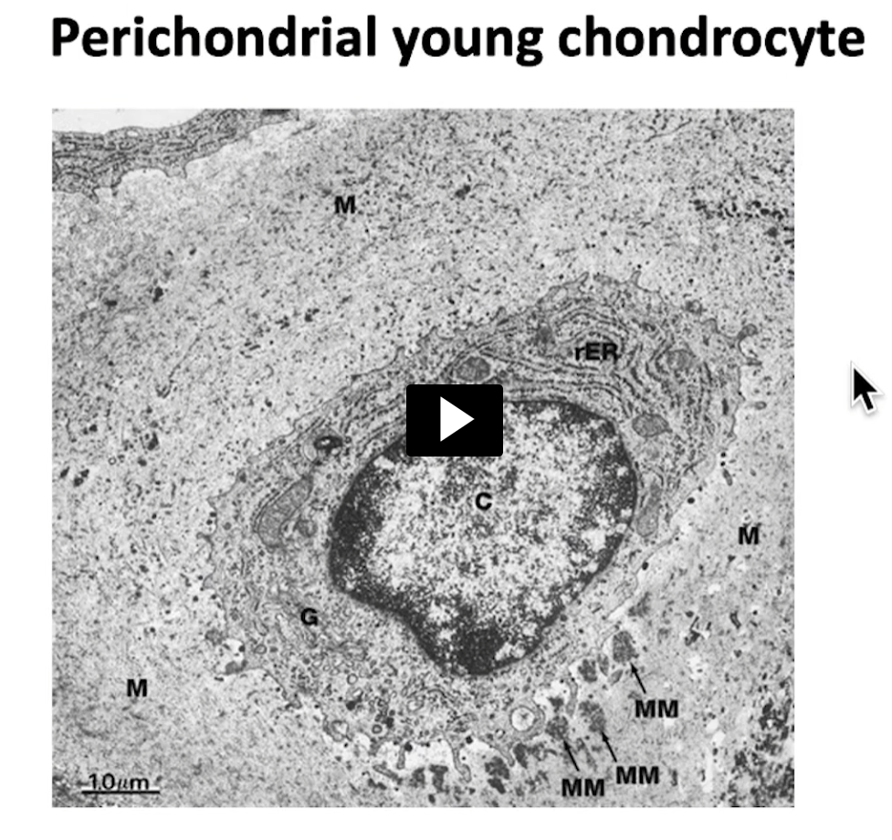
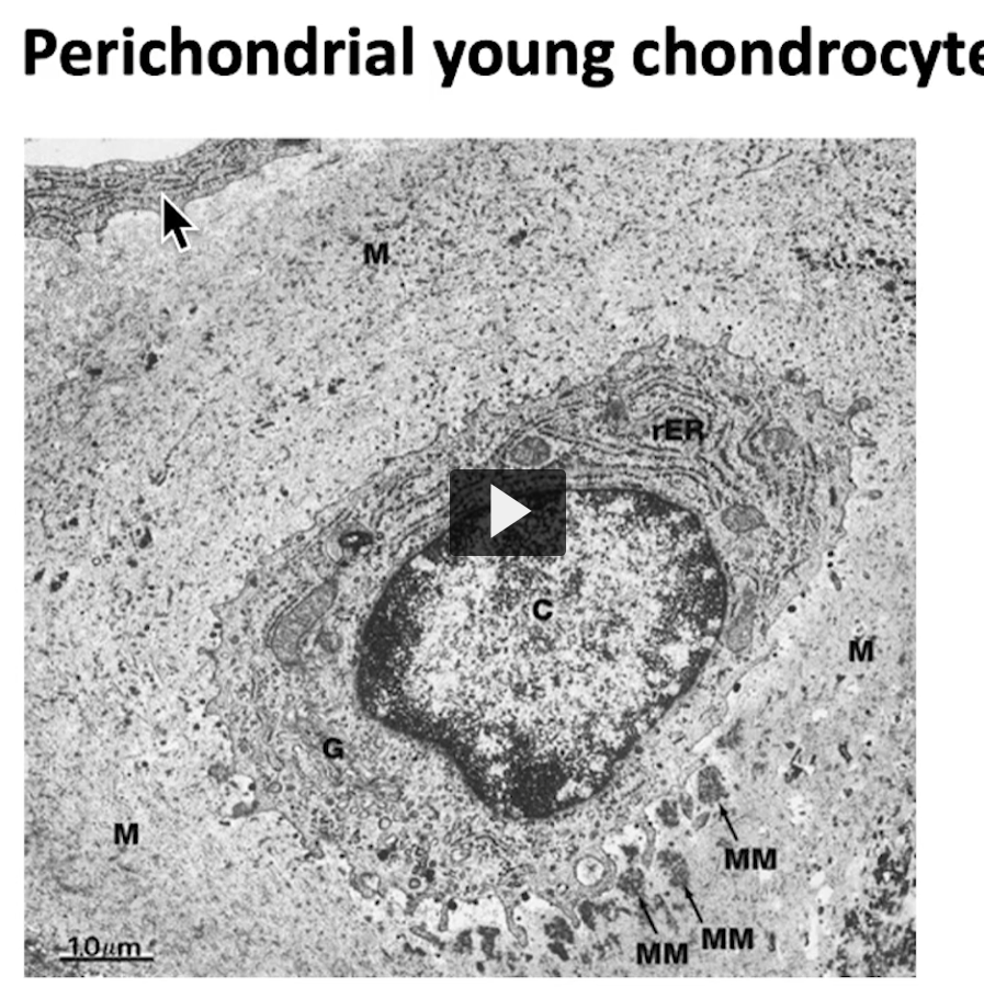
It’s interesting because if I go to this image, which is an electron micrograph, we can see this small cell, where I’m pointing at with the mouse cursor, this cell is a fibroblast. This means that this is a young chondrocyte, it is a product of that appositional growth mechanism we discussed earlier, sourced from perchondrium. In a similar manner, another cell, is a chondroblast
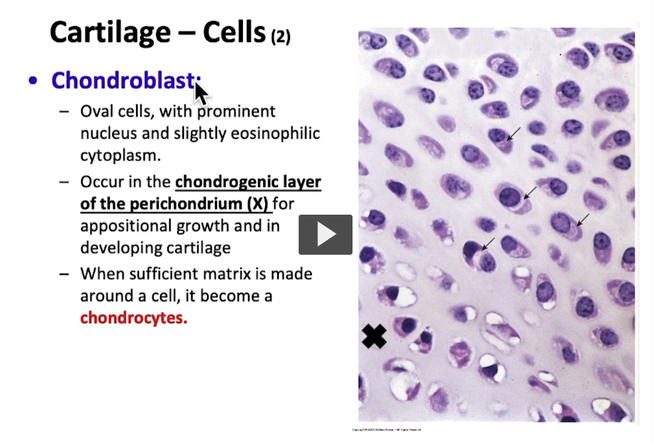
<p>It’s interesting because if I go to this image, which is an electron micrograph, we can see this small cell, where I’m pointing at with the mouse cursor, this cell is a fibroblast. This means that this is a young chondrocyte, it is a product of that appositional growth mechanism we discussed earlier, sourced from perchondrium. In a similar manner, another cell, is a chondroblast </p>
7
New cards
<p>If I wish to see a chondroblast, I must go direclty to the perichondrium, specifically the chondrogenic layer. </p>

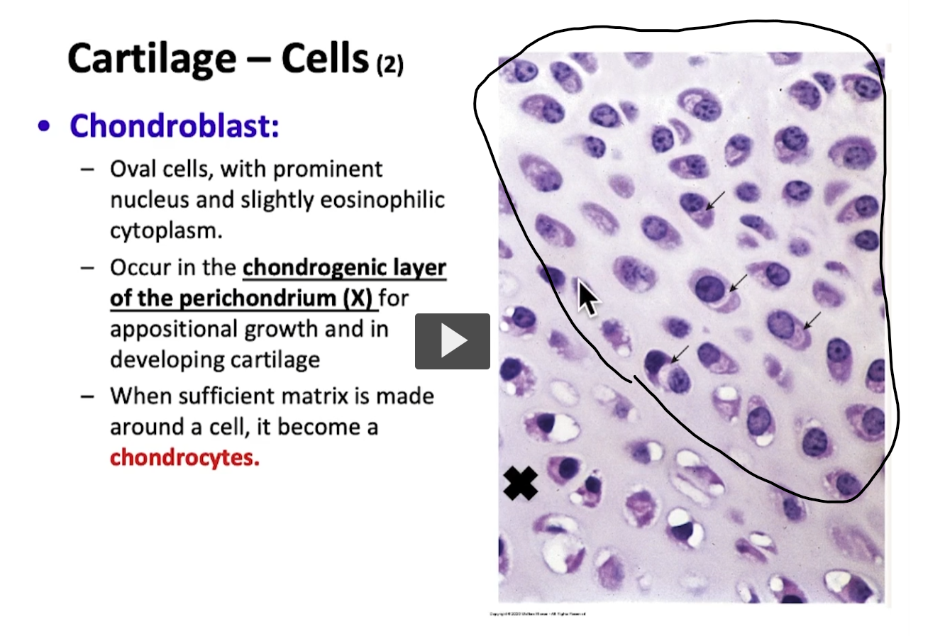
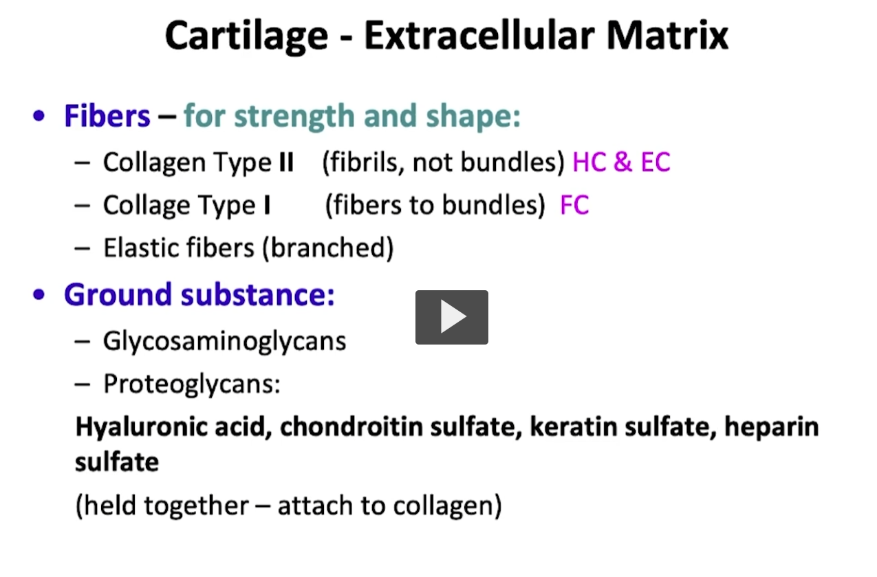
If I wish to see a chondroblast, I must go direclty to the perichondrium, specifically the chondrogenic layer.

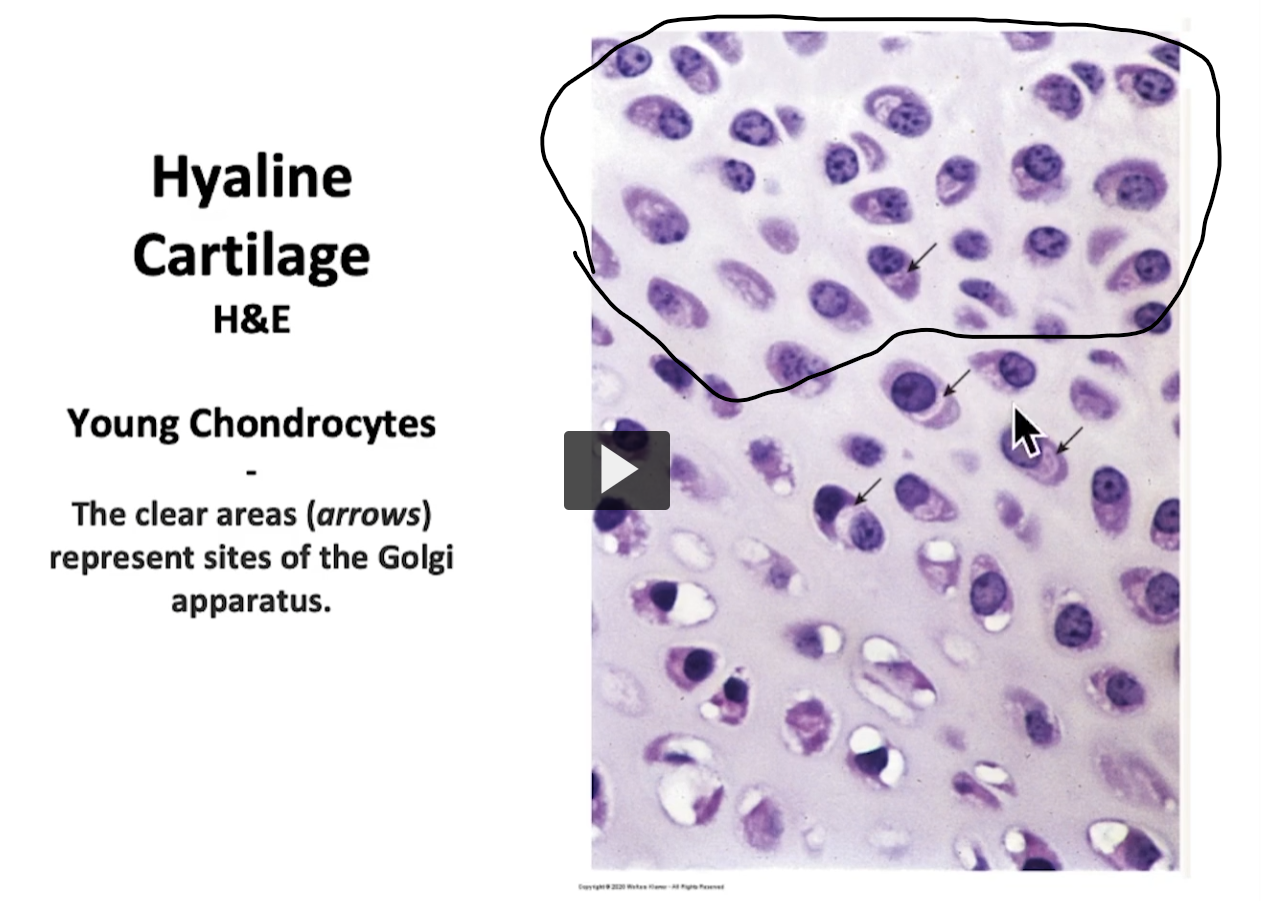
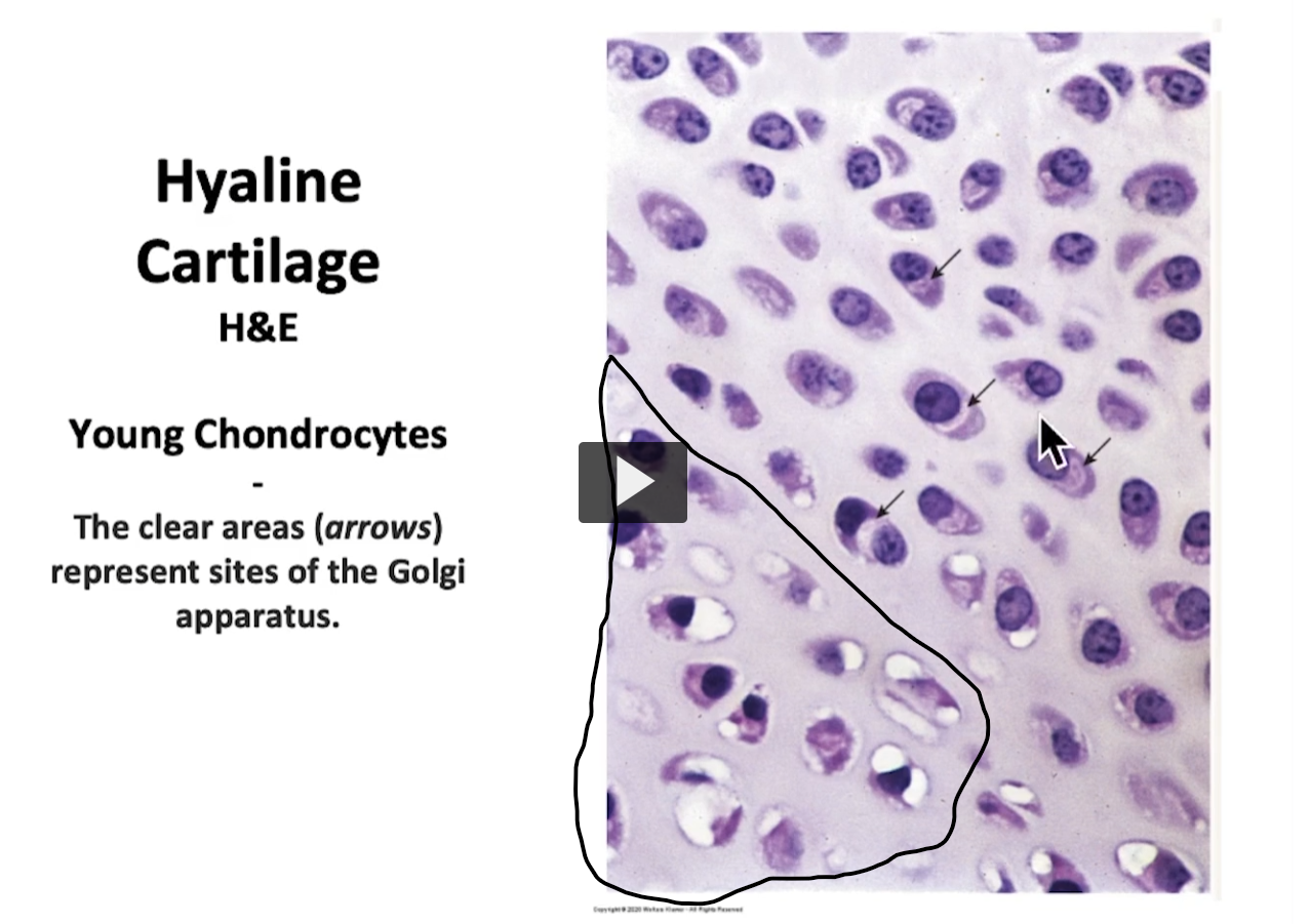
This is the area of mature chondrocytes.

As we can this photomicrograph, This area that Im pointing with, the arrow is the area of mature cartilage. Here we have mature chondrocytes. Interestingly, we can observe these small arrows indicating a clear area near the nucleus which is negative reaction from Golgi that is seen in cells.

The area demarcated by the black x is part of the perichondrium.

Possibly, if I wish to see chondroblasts, I must go to the area of the perichondrium where the cells being developed are chondroblasts that eventually become chondrocytes once surrounded by a matrix and produce cartilage mass.
<p>This is the area of mature chondrocytes. </p><p>As we can this photomicrograph, This area that Im pointing with, the arrow is the area of mature cartilage. Here we have mature chondrocytes. Interestingly, we can observe these small arrows indicating a clear area near the nucleus which is negative reaction from Golgi that is seen in cells. </p><p>The area demarcated by the black x is part of the perichondrium. </p><p>Possibly, if I wish to see chondroblasts, I must go to the area of the perichondrium where the cells being developed are chondroblasts that eventually become chondrocytes once surrounded by a matrix and produce cartilage mass. </p>
8
New cards
<p>When we refer to the cartilage extracellular matrix, remember that the matrix is an element made up of collagen fibers. </p><p>I mentioned earlier that if it is collagen type II fibers, it is typically found in elastic fibers and hyaline cartilage. </p><p>If it is collagen type I, you will find it in fibrocartilage. In the same manner, another type of fiber that is present in the matrix of cartilage tissue are elastic fibers. These fibers are important to maintain the strength, the flexibility and consistency of this tissue. </p><p>These fibers are going to be surrounded by grounded substance which is normally composed of glycosaminoglycans and proteoglycans that are important substances for the adhesion of components of the matrix. </p><p>They provide strength and rigidity to the matrix. Basically, within the group of proteoglycans, when speaking of cartilage, we can metnioned a few hyaluronic acid, chondroitin sulfate, keratin sulfate, and heparin sulfate. </p><p>It is interesting because these types of proteoglycans are important to maintain a good union of the collagen within the matrix other components of the same matrix in order provide resistance to that cartilage. </p>

When we refer to the cartilage extracellular matrix, remember that the matrix is an element made up of collagen fibers.

I mentioned earlier that if it is collagen type II fibers, it is typically found in elastic fibers and hyaline cartilage.

If it is collagen type I, you will find it in fibrocartilage. In the same manner, another type of fiber that is present in the matrix of cartilage tissue are elastic fibers. These fibers are important to maintain the strength, the flexibility and consistency of this tissue.

These fibers are going to be surrounded by grounded substance which is normally composed of glycosaminoglycans and proteoglycans that are important substances for the adhesion of components of the matrix.

They provide strength and rigidity to the matrix. Basically, within the group of proteoglycans, when speaking of cartilage, we can metnioned a few hyaluronic acid, chondroitin sulfate, keratin sulfate, and heparin sulfate.

It is interesting because these types of proteoglycans are important to maintain a good union of the collagen within the matrix other components of the same matrix in order provide resistance to that cartilage.

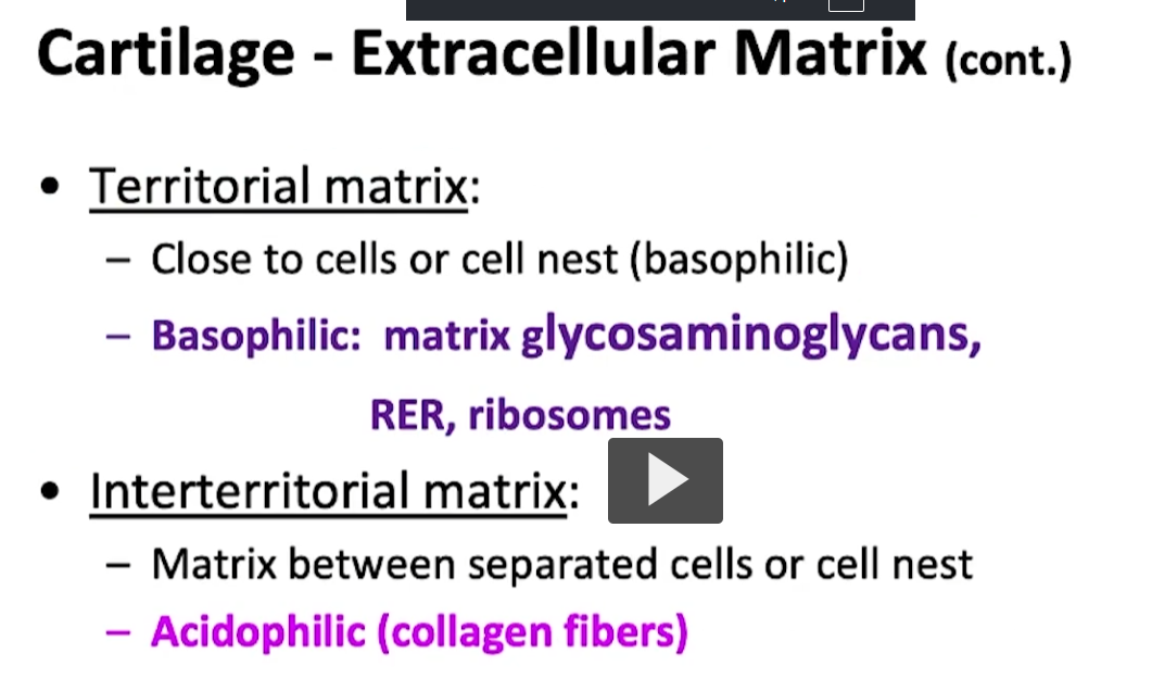
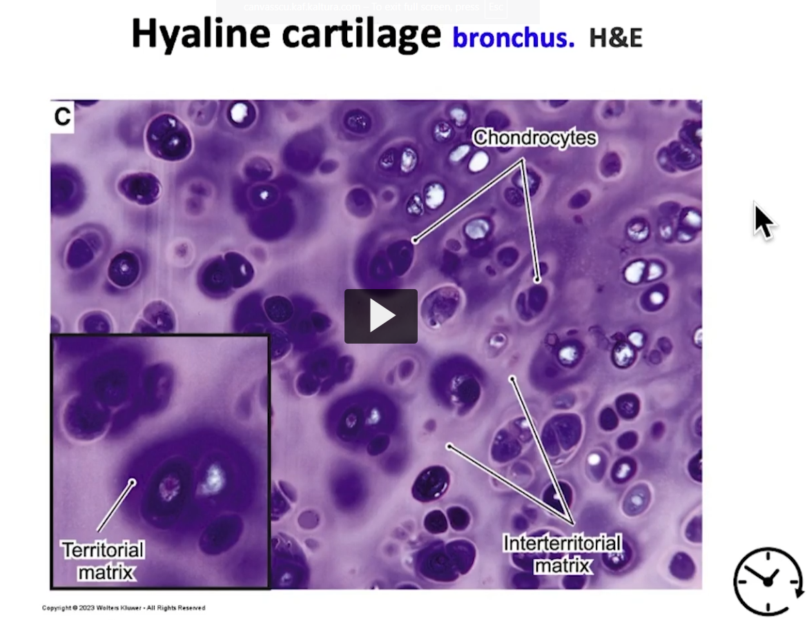
When you see photos of cartilage, you need to also focus on what is known as territorial matrix and interterritorial matrix. Remember that the extracellular is composed of fibers and ground substance. There is a matrix that is proximal to the chondrocytes.

Basically, you’ll see many isogenous groups or isolated chondrocytes surrounded by a matrix, essentially a very basophilic area, which is known as the territorial matrix, The basophilia is due to the presence of the rough endoplasmic reticulum, free ribosomes, and glycosaminoglycans found in the territorial matrix, as well as from the chondrocyte, while carrying out its function of synthesizing that matrix. It is a dark area, literally, surrounding the periphery proximal to the chondrocyte or the group of chondrocytes.

As you will see, the interterritorial matrix, which is separate from the isogenous groups, between those isogenic groups or between those chondrocyte groups, is more of an acidophilic area due to the abundant presence of collagen fibers. It is our responsibility to identify those components of the matrix in what is supposed to be the territorial and the interterritorial matrix.
<p>When you see photos of cartilage, you need to also focus on what is known as territorial matrix and interterritorial matrix. Remember that the extracellular is composed of fibers and ground substance. There is a matrix that is proximal to the chondrocytes. </p><p>Basically, you’ll see many isogenous groups or isolated chondrocytes surrounded by a matrix, essentially a very basophilic area, which is known as the territorial matrix, The basophilia is due to the presence of the rough endoplasmic reticulum, free ribosomes, and glycosaminoglycans found in the territorial matrix, as well as from the chondrocyte, while carrying out its function of synthesizing that matrix. It is a dark area, literally, surrounding the periphery proximal to the chondrocyte or the group of chondrocytes. </p><p>As you will see, the interterritorial matrix, which is separate from the isogenous groups, between those isogenic groups or between those chondrocyte groups, is more of an acidophilic area due to the abundant presence of collagen fibers. It is our responsibility to identify those components of the matrix in what is supposed to be the territorial and the interterritorial matrix. </p>
9
New cards
<p>As she said, there are three types of cartilage tissues. Hyaline, elastic, and fibrocartilage. We will begin with hyaline cartilage, the most common type. </p>

As she said, there are three types of cartilage tissues. Hyaline, elastic, and fibrocartilage. We will begin with hyaline cartilage, the most common type.

Where can I find hyaline cartilage? As she mentioned before, you can find it in the anterior portion of the nose, in costal cartilage, the ends of long bones, in the articular surfaces (known as articular cartilage), as well as in the rings of the trachea and the walls of the larynx.

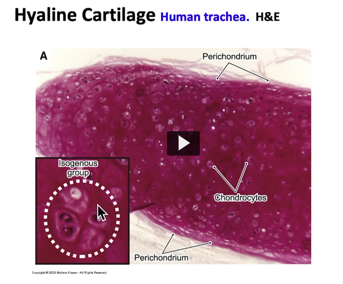
This cartilage, if I use this image (the one on the slide) as a reference, we can see the component of perichondrium with a fibrous layer (yellow stuff at the top), and with a chondrogenic layer. Here we can see the mass of mature chondrocytes. Some are isolated chondrocytes in their lacunae, while others are forming isogenous groups, as demarcated by this separate group within the circle.
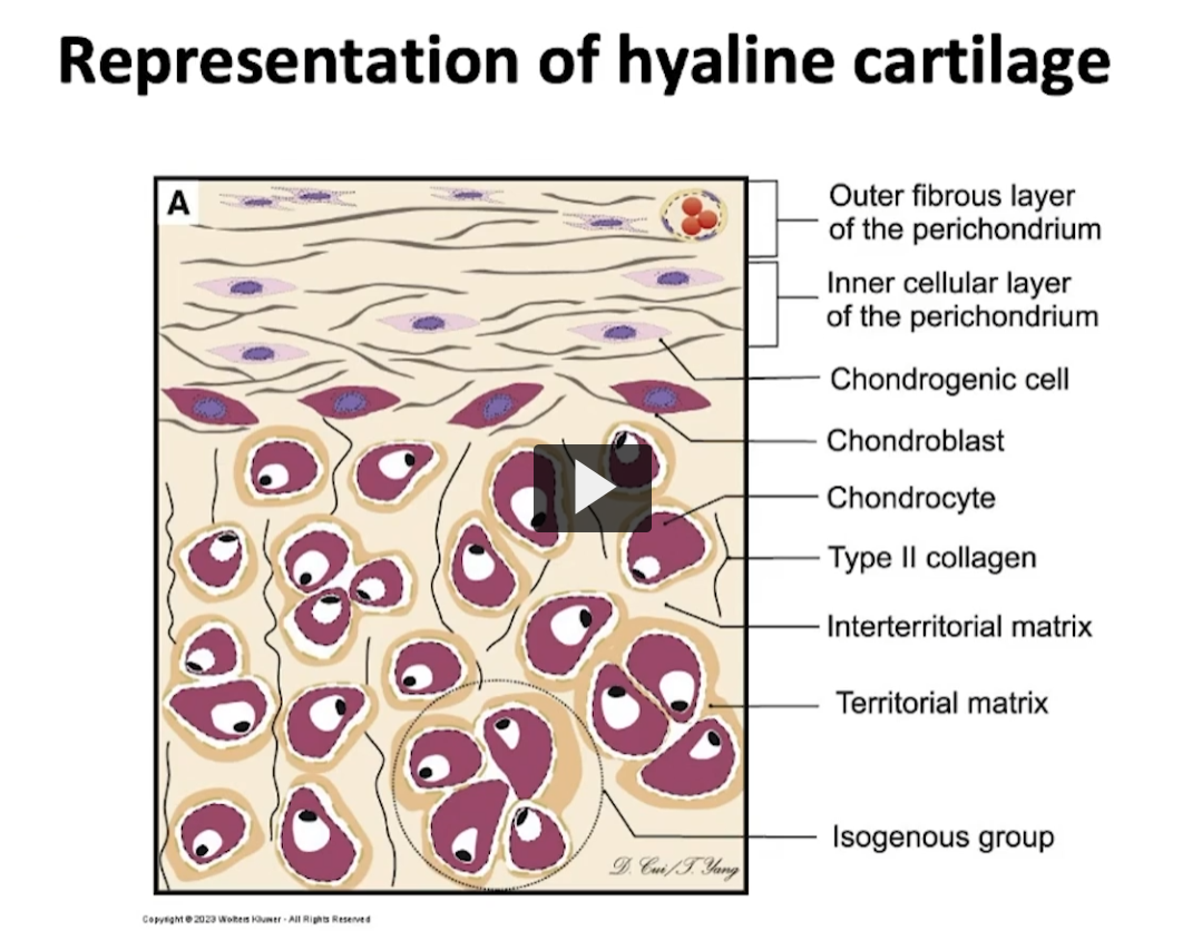
<p>Where can I find hyaline cartilage? As she mentioned before, you can find it in the anterior portion of the nose, in costal cartilage, the ends of long bones, in the articular surfaces (known as articular cartilage), as well as in the rings of the trachea and the walls of the larynx. </p><p>This cartilage, if I use this image (the one on the slide) as a reference, we can see the component of perichondrium with a fibrous layer (yellow stuff at the top), and with a chondrogenic layer. Here we can see the mass of mature chondrocytes. Some are isolated chondrocytes in their lacunae, while others are forming isogenous groups, as demarcated by this separate group within the circle. </p>
10
New cards
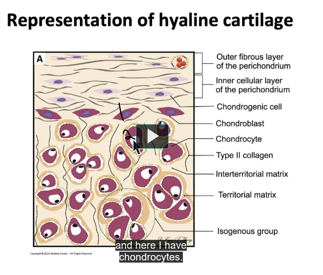
<p>Observe in this diagram the difference between this first cell and the second cell. Here I have chondroblasts (1st cell) and here I have chondrocytes (2nd cell). </p>

Observe in this diagram the difference between this first cell and the second cell. Here I have chondroblasts (1st cell) and here I have chondrocytes (2nd cell).

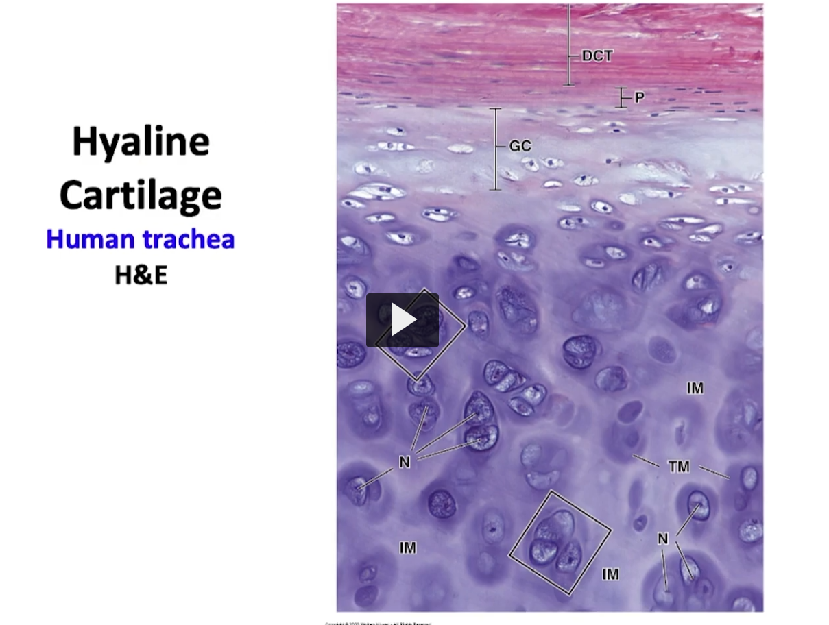
Observe this slide. Here we have a slice of a tracheal ring. The rings of the trachea are pure hyaline cartilage. This is hyaline cartilage (pointing to the bottom purple).

The reddish--pink at the top if perichondrium, where we have the chondrogenic layer and the fibrous layer. These developing small cells here are chondroblasts (where the GC is located). Over here (going to the purple) with the mature cartilage we have chondrocytes in their lacunae.

Notice the dark area I mentioned previously that surrounds the chondrocytes known as the territorial matrix, and between them the clear areas which are more acidophilic (the lighter purple between the dark purple) are the interterritorial matrix portion.

Where are the isogenous groups? We have two isogenous groups, demarcated by the squares and they are a group of chondrocytes which were the products of cellular division, interstitial growth.
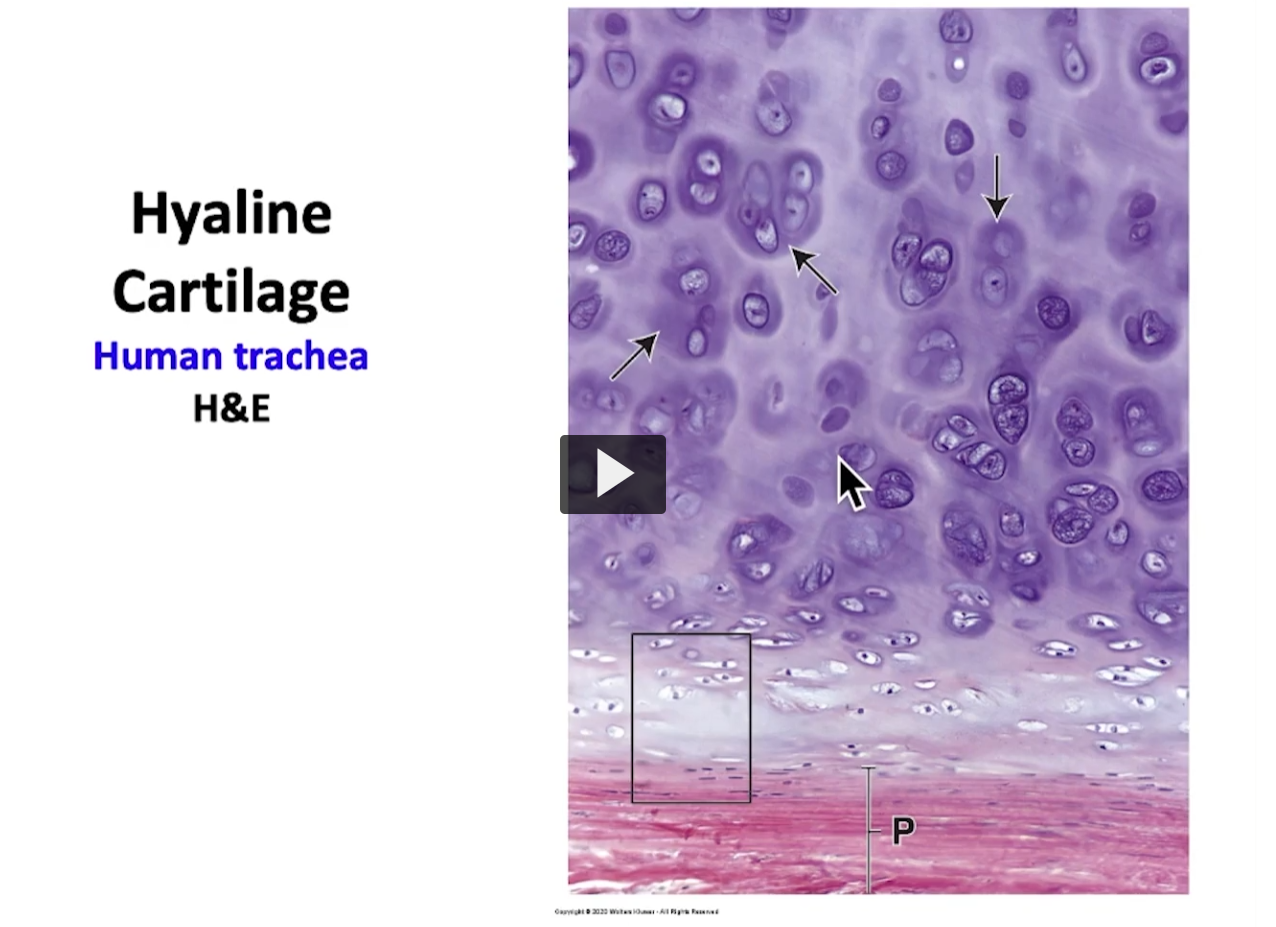
<p>Observe this slide. Here we have a slice of a tracheal ring. The rings of the trachea are pure hyaline cartilage. This is hyaline cartilage (pointing to the bottom purple). </p><p>The reddish--pink at the top if perichondrium, where we have the chondrogenic layer and the fibrous layer. These developing small cells here are chondroblasts (where the GC is located). Over here (going to the purple) with the mature cartilage we have chondrocytes in their lacunae. </p><p>Notice the dark area I mentioned previously that surrounds the chondrocytes known as the territorial matrix, and between them the clear areas which are more acidophilic (the lighter purple between the dark purple) are the interterritorial matrix portion. </p><p>Where are the isogenous groups? We have two isogenous groups, demarcated by the squares and they are a group of chondrocytes which were the products of cellular division, interstitial growth. </p>
11
New cards
<p>Observe the big areas pointing at the territorial matrix and the clear areas between the individual chondrocytes or isogenous groups is the interterritorial matrix </p><p>Dark purple at the top: isogenous group</p><p>isogenous group (I think its groups that are formed from the same cellular division)</p><p>If I go to the periphery (P), then I am in the chondrogenic layer of the perichondrium. This area is a perichondrium (the pink at the bottom). </p><p>This square demarcates the chondrogenic layer and the cells within that area are the chondroblasts. </p><p>If I magnifiy this area of the perichondrium, this is what I can observe (next slide) </p>

Observe the big areas pointing at the territorial matrix and the clear areas between the individual chondrocytes or isogenous groups is the interterritorial matrix

Dark purple at the top: isogenous group

isogenous group (I think its groups that are formed from the same cellular division)

If I go to the periphery (P), then I am in the chondrogenic layer of the perichondrium. This area is a perichondrium (the pink at the bottom).

This square demarcates the chondrogenic layer and the cells within that area are the chondroblasts.

If I magnifiy this area of the perichondrium, this is what I can observe (next slide)

If I magnifiy this area of the perichondrium, this is what I can observe (next slide)

Observe here the area of perichondrium, chondrogenous layer (Ch) and a fibrous layer (Fib). These are fibroblasts (F) and these are fibroblasts that are being converted to chondroblasts (FCh), and are proximal to the chondrogenic layer where they are recently matured chondrocytes.

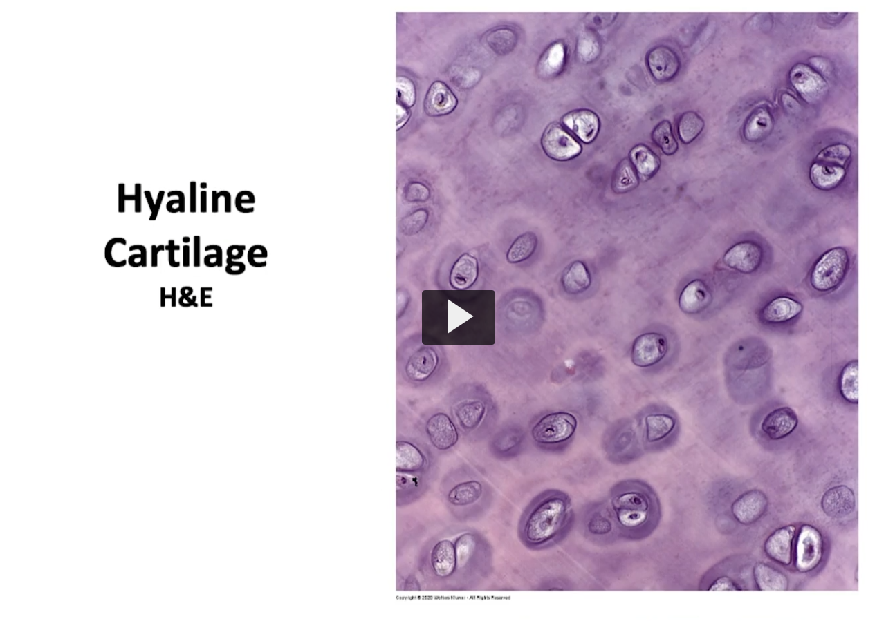
If I observe the mass of tissue cartilage, this is what I observe.
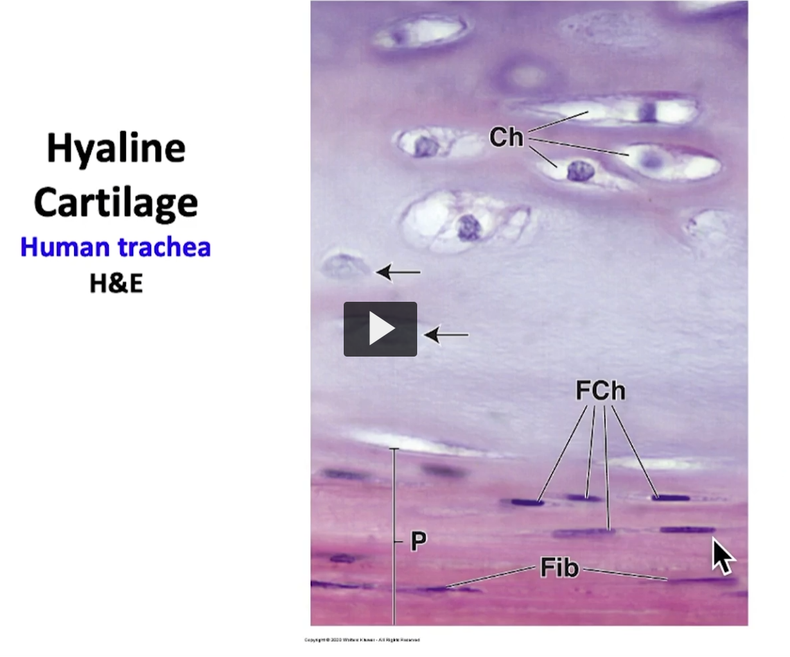
<p>If I magnifiy this area of the perichondrium, this is what I can observe (next slide) </p><p>Observe here the area of perichondrium, chondrogenous layer (Ch) and a fibrous layer (Fib). These are fibroblasts (F) and these are fibroblasts that are being converted to chondroblasts (FCh), and are proximal to the chondrogenic layer where they are recently matured chondrocytes. </p><p>If I observe the mass of tissue cartilage, this is what I observe. </p>
12
New cards
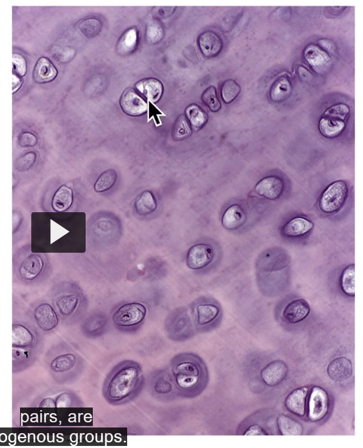
<p>If I observe the mass of tissue cartilage, this is what I observe. Since this is the center of the mass of mature cartilage, Observe the chondrocytes in their lacunae. </p>

If I observe the mass of tissue cartilage, this is what I observe. Since this is the center of the mass of mature cartilage, Observe the chondrocytes in their lacunae.

Observe the chondrocytes in their lacunae. These chondrocytes are in pairs, are isogenous groups. Observe territorial matrix surrounding groups and come from the same cellular division.

Remember if I wish to see chondrocytes or mature chondrocytes, I have to go near the perichondrium area.
<p>Observe the chondrocytes in their lacunae. These chondrocytes are in pairs, are isogenous groups. Observe territorial matrix surrounding groups and come from the same cellular division. </p><p>Remember if I wish to see chondrocytes or mature chondrocytes, I have to go near the perichondrium area. </p>
13
New cards
<p>For example, in this slide seen previously, where we saw the C of mature chondrocytes forming part of mature cartilage tissue (at the top) </p>

For example, in this slide seen previously, where we saw the C of mature chondrocytes forming part of mature cartilage tissue (at the top)

Here we have an area of perichondrium, which is where we have chondroblasts differentiating into mature chondrocytes and eventually incorporating as mature cartilage tissue.
<p>Here we have an area of perichondrium, which is where we have chondroblasts differentiating into mature chondrocytes and eventually incorporating as mature cartilage tissue. </p>
14
New cards
<p>I placed this interesting image for you to review. </p><p>Observe how the asterisk indicates intraterritorial matrix. The L is for lacunae, which is where every chondrocyte is. </p><p>Basically, we have a dark area surrounding this group of chondrocytes, which is the territorial matrix. This is an isogenous group. </p><p>Over here, I have the part that correspondss to the perichhondrium A. A good image to review. </p>

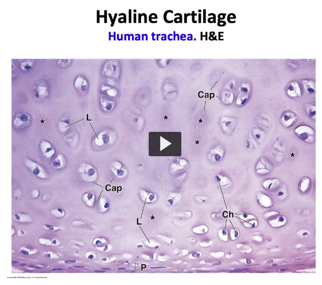
I placed this interesting image for you to review.

Observe how the asterisk indicates intraterritorial matrix. The L is for lacunae, which is where every chondrocyte is.

Basically, we have a dark area surrounding this group of chondrocytes, which is the territorial matrix. This is an isogenous group.

Over here, I have the part that correspondss to the perichhondrium A. A good image to review.

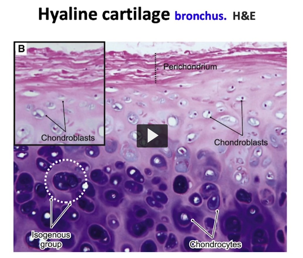
In the same way, in the second image already identified, we can review the components of the hyaline cartilage.

We have the perichondrium (it’s labeled on the image), chondroblasts that form part of the chondrogenic layer, (it’s labeled on the image), over here we have mature chondrocytes as part of the mature hyaline cartilage (outlined in white at the bottom), isogenous groups, observe the territatorial matrix covering the areas close to those chondrocytes and between them the interterritorial matrix.

If I increase the magnification of this mature cartilage tissue to observe those chondrocytes, we can appreciate the following.
<p>In the same way, in the second image already identified, we can review the components of the hyaline cartilage. </p><p>We have the perichondrium (it’s labeled on the image), chondroblasts that form part of the chondrogenic layer, (it’s labeled on the image), over here we have mature chondrocytes as part of the mature hyaline cartilage (outlined in white at the bottom), isogenous groups, observe the territatorial matrix covering the areas close to those chondrocytes and between them the interterritorial matrix. </p><p>If I increase the magnification of this mature cartilage tissue to observe those chondrocytes, we can appreciate the following. </p>
15
New cards
<p>Observe the chondrocytes in their lacunae, the interterritorial matrix, and the territorial matrix, as well as the isogenous groups. This is a very good image to use as a review. </p>

Observe the chondrocytes in their lacunae, the interterritorial matrix, and the territorial matrix, as well as the isogenous groups. This is a very good image to use as a review.

Here I am magnifying the isogenous group. The rings of the trachea is normally a classic model to observe hyaline cartilage. How do you visualize an isogenous group? What do chondrocytes look like in electron microscopy?
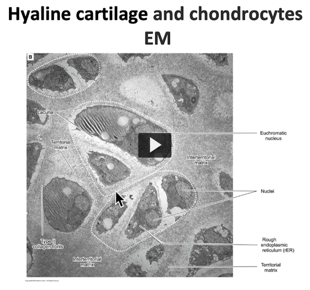
<p>Here I am magnifying the isogenous group. The rings of the trachea is normally a classic model to observe hyaline cartilage. How do you visualize an isogenous group? What do chondrocytes look like in electron microscopy? </p>
16
New cards
<p>Let us see in this last slide where you will see a group of chondrocytes in their lacunae. </p><p>See the territorial matrix, the one that will surround the group of chondrocytes, forming an isogenous group. </p>

Let us see in this last slide where you will see a group of chondrocytes in their lacunae.

See the territorial matrix, the one that will surround the group of chondrocytes, forming an isogenous group.

Over here we can observe the interterritorial matrix and every group. Here we have an isogenous group, a second isogenous group, and a third isogenous group. Basically, you can see isogenous groups in any type of cartilage but they are better visualized in hylaline cartilage. It is more difficult to visualize elastic cartilage due to presence of elastic fibers, and it’s also difficult to visualize in fibrocartilage because it contains an abundant amount of collagen type I.
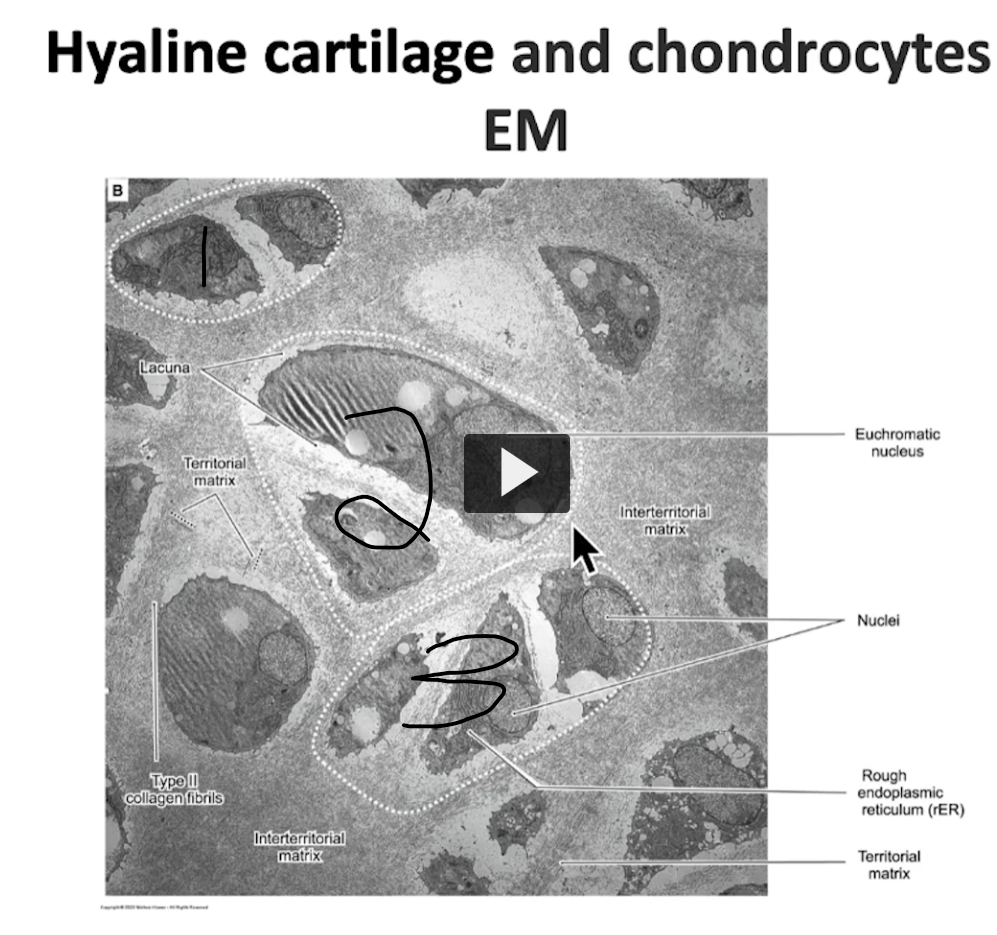
<p>Over here we can observe the interterritorial matrix and every group. Here we have an isogenous group, a second isogenous group, and a third isogenous group. Basically, you can see isogenous groups in any type of cartilage but they are better visualized in hylaline cartilage. It is more difficult to visualize elastic cartilage due to presence of elastic fibers, and it’s also difficult to visualize in fibrocartilage because it contains an abundant amount of collagen type I. </p>
17
New cards
<p><u>Cartilage Tissue Part 2</u></p><p></p>

Cartilage Tissue Part 2

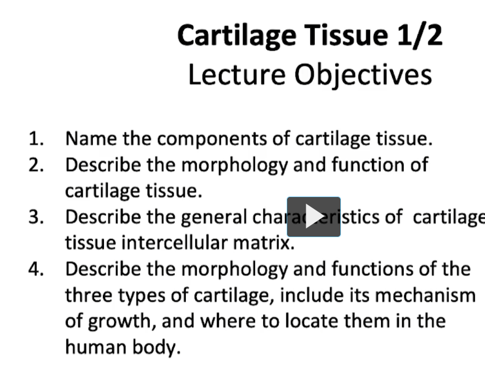
same objectives as the first video.
18
New cards
<p>Let us review the components and the cells of cartilage tissue. The cells are the chondrocytes and chondroblasts, and as a reminder, the chondroblast can only be seen if there’s perichondrium. </p><p>Remember that cartilage contains a matrix that’s composed of fibers and ground substance. Fibrocartilage will contain collagen type I fibers in it's matrix vs elastic cartilage which will contain type II collagen and elastic fibers. Both contain ground substance Remember that cartilage is an avascular tissue and it doesn’t contain nerve or lymphatic vessels, and it functions as a strong and resilient support that can handle compression well. </p>

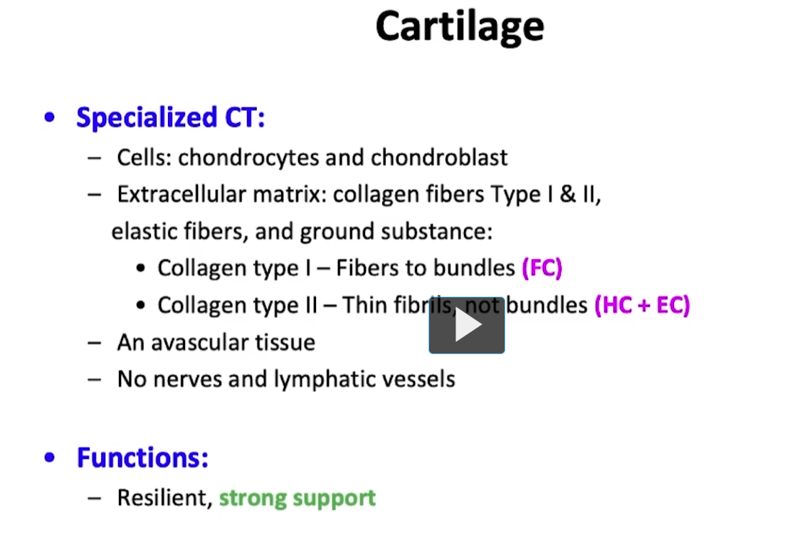
Let us review the components and the cells of cartilage tissue. The cells are the chondrocytes and chondroblasts, and as a reminder, the chondroblast can only be seen if there’s perichondrium.

Remember that cartilage contains a matrix that’s composed of fibers and ground substance. Fibrocartilage will contain collagen type I fibers in it's matrix vs elastic cartilage which will contain type II collagen and elastic fibers. Both contain ground substance Remember that cartilage is an avascular tissue and it doesn’t contain nerve or lymphatic vessels, and it functions as a strong and resilient support that can handle compression well.

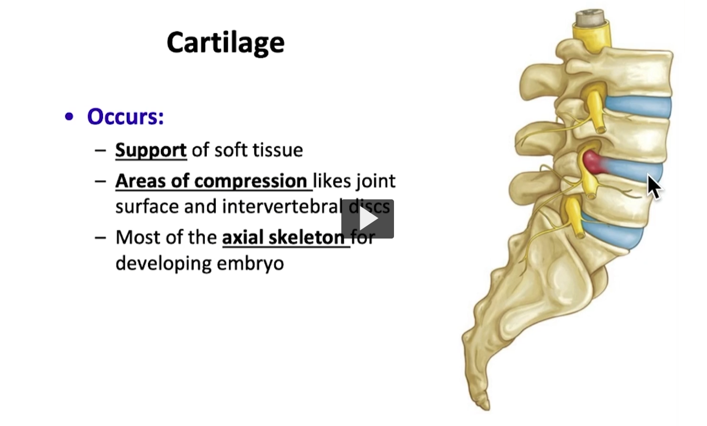
A good classic example is the intervertebral disc, which is made up of fibrocartilage and can handle areas of compression and resist against movements of the vertebral column.
<p>A good classic example is the intervertebral disc, which is made up of fibrocartilage and can handle areas of compression and resist against movements of the vertebral column. </p>
19
New cards
<p>Cartilage is made of up chondrocytes and chondroblasts. The principal cell of mature cartilage tissue is the chondrocyte. </p><p>Because of its function to synthesize the extracelluar matrix of cartilage tissue </p>

Cartilage is made of up chondrocytes and chondroblasts. The principal cell of mature cartilage tissue is the chondrocyte.

Because of its function to synthesize the extracelluar matrix of cartilage tissue

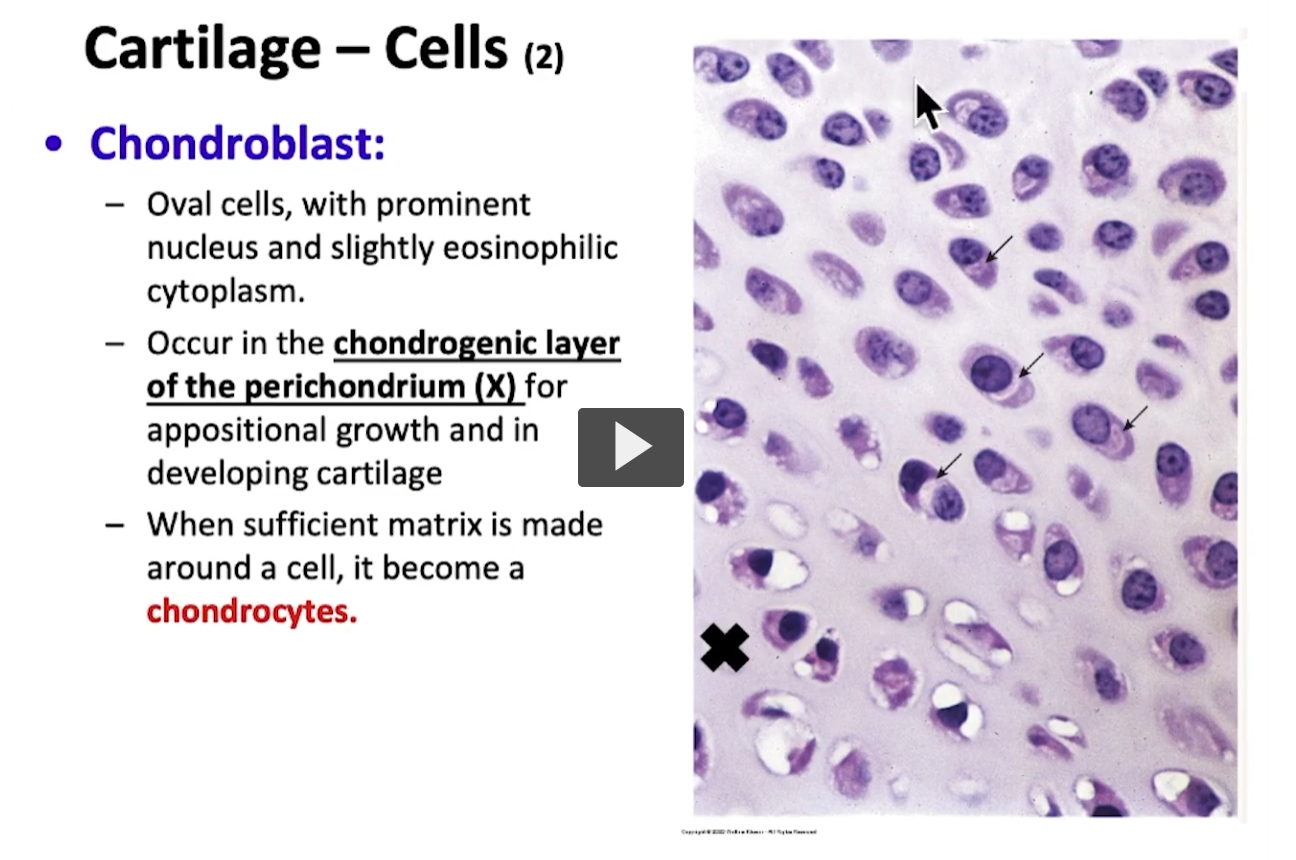
Developing chondroblasts are found near the perichondrium demarcated by the X on the image, in what is known as the chondrogenic layer.

These chondroblasts then differentiate into chondrocytes, which can be found in the mature cartilage tissue.

stopped at 1:50 of the video.
<p>Developing chondroblasts are found near the perichondrium demarcated by the X on the image, in what is known as the chondrogenic layer. </p><p>These chondroblasts then differentiate into chondrocytes, which can be found in the mature cartilage tissue. </p><p>stopped at 1:50 of the video. </p>