muscles & nerves
1/37
There's no tags or description
Looks like no tags are added yet.
Name | Mastery | Learn | Test | Matching | Spaced | Call with Kai |
|---|
No analytics yet
Send a link to your students to track their progress
38 Terms
general features of muscle tissue
elongated cells (muscle fibres/myocytes)
use ATP to generate force
contain actin and myosin filaments
types of muscle tissue
skeletal
cardiac
smooth
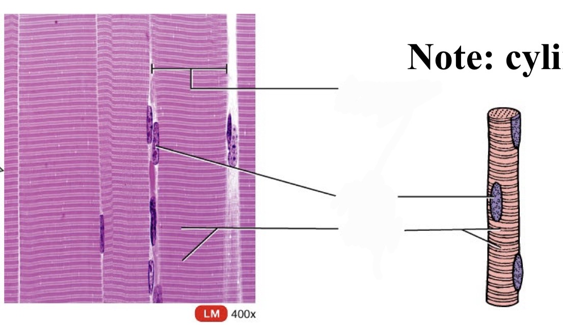
skeletal muscle (location, structure, function)
attached to bones via tendons;
long cylindrical fibres, striated, multinucleated (peripheral nuclei).
voluntary control
functions: movement, posture, heat production, protection
internal structure of skeletal muscle (hierarchy)
muscles
fascicles
myocyte (cell)
myofibrils
myofilaments (actin and myosin)
sarcomeres
connective tissue of skeletal muscle (hierachy)
epimysium (surrounds muscle)
perimysium (around fascicles)
endomysium (inside fascicle, wraps around sarcolemma)
sarcolemma (actual cell membrane)
sarcoplasm
key components of skeletal muscle
sarcolemma (cell membrane)
sarcoplasm (cytoplasm)
actin (thin)
myosin (thick)
myofibrils
bundles of actin and myosin in sarcomeres
causes striations
sarcomere structure
A band, I band, H zone, M line, Z disc
= contains thick and thin
= two at each end, thin only. (titin filament)
= middle, thick only
= centre
= boundary of sarcomere
sarcomere changes during contraction
I band shortens , H zone reduces, A band constant
function of fascicles
bundle of muscle fibres, coordinated contraction
titin filament
located in I band of sarcomere
stabilises & assists during contraction
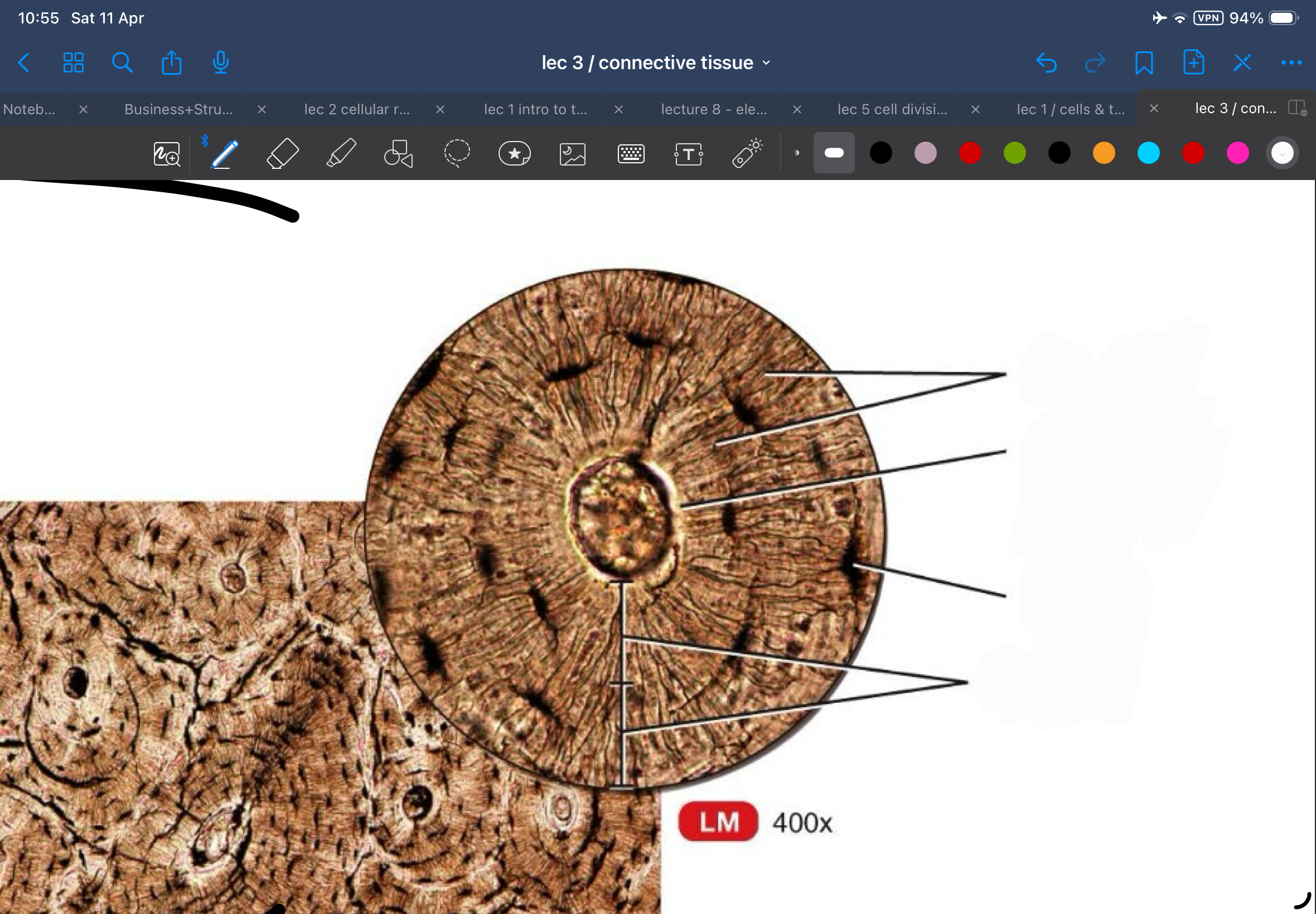
cardiac muscle
found in heart; striated, branched fibres
single central nucleus
coordinated pump
intercalated discs
purkinje fibres
intercalated discs
junctions connecting cardiac cells
contain desmosomes and gap junctions
purkinje fibres
cardiac muscle cells for electrical conduction
few myofibrils, more gap junctions
smooth muscle (location, structure, function)
walls of hollow organs (intestines, blood vessels, uterus, bladder)
non-striated, spindle-shaped cells
single nucleus
involuntary
functions in movement of substances (peristalsis) and control of lumen
internal structure of smooth muscle
actin and myosin
no sacromeres
filaments attach to dense bodies
general features of nervous tissue
specialised for communication
detects and responds to stimuli
maintains homeostasis
divisions of nervous system
CNS - brain, spinal cord, optic nerve
PNS - all nervous tissue outside CNS
functional divisions of PNS
afferent (sensory) to PNS
efferent (motor) from CNS to effectors (muscles and glands)
Afferent arrives, efferent exits
cells in nervous tissuee
neurons (signal transmissions) neuroglia (support cells)
large, excitable cells
action potentials
does not divide
high metabolic rate
neurons
structure of neuron
dendrites
cell body (soma)
trigger zone
axon
myelin sheath
axon terminals
receive input in a neuron
dendrites
link together , form blood brain barrier
astrocytes
form myelin sheath in CNS
myelinate multiple axons
increase action potential
oligodendrocytes
phagocytes of CNS
microglia
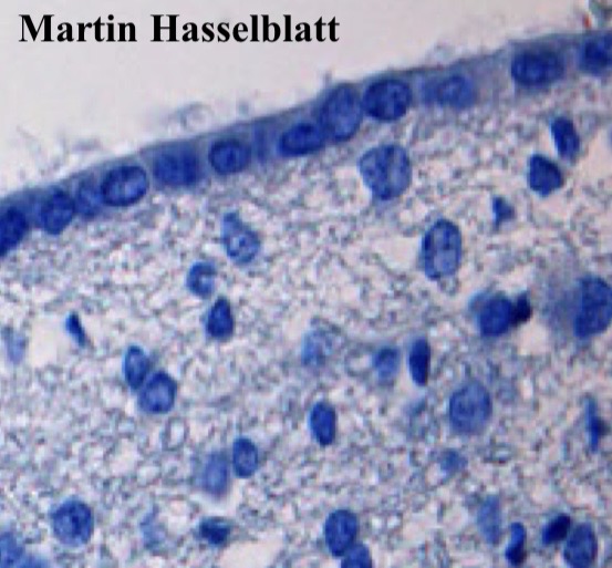
ependymal CELLS
line brain ventricle and spinal canal where cerebrospinal fluid circulates.
cilia for movement, microvilli samples fluid.
schwann cell
pns neuroglia
myelinates one axon at a time, supports others
satellite cells
pns neuroglia
like cns astrocytes
periastalsis
involuntary contractions that move food, liquids, waste through digestive tract