Valvular diseases exam
1/96
There's no tags or description
Looks like no tags are added yet.
Name | Mastery | Learn | Test | Matching | Spaced | Call with Kai |
|---|
No analytics yet
Send a link to your students to track their progress
97 Terms
What are the three main types of mitral valve disease?
Mitral Stenosis
Mitral Regurgitation
Mitral Valve Prolapse
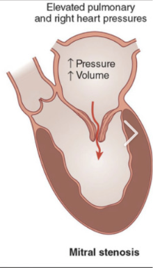
What is mitral stenosis?
The narrowing of the mitral valve orifice, impeding diastolic blood flow from the left atrium to the left ventricle.
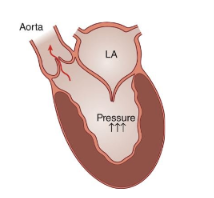
What happens in acute mitral regurgitation?
Blood flows backward from the left ventricle into the left atrium during systole, causing high left atrial pressure and pulmonary edema.
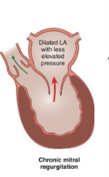
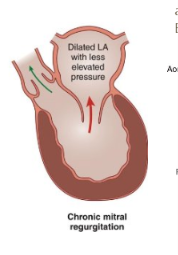
What happens in chronic mitral regurgitation?
The left atrium becomes dilated due to regurgitant blood flow, with less elevated pressure compared to acute cases.
What is mitral valve prolapse (MVP)?
It’s characterized by abnormal billowing of one or both mitral leaflets into the left atrium during ventricular systole, often accompanied by mitral regurgitation. Also known as Barlow Syndrome.
What are the main causes (etiology) of mitral stenosis?
Rheumatic fever (most common) and infective endocarditis.
Describe the pathophysiology of mitral stenosis.
Pressure builds up in the left atrium, causing dilation, fluid buildup in lungs, Stroke due to atrial fibrillation, stroke risk, and right heart failure (leg swelling, enlarged liver, ascites).
What are the symptoms of early mitral stenosis?
Shortness of breath during exercise and feeling unusually tired.
What are the symptoms of mild mitral stenosis?
Shortness of breath even at rest, waking up breathless at night, and swelling.
What are the symptoms of severe mitral stenosis?
Signs of right heart failure—neck vein bulging, liver swelling, leg swelling, and fluid in belly.
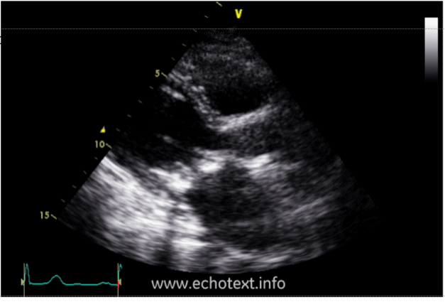
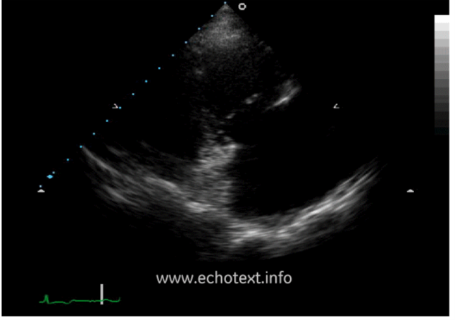
What is this?
Mitral stenosis
What is mitral regurgitation in etiology?
The valve doesn’t close properly, causing blood to leak backward into the left atrium during ventricular systole.
What are the two types of mitral regurgitation?
Primary MR, Secondary MR
What is Primary mitral regurgitation?
The valve itself is damaged.
What is in Primary mitral regurgitation?
Acute primary mitral regurgitation, Chronic primary mitral regurgitation
What are causes of acute in primary mitral regurgitation?
Papillary muscle rupture after a heart attack; torn chordae from infection, chest injury, or Marfan syndrome.
What are causes of chronic in primary mitral regurgitation?
Mitral valve prolapse
rheumatic heart disease
calcium buildup around the valve
What is Secondary MR?
The valve is normal, but the left ventricle is enlarged or weak, stretching the valve.
What happens in the pathophysiology of mitral regurgitation?
Pressure increases in the left atrium, causing it to dilate. This leads to lower cardiac output and volume overload in the left ventricle.
What does “pathophysiology” mean?
How a disease changes how the body works.
What does etiology mean?
The cause of a disease.
What are the symptoms of acute mitral regurgitation?
Sudden shortness of breath and pulmonary edema.
What are the symptoms of chronic mitral regurgitation?
Fatigue, shortness of breath with exertion (physical activity), swelling (if right heart failure develops), and atrial fibrillation.
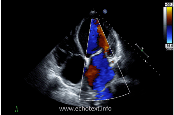
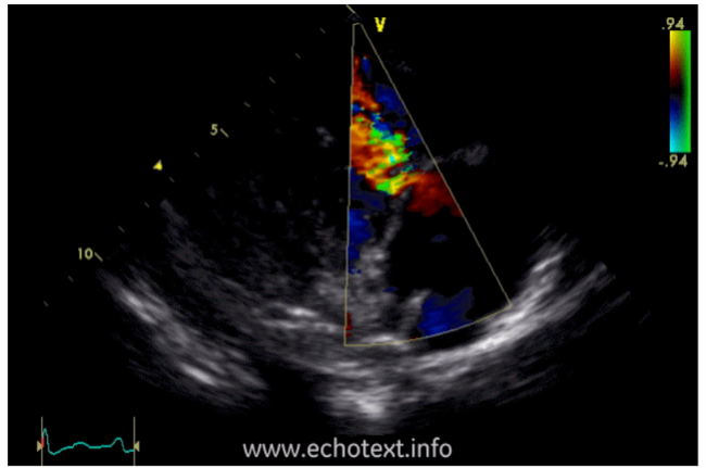
What is this?
Mitral rgurgitation.
What is the treatment for acute mitral regurgitation?
Surgery is usually required right away; medicines only stabilize temporarily.
What is the treatment for chronic mitral regurgitation?
Surgery if symptoms develop or if the left ventricle starts weakening; repair preferred, if possible, replacement if necessary; medicines are less effective long‑term.
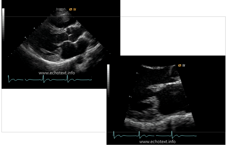
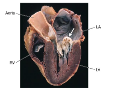
What is this?
Mitral valve Prolapse
What are the causes (etiology) of mitral valve prolapse?
It can be inherited or occur as part of connective tissue disorders such as Marfan syndrome.
What happens to the valve leaflets in mitral valve prolapse?
The leaflets—especially the posterior one—become enlarged and floppy.
What happens in severe cases for mitral valve prolapse?
The chordae may stretch or tear, the valve ring (annulus) may enlarge, and the leaflets may thicken.
What are common symptoms of mitral valve prolapse?
Often no symptoms; some people may feel chest pain or palpitations.
What are rare symptoms for MVP?
Sudden severe MR if chordae rupture → pulmonary edema, infective endocarditis, blood clots/emboli, and arrhythmias (atrial or ventricular).
What are the two main types of aortic valve disease?
Aortic stenosis and aortic regurgitation.
What is aortic stenosis?
The narrowing of the aortic valve orifice, which impedes the systolic flow of blood from the left ventricle (LV) into the aorta.
What are the main causes (etiology) of aortic stenosis?
Degenerative calcification, congenital bicuspid valve, and rheumatic disease.
What is degenerative calcification in aortic stenosis?
Calcium buildup on a normal valve as people age, causing narrowing.
How does a congenital bicuspid valve lead to aortic stenosis?
Having only two leaflets instead of three causes faster wear and earlier calcification.
What does rheumatic disease cause aortic stenosis?
Scarring from past rheumatic fever damages the valve, making it less flexible. (less common)
What happens in the pathophysiology of Aortic Stenosis?
Left atrium (LA) and left ventricle (LV) thicken — hypertrophy due to increased pressure load
Aortic valve narrows, obstructing blood flow from LV to aorta
What are the main symptoms of aortic stenosis?
Chest pain, fainting (syncope) especially with exercise, and heart failure symptoms.
What are the signs of heart failure in aortic stenosis?
Shortness of breath, pulmonary congestion, and swelling (edema).
Why does fainting occur in aortic stenosis?
Reduced blood flow through the narrowed valve limits oxygen delivery during exertion.
What are the treatments for Aortic Stenosis?
No medicine can stop progression
Surgical Aortic Valve Replacement (AVR)
Balloon Valvuloplasty
TAVR (Transcatheter Aortic Valve Replacement)
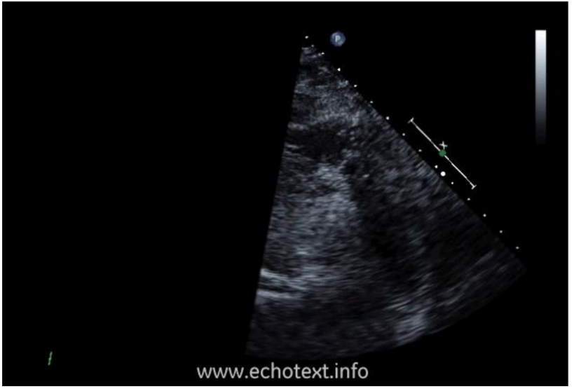
What is this?
Aortic Stenosis
What is aortic regurgitation?
The backward flow of blood into the left ventricle (LV) during ventricular diastole.
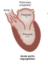
Why does pressure rise in the left ventricle during acute aortic regurgitation?
The LV fills with both normal incoming blood and the backward leak, causing a rapid pressure increase.
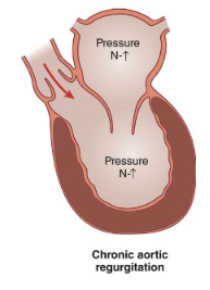
What happens in chronic aortic regurgitation?
Blood leaks backward from the aorta into the left ventricle during diastole over time.
How does the left ventricle respond in chronic aortic regurgitation?
It gradually enlarges (dilates) to handle the extra volume, keeping pressure near normal.
What are the two main categories of causes for aortic regurgitation?
Valve problems and aortic root problems.
What are the valve problems that cause aortic regurgitation?
Bicuspid valve, infection (endocarditis), and rheumatic heart disease.
What are the aortic root problems that cause aortic regurgitation?
The valve is normal, but the aortic root is too wide
What causes the aortic root to widen in aortic regurgitation?
Age‑related enlargement, aortic aneurysm, and aortic dissection (tear in the aortic wall).
What is the pathophysiology of aortic regurgitation?
Volume overload of the left ventricle (LV) leads to pulmonary edema (acute), heart failure (chronic), and angina (chronic).
What are the chronic symptoms of aortic regurgitation?
Shortness of breath with exertion and fatigue.
What are the acute symptoms of aortic regurgitation?
Sudden severe shortness of breath and pulmonary edema.
What are the treatments for aortic regurgitation?
ACE inhibitors and surgery (valve replacement).
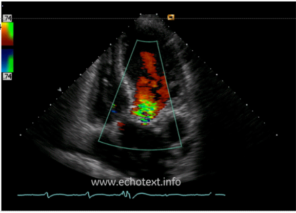
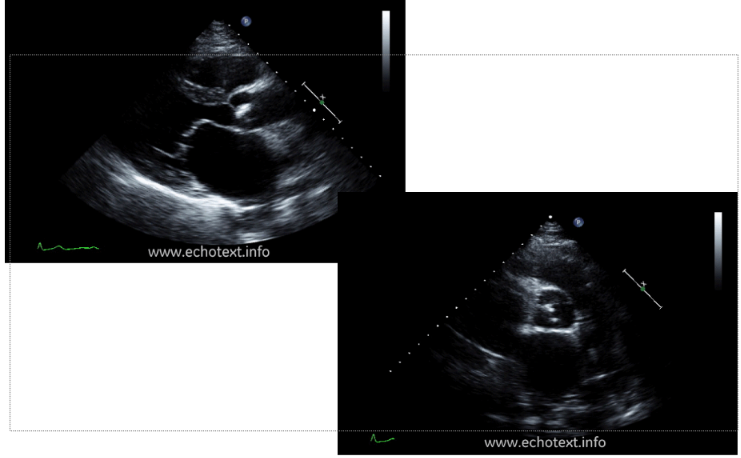
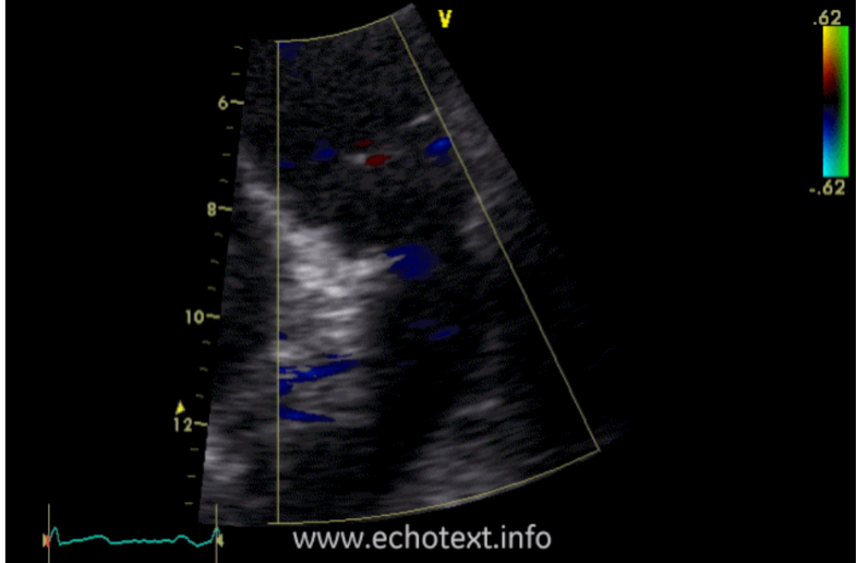
What is this?
Aortic regurgitation.
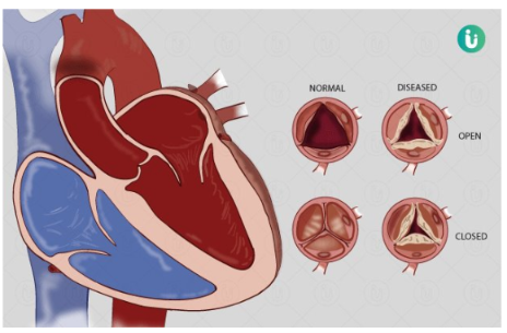
What are the two main types of tricuspid valve disease?
Tricuspid stenosis and tricuspid regurgitation.
What is tricuspid stenosis?
The narrowing of the tricuspid valve orifice, impeding the diastolic flow of blood from the right atrium into the right ventricle.
What are the causes (etiology) of tricuspid stenosis?
Rheumatic fever (most common), carcinoid heart disease, and endomyocardial fibrosis.
What are the symptoms of tricuspid stenosis?
Neck vein swelling (JVD), abdominal bloating, hepatomegaly, edema, fatigue/weakness, and cool skin from low cardiac output.
What are the treatments for Tricuspid Stenosis?
Balloon valvuloplasty – stretches the valve open to improve blood flow
Valve repair or replacement – performed if the condition is severe
What surgical options exist for severe tricuspid stenosis?
Valve repair or valve replacement for tricuspid stenosis.
What is this?
Tricuspid Stenosis
What is tricuspid regurgitation?
The backward flow of blood into the right atrium during ventricular systole.
What are the causes (etiology) of tricuspid regurgitation?
Functional (right ventricle enlargement) and carcinoid syndrome (rare).
What are the symptoms of tricuspid regurgitation?
Neck veins bulge with prominent v waves, pulsating liver, and systolic murmur at the lower left sternal border.
Why does the liver pulsate in tricuspid regurgitation?
Because blood regurgitates back into systemic veins, causing liver pulsations.
What murmur is heard in tricuspid regurgitation?
A systolic murmur at the lower left sternal border.
What are the treatments for tricuspid regurgitation?
use diuretics for fluid overload, and surgery (valve repair or replacement).
How is functional tricuspid regurgitation treated?
By treating the underlying problem (e.g., pulmonary hypertension or RV enlargement).
What is this?
tricuspid regurgitation
What are the two main types of pulmonic valve disease?
Pulmonic stenosis and pulmonic regurgitation.
What is Pulmonic Stenosis?
Narrowing of the pulmonic valve orifice that impedes systolic blood flow from the right ventricle (RV) into the main pulmonary artery (MPA).
What is the etiology of Pulmonic Stenosis?
It is rare and usually congenital (present at birth). Very rarely, it can be caused by carcinoid syndrome, where plaques stiffen the valve.
What are the symptoms of mild Pulmonic Stenosis?
No symptoms.
What are the symptoms of moderate Pulmonic Stenosis?
Shortness of breath, especially with exertion
Fatigue
Chest pain or discomfort
Palpitations
Syncope (fainting)
What is the treatment for Pulmonic Stenosis?
Balloon valvuloplasty — a catheter with a balloon is used to stretch open the narrowed pulmonic valve.
What is this?
Pulmonic Stenosis
What is Pulmonic Regurgitation?
The backward flow of blood from the pulmonary artery into the right ventricle (RV) during ventricular diastole.
Which underlying cause (etiology) results in Pulmonic Regurgitation?
Severe pulmonary hypertension
What are the symptoms of Pulmonic Regurgitation?
Shortness of breath
Fatigue
Palpitations
Swelling in ankles and feet
Jugular venous distention
Hepatomegaly (enlarged liver)
What is the treatment for mild Pulmonic Regurgitation?
Mild PR doesn’t need treatment.
How is Severe Pulmonic Regurgitation treated?
is treated by managing the underlying cause (usually pulmonary hypertension) and using diuretics to relieve fluid overload.
What is the treatment if the right ventricle becomes weak or symptoms are significant in Pulmonic Regurgitation?
Valve repair or replacement may be needed for pulmonic regurgitation.
What is this?
Pulmonic Regurtation.
What is Infective Endocarditis (IE)?
It’s an infection caused by bacteria entering the bloodstream and settling in the heart lining, valve, or blood vessel. It’s also called bacterial endocarditis.
Is Infective Endocarditis common?
No, IE is uncommon, but people with certain heart conditions have a greater risk of developing it.
What are the risk factors for developing Infective Endocarditis?
Heart valve disease
Previous heart valve surgery
Congenital heart disease
Intravenous drug use
Previous history of IE
Hypertrophic cardiomyopathy
When is the risk of Infective Endocarditis higher?
The risk is higher after procedures that allow bacteria access to the bloodstream.
What procedures increase the risk of Infective Endocarditis?
Dental procedures involving the gums
Insertion of catheters or needles
Procedures to treat infections of the oral mucosa
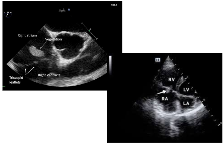
What is this?
Infective Endocarditis
What is Rheumatic Heart Disease?
Rheumatic Heart Disease is heart valve damage caused by rheumatic fever, which is the body’s inflammatory response to a bacterial infection.
What causes Rheumatic Heart Disease?
It results from valvular damage caused by an abnormal immune response to Streptococcus pyogenes infection (group A streptococcus) that leads to acute rheumatic fever.
When does acute rheumatic fever occur?
It usually develops about three weeks after group A streptococcal pharyngitis and can affect the joints, skin, brain, and heart.
How does rheumatic fever lead to Rheumatic Heart Disease?
Heart valve inflammation from rheumatic fever causes damage that may occur immediately or develop over time from repeated strep infections.
What happens to the heart valves in Rheumatic Heart Disease?
Continuing inflammation leads to scarring and narrowing of the valves, damaging mainly the mitral and aortic valves.