CASSY'S CARDS - Exam 4 Bio 223
1/130
There's no tags or description
Looks like no tags are added yet.
Name | Mastery | Learn | Test | Matching | Spaced | Call with Kai |
|---|
No analytics yet
Send a link to your students to track their progress
131 Terms
Functions of the nervous system
Controls perception and experience of the world, directs voluntary movement, and regulates homeostasis (respiration, blood pressure, body temperature, pH, and sleep-wake cycles).
two structural divisions of the nervous system
Central Nervous System (CNS) and Peripheral Nervous System (PNS).
What structures are included in the CNS?
The brain and spinal cord.
CNS
Integration—receiving sensory input and sending motor output.
What structures are included in the PNS?
Cranial nerves and spinal nerves.
How many pairs of cranial and spinal nerves are there?
12 pairs of cranial nerves; 31 pairs of spinal nerves.
PNS
Carries sensory information to the CNS and motor commands from the CNS to the body.
What are the three functional categories of the nervous system?
Sensory (afferent), integrative, and motor (efferent).
What are the two divisions of the sensory (afferent) system?
Somatic sensory and visceral sensory.
somatic sensory division
Detects signals from skin, skeletal muscles, bones, joints, and special senses (vision, hearing, taste, smell, balance).
visceral sensory division
Detects signals from internal organs (heart, lungs, kidneys, bladder, stomach, etc.).
What are the two divisions of the motor (efferent) system?
Somatic motor (voluntary) and autonomic motor (involuntary).
somatic motor division
Controls skeletal muscles it's a voluntary action
autonomic motor division
Controls smooth muscle, cardiac muscle, and glands. It's an involuntary action
What are the two branches of the autonomic nervous system?
Sympathetic ("fight or flight") and parasympathetic ("rest and digest").
gray matter
Made of neuron cell bodies, dendrites, unmyelinated axons, neuroglia, synapses.
white matter
Made of myelinated axons (plus some unmyelinated axons and glia).
Where is gray vs white matter in the brain?
Cortex (outer gray) + deep nuclei (gray) with underlying white matter tracts.
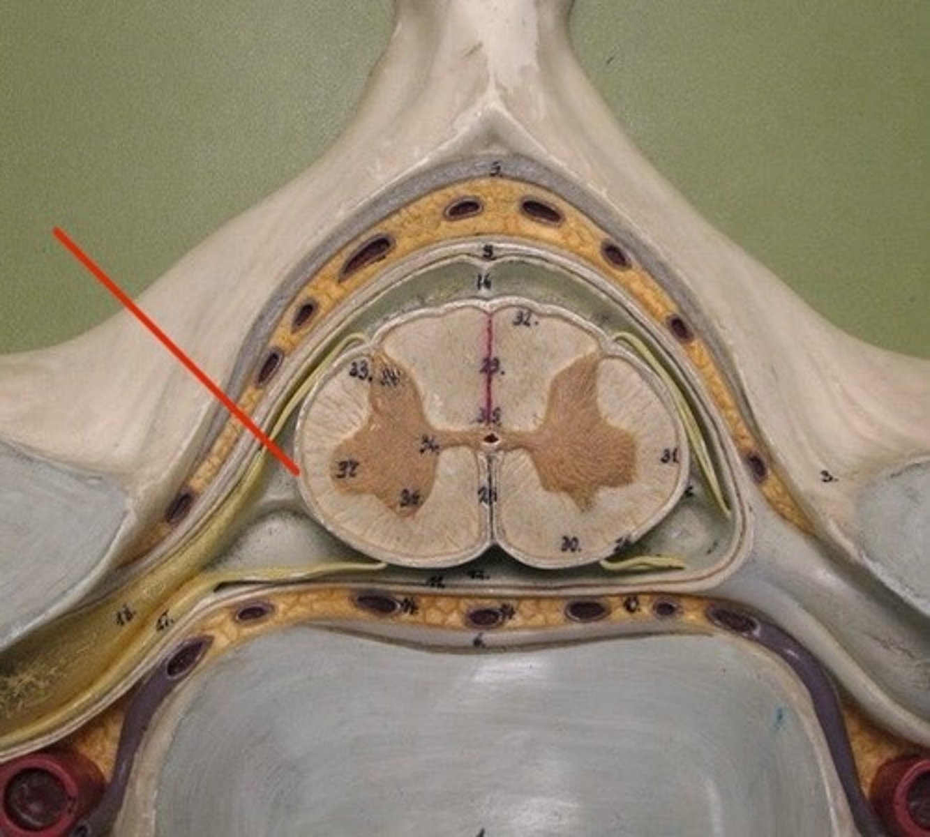
Where is gray vs white matter in the spinal cord?
Inner "H" of gray matter surrounded by outer white matter columns.
Nucleus
cluster of neuron cell bodies in CNS
Ganglion
cluster of neuron cell bodies in PNS
Tract
bundle of axons in CNS
Nerve
bundle of axons in PNS
How do skull and vertebral column protect the CNS?
Provide rigid bony encasement to resist mechanical injury.
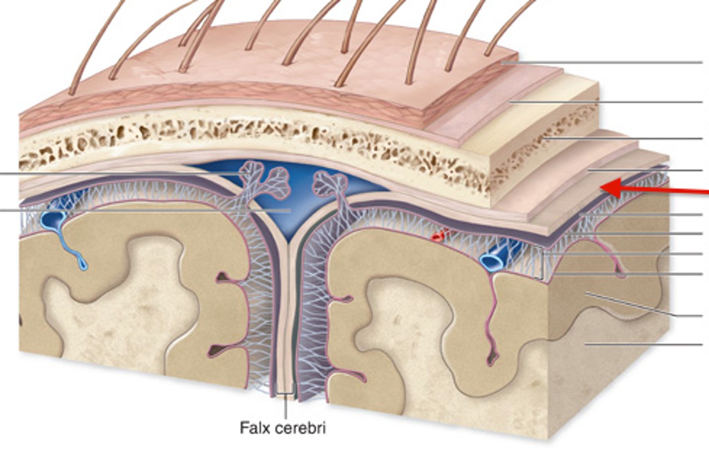
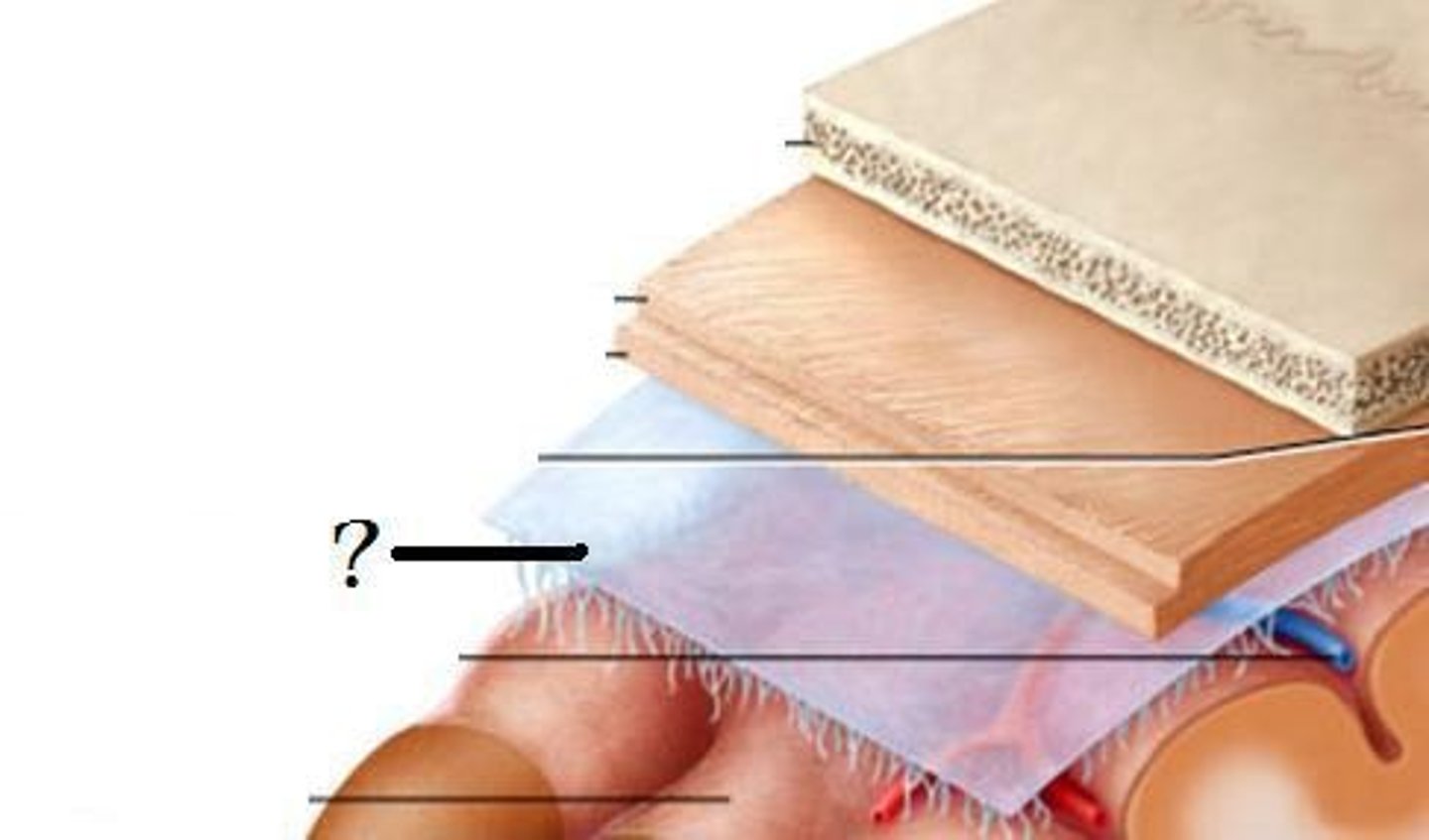
List the meninges from superficial to deep.
Dura mater - tough outer layer
Arachnoid mater - thin, web-like middle layer
Pia mater - delicate inner layer that adheres directly to the brain and spinal cord
Describe how the Dura Mater layer relates anatomically and functionally to the CNS.
Forms a durable protective covering.
-In the cranial cavity it has two layers: periosteal (dural sinuses) + meningeal.
-In the spinal cord it has one meningeal layer and an epidural space filled with fat and veins.
Describe how the Arachnoid mater layer relates anatomically and functionally to the CNS.
Lies beneath dura; spider-web trabeculae bridge the subarachnoid space which holds CSF and blood vessels.
Describe how the Pia mater layer relates anatomically and functionally to the CNS.
Thin, vascular layer that clings tightly to the surface of the brain and spinal cord, following all folds (gyri and sulci), supplying oxygen/nutrients, and helping form the choroid plexus with ependymal cells.
How do the meninges protect and support the CNS?
They provide a triple-layered protective barrier, anchor the CNS within the skull/spinal canal, circulate CSF, and stabilize blood vessels serving neural tissue.
How does cranial dura differ from spinal dura?
Cranial dura has two layers (periosteal + meningeal) and forms dural sinuses; spinal dura is single meningeal layer with an epidural space.
Epidural space
Lies between the vertebral canal and the dura mater in the spinal cord. It contains fat and blood vessels that cushion and protect the spinal cord. (Note: there is no true epidural space around the brain — only potential.)
Subdural space
It is a potential space between the dura mater and the arachnoid mater. Under normal conditions, the layers are close together, but the space can fill with blood or fluid in injury (subdural hematoma).
Subarachnoid space
Lies between the arachnoid mater and the pia mater. It contains cerebrospinal fluid (CSF) and blood vessels, providing cushioning and nutrient exchange for the CNS.
General functions of cerebrospinal fluid (CSF)?
Cushions (buoyancy), protects (shock absorption), transports nutrients/waste, maintains chemical stability.
CSF production site?
Choroid plexus (ependymal cells + capillaries) in ventricles.
CSF flow (start → finish)?
Lateral ventricles → interventricular foramina → 3rd ventricle → cerebral aqueduct → 4th ventricle → median/lateral apertures → subarachnoid space → arachnoid granulations → dural venous sinuses.
How is CSF reabsorbed?
Via arachnoid villi/granulations into dural venous sinuses.
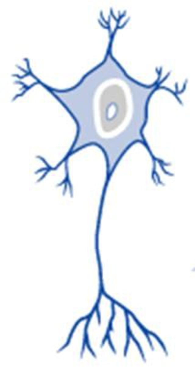
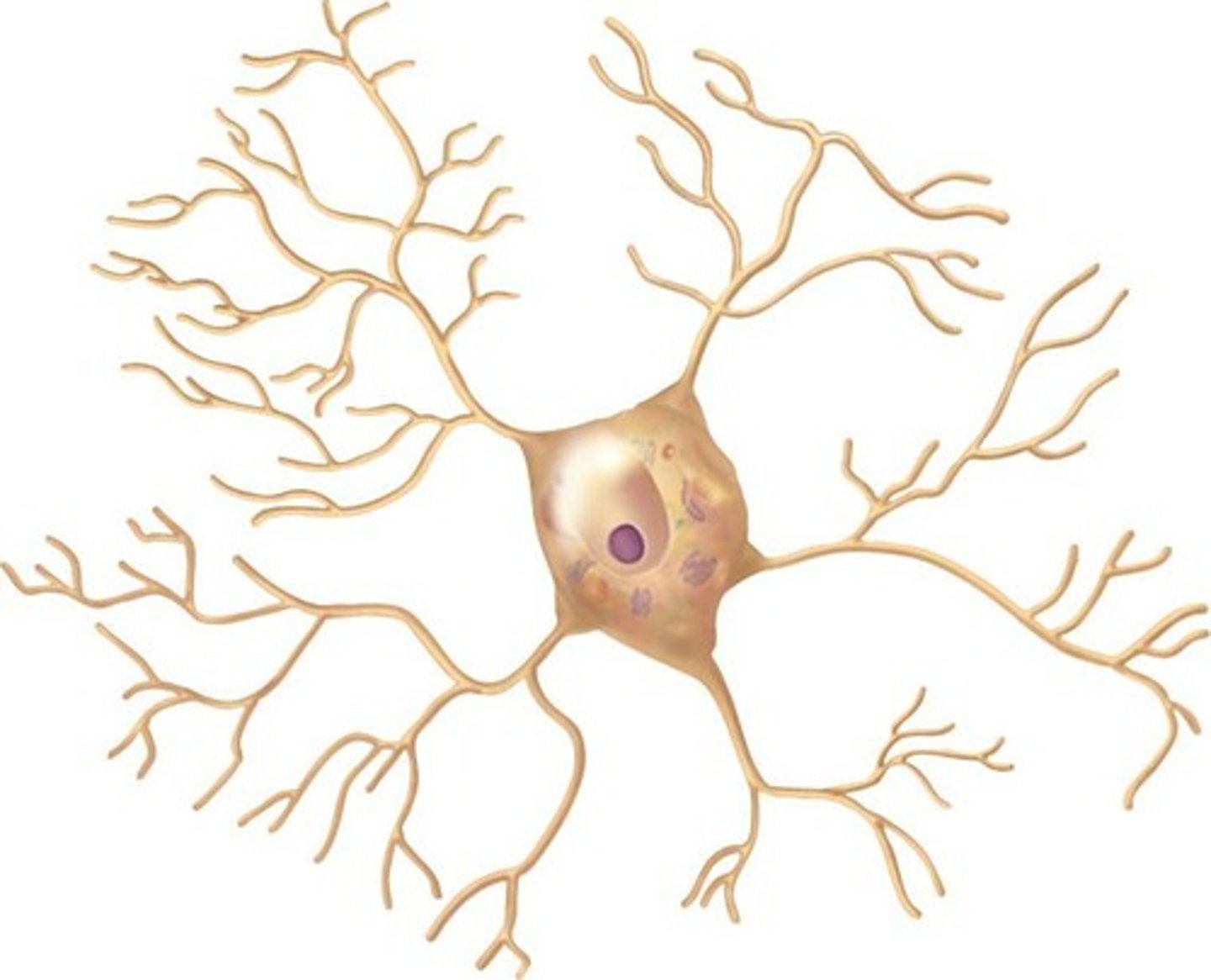
What are the main parts of a neuron?
cell body, axon, dendrites
dendrites
receive input signals from other neurons and transmit those signals toward the cell body.
soma (cell body)
integrates incoming signals and contains organelles like the nucleus and Nissl bodies (rough ER) that make proteins for the neuron.
axon hillock
It is the trigger zone where the action potential begins.
axon
conducts the action potential away from the cell body.
axon terminals (synaptic knobs)
they release neurotransmitters (NTs) to communicate with other neurons, muscles, or glands.
Which regions are receptive, conducting, secretory?
Receptive = dendrites/soma; conducting = axon; secretory = terminals.
Which structures receive input vs send output?
Input: dendrites/soma; Output: axon terminals (via AP down axon).
Structural neuron types
multipolar, bipolar, pseudounipolar and anaxonic.
multipolar neurons
They have one axon and multiple dendrites. They are the most common type, found in the CNS and as motor neurons.
bipolar neurons
They have one axon and one dendrite with the cell body between them. They are found in special sense organs like the retina (eye) and olfactory epithelium (smell).

pseudounipolar neurons
They have one process that splits into two branches (peripheral and central). They function as sensory neurons that carry information such as touch, pain, and pressure from the skin and joints.

anaxonic neurons
They have no distinct axon—only dendrite-like processes. They act as interneurons in the CNS, especially in the brain.
Functional types of neurons
sensory, motor, interneurons
sensory (afferent) neurons
Carry information from sensory receptors to the CNS.
interneurons (association neurons)
Connect sensory and motor neurons within the CNS and make up most of the neurons in the body.
motor (efferent) neurons
Carry information away from the CNS to muscles and glands (effectors) to produce a response.
What are the CNS glial cells?
-Astrocytes
-Oligodendrocytes
-Microglia
-Ependymal cells
astrocytes
They form the blood-brain barrier (BBB), provide structural support, regulate the extracellular environment, and repair damaged CNS tissue.
oligodendrocytes
They form myelin sheaths in the CNS. Each one myelinates multiple axon segments (many internodes).
microglia
They are small phagocytic cells that remove debris, dead cells, and pathogens in the CNS ("cleanup crew").
ependymal cells
They line ventricles and the central canal, are ciliated, and produce and circulate cerebrospinal fluid (CSF).
What are the PNS glial cells?
Schwann cells and satellite cells
Schwann cells
They form myelin sheaths around axons in the PNS; each Schwann cell myelinates one segment of one axon.
satellite cells
They surround and support neuron cell bodies in PNS ganglia, helping regulate their environment.
myelination
It is the wrapping of an axon with multiple layers of glial plasma membrane, providing electrical insulation and greatly increasing conduction speed.
How does CNS myelination differ from PNS myelination?
-CNS: Oligodendrocyte myelinates many axons.
-PNS: Schwann cell myelinates only one segment of one axon.
Nodes of Ranvier
Gaps between myelin segments (internodes) where voltage-gated Na⁺ channels are concentrated; allow saltatory conduction of action potentials.
What's the difference between white matter and gray matter?
-White matter: Myelinated axons.
-Gray matter: Neuron cell bodies, dendrites, and synapses.
What is required for PNS axon regeneration (Wallerian degeneration)?
The soma must remain intact and the axon must be myelinated; Schwann cells form a regeneration tube, guide regrowth, and remyelinate the new axon.
Major neuronal ion channel types?
Leak (always open), ligand-gated (chemically gated), voltage-gated, mechanically gated.
Typical channel locations?
-Ligand-gated on dendrites/soma
-voltage-gated Na⁺/K⁺ on axon/initial segment
-voltage-gated Ca²⁺ at terminals; leak throughout.
What creates the resting membrane potential (about −70 mV)?
K⁺ leak out > Na⁺ leak in, intracellular anions, and Na⁺/K⁺ ATPase maintaining gradients.
Role of Na⁺/K⁺ ATPase in RMP?
Pumps 3 Na⁺ out / 2 K⁺ in to preserve gradients and help keep inside negative.
Depolarization
It is when the membrane potential becomes less negative (moves closer to zero) due to positive ions entering the cell, usually Na⁺.
Repolarization
It is the process of the membrane returning toward the resting membrane potential (RMP) after depolarization, caused by K⁺ ions leaving the cell.
Hyperpolarization
It is when the membrane potential becomes more negative than the resting potential, often due to continued K⁺ efflux or Cl⁻ influx.
Threshold
It is the critical membrane voltage (about −55 to −60 mV) that must be reached at the axon hillock to trigger an action potential.
Graded potential
It is a small, local change in membrane potential that occurs on dendrites or the soma and varies in strength depending on the stimulus.
What ions and channels are involved in graded potentials?
They use ligand-gated or mechanically gated channels that allow ions like Na⁺, K⁺, or Cl⁻ to move across the membrane.
Are graded potentials reversible or all-or-none?
They are reversible and not all-or-none; they can fade (decremental) and return to resting potential when the stimulus ends.
Action Potential (AP)
It is a rapid, uniform depolarization and repolarization of the membrane that travels along the axon to transmit a signal.
What ions and channels are involved in action potentials?
Involve voltage-gated Na⁺ and K⁺ channels that open and close in sequence to produce depolarization and repolarization.
Are action potentials graded or all-or-none?
They follow the all-or-none principle—once threshold is reached, the full AP occurs; if not, no AP happens.
Do graded potentials or action potentials travel farther?
Action potentials travel long distances (axon), while graded potentials are local and decay over short distances.
Phases of an action potential
-Depolarization: v-gated Na⁺ open → Na⁺ in
-Repolarization: Na⁺ inactivate, v-gated K⁺ open → K⁺ out
-Hyperpolarization: K⁺ channels slow to close.
Why are voltage-gated channels essential for AP propagation?
Their sequential opening along the axon regenerates the action potential at each segment, preventing decay of the signal.
Absolute refractory period
The time during which no new action potential can occur because Na⁺ channels are already open or inactivated.
Relative refractory period
A period following the absolute refractory period when the membrane is hyperpolarized and a stronger-than-normal stimulus is needed to trigger another AP.
How do axon diameter and myelination affect conduction speed?
Larger diameter and myelinated axons conduct action potentials faster because resistance is lower and insulation reduces ion leakage.
saltatory conduction
In myelinated axons, the action potential jumps between Nodes of Ranvier, making transmission fast.
continuous conduction
In unmyelinated axons, the action potential is regenerated at every patch of membrane, making it slower.
What are the three fiber classes and their characteristics?
Synapse
The junction where a neuron communicates with another neuron, muscle, or gland.
What's the difference between an electrical and chemical synapse?
-Electrical: Connected by gap junctions, bidirectional, very fast.
-Chemical: Uses neurotransmitters, unidirectional, most common type.
What are the parts of a chemical synapse?
1. Presynaptic terminal - has voltage-gated Ca²⁺ channels and vesicles.
2. Synaptic cleft - space between cells.
3. Postsynaptic membrane - contains receptors and ligand-gated ion channels.
What are the steps of chemical synaptic transmission?
1. Action potential arrives.
2. Voltage-gated Ca²⁺ channels open.
3. Ca²⁺ enters axon terminal.
4. Vesicles fuse and release neurotransmitter.
5. Neurotransmitter binds to postsynaptic receptors.
6. Ion channels open → postsynaptic potential.
7. Signal ends by enzyme breakdown, diffusion, or reuptake.
What is the difference between a neurotransmitter and a neuromodulator?
Neurotransmitters cause fast, direct synaptic signaling; neuromodulators cause slower, longer-lasting changes in synaptic strength or excitability.
What are the main excitatory and inhibitory neurotransmitters in the CNS?
Excitatory: Glutamate (opens Na⁺ or Ca²⁺).
Inhibitory: GABA and glycine (open Cl⁻ channels).
How can one neurotransmitter cause different effects?
Its effect depends on the receptor type (ionotropic = direct channel; metabotropic = G protein/2nd messenger).
EPSPs
Excitatory Postsynaptic Potential: Depolarizing, brings neuron closer to threshold.
When a neurotransmitter causes an EPSP, what kind of effect does it have?
Excitatory effect — it depolarizes the postsynaptic membrane and increases the likelihood of an action potential.
IPSPs
Inhibitory Postsynaptic Potential: Hyperpolarizing, makes neuron less likely to fire.