2.1 Introduction to cells
1/20
There's no tags or description
Looks like no tags are added yet.
Name | Mastery | Learn | Test | Matching | Spaced | Call with Kai |
|---|
No analytics yet
Send a link to your students to track their progress
21 Terms
Define magnification
Magnification is the ratio between the size of an image (drawing or photograph) and the actual size of the object being viewed. It shows how many times larger the image is compared to the real specimen.
Define resolution
Resolution is the ability of a microscope or eye to distinguish two points as separate objects. Higher resolution means finer detail can be seen, and it is limited by the wavelength of light in light microscopy.
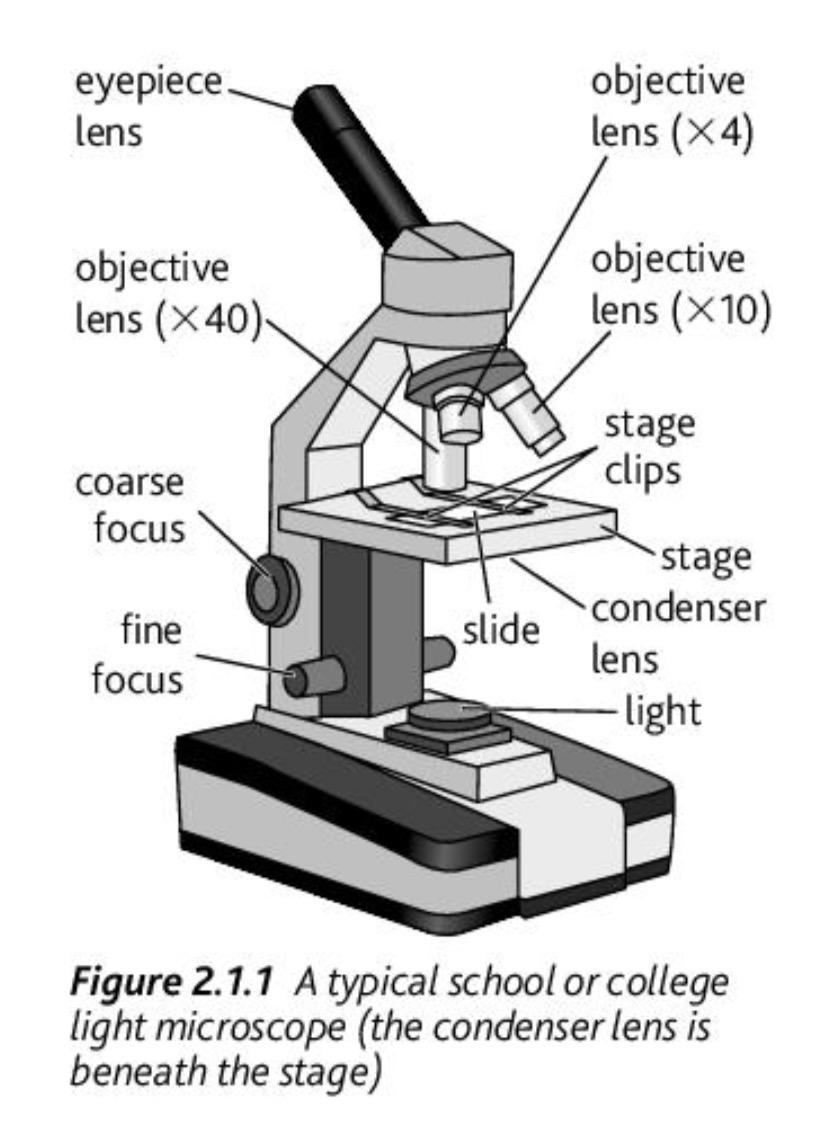
What are the main parts of a light microscope and their functions?
Eyepiece lens: magnifies image (usually ×10)
Objective lenses: provide different magnifications (×4, ×10, ×40)
Stage: holds slide
Coarse focus: brings image roughly into focus
Fine focus: sharpens image
Condenser lens: focuses light onto specimen
What are the typical total magnifications of a light microscope?
Low power: ×40 (×4 objective ×10 eyepiece)
Medium power: ×100 (×10 ×10)
High power: ×400 (×40 ×10)
Why is a condenser lens important?
It focuses and concentrates light onto the specimen, improving image brightness and clarity for better viewing.
What limits the resolution of a light microscope?
The resolution is limited by the wavelength of visible light (400–700 nm), meaning structures smaller than about 200 nm cannot be distinguished as separate objects.
Why can’t cell membranes be seen with a light microscope?
Cell membranes are too thin (about 7–10 nm), which is far below the 200 nm resolution limit of light microscopes.
Difference between magnification and resolution
Magnification increases the size of the image, while resolution determines how clearly fine detail can be seen. A high magnification without good resolution results in a blurry image.
How do you prepare a temporary plant slide?
Place thin plant tissue (e.g. Elodea leaf) on a slide in water
Add iodine stain
Carefully place a coverslip to avoid air bubbles
Blot excess liquid
Observe under low power, then increase magnification
How do you prepare an animal cell slide?
Place sample (e.g. liver tissue) on slide
Add methylene blue stain
Add coverslip
Observe under low power, then increase magnification
Rules for biological drawings
Use sharp pencil (no pen)
Draw clear, continuous lines
No shading or colouring
Show correct proportions
Label clearly
Only draw what is seen, not what is expected
Why must biological drawings show proportions accurately?
To ensure the drawing correctly represents the actual structure of the cells, allowing valid comparison and analysis.
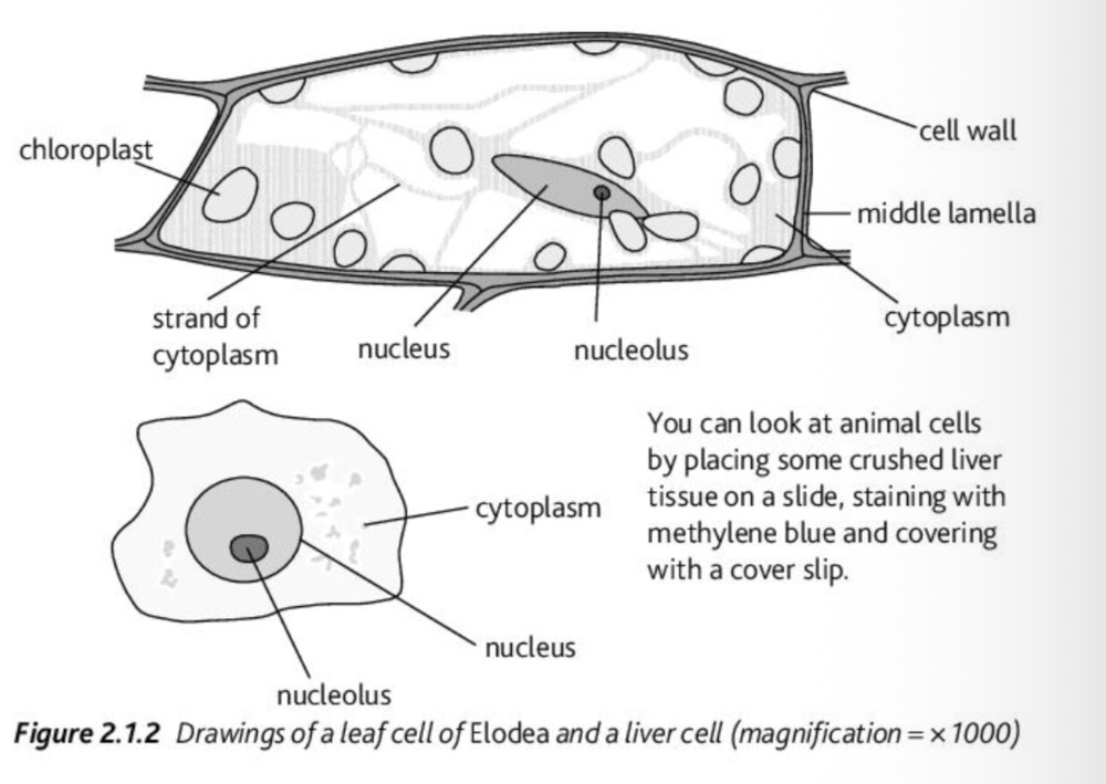
Differences between plant and animal cells (light microscope)
Plant cells: have cell wall, chloroplasts, large permanent vacuole, regular shape
Animal cells: no cell wall, no chloroplasts, small or no vacuole, irregular shape
Similarities between plant and animal cells
Both contain nucleus, cytoplasm, and cell membrane, and are similar in basic eukaryotic structure.
Approximate size difference between plant and animal cells
Plant cells are generally larger (~40 µm) than animal cells (~20 µm).
Why are cell membranes labelled in diagrams even if not visible?
Cell membranes are too thin to be seen with a light microscope, but they are included in diagrams to show the theoretical boundary of the cell.
Why is iodine used for plant cells?
Iodine stains starch and improves contrast, making structures like chloroplasts and cell walls easier to see.
Why is methylene blue used for animal cells?
It stains acidic structures such as the nucleus, increasing contrast and making cell components easier to observe.
How do you focus a microscope properly?
Start with low power, use coarse focus to find image, then switch to medium/high power and use fine focus to sharpen the image.
Why must you start with low power on a microscope?
Low power gives a wider field of view, making it easier to locate the specimen before increasing magnification.
Difference between plant and animal cells (summary exam answer)
Plant cells have a cell wall, chloroplasts, and a large vacuole giving a fixed shape, while animal cells lack these features and are more irregular in shape.