Thorax, Chest, CP imaging
1/123
There's no tags or description
Looks like no tags are added yet.
Name | Mastery | Learn | Test | Matching | Spaced | Call with Kai |
|---|
No analytics yet
Send a link to your students to track their progress
124 Terms
T-spine imaging is used to identify/exclude what disease processes
osteoporosis
tuberculosis osteomyelitis
Scheuermann’s disease
Neoplasm
metabolic disorders
T-spine imaging is used to identify/exclude what anatomic abnormalities
scoliosis
rib fx
spine fx
ligamentous injury
dislocations
describe scheuermann’s disease
back ache/kyphosis of the lumbar spine that leads to osteochondrosis of secondary ossification centers
in adolecents
routine XR of t-spine
AP
lateral
What is 1

rib
What is 2

vertebral body
What is 3
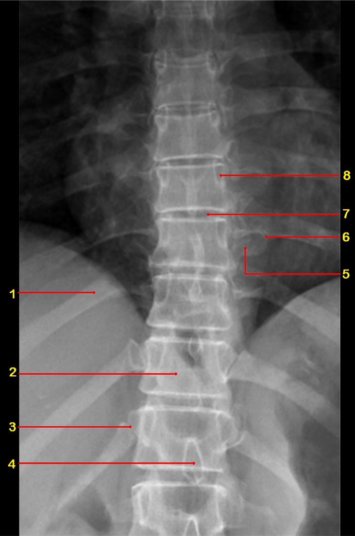
costovertebral joint
What is 4

spinous process
What is 5

transverse process
What is 6

costotransverse joint
What is 7

intervertebral disc
What is 8

pedicle
What view is this

AP
What is 9

facet joint
What is 10

inferior articular process
What is 11
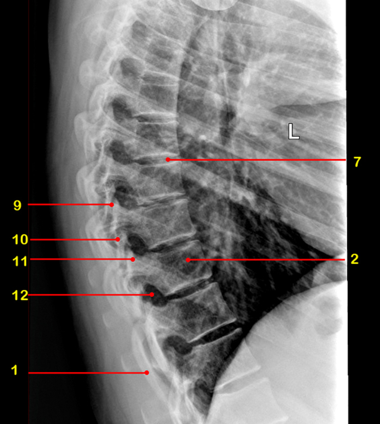
superior articular process
What is 12

intervertebral foramen
What view is this

sagittal
What view is this
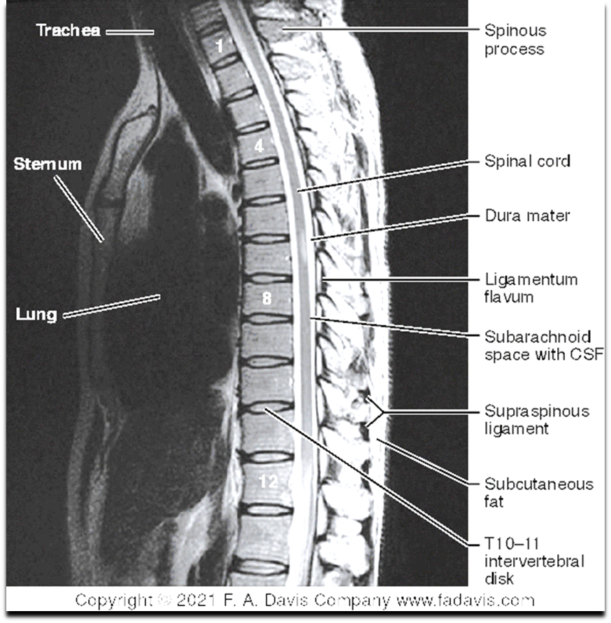
sagittal MRI
What view is this
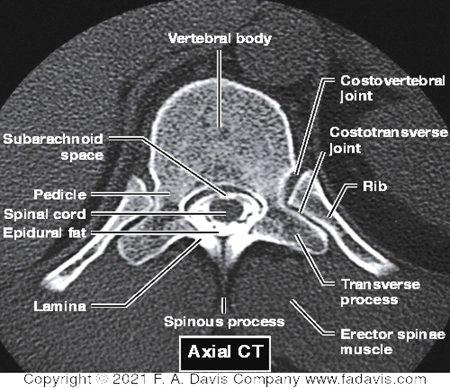
axial
What view is this
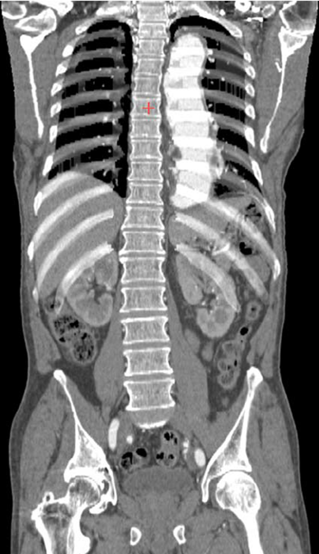
coronal
what does the anterior column consist of
anterior longitudinal ligament and ant 2/3 of vertebral body and annulus fibrosis
what does the middle column consist of
posterior 1/3 of the body and annulus, posterior longitudinal lig
what does the posterior column consist of
posterior lig complex and the vertebral arch structures
indications of CT for t-spine
acute trauma (adults)
degenerative conditions
postop eval
infectious processes
image guidance
neoplastic conditions
inflammatory lesions
spine abnormalities
spinal cord assessment (if MRI contraindicated)
steps of CT interpretation for t-spine
Alignment
Bone density
Canal space
Disk integrity
Soft tissues
indications of MRI for t-spine
spinal cord malformations
inflammatory/autoimmune diseases
infectious conditions
vascular disorders
degenerative conditions
trauma
neoplastic abnormalities
misc. fluid leaks, procedural check ups, or a combo of above
MRI contraindications
pacemakers
ferromagnetic intracranial aneurysm, clips, foriegn bodies, electronic devices
certain neurostimulators
certain cochlear implants
extensive tattoos
nonremovable body piercings
steps of MRI interpretation for t-spine
Alignment
Bone density
Canal space/CNS
Disk integrity
Soft tissues
range for right thoracic scoliosis
T4/6-T11/L1
range for right thoracolumbar scoliosis
T4/6-L2/4
range for Left lumbar scoliosis
T11/12 - L5
describe a double major scoliosis curve
thoracic one way and a lumbar the opposite with equal prominence
What curve is this
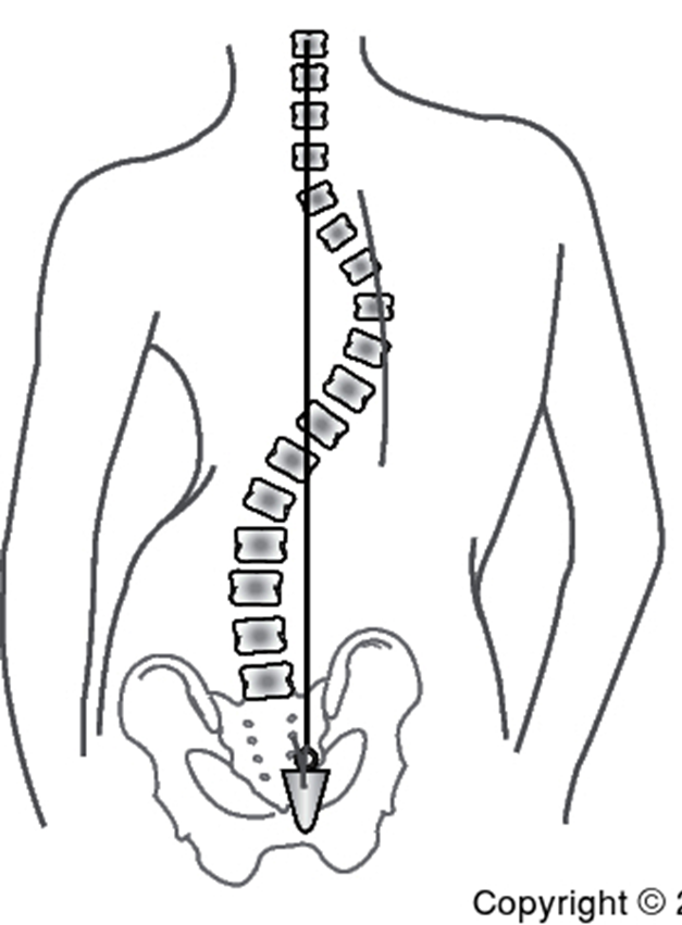
right thoracic
What curve is this
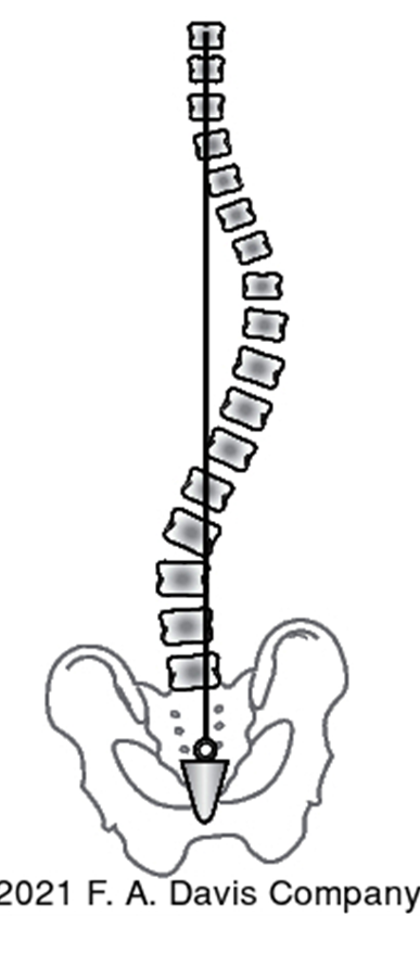
right thoracolumbar
purpose of scoliosis SB test
to determine if the curve is structural (rigid) or flexible
describe a structural cure results
R curve stays when SB to R
describe a flexible cure results
L curve disappears when SB to L
describe the pedicle method for measuring curve size
identifying how far the convex side pedicle has rotated toward midline
score of 0 on the pedicle method
no rotation
score of +1 on the pedicle method
pedicle toward midline
score of +2 on the pedicle method
pedicle 2/3 to midline
score of +3 on the pedicle method
pedicle in midline
score of +4 on the pedicle method
pedicle beyond midline
describe the Cobb method for measuring curve size
find the upper and lower most vertebrae involved
last/first vertebrae who’s pedicles align
draw a line along that angle
create a 90 degree line to original lines
measure the angle of intersection
What view is used for Cobb method
frontal plane of AP projection
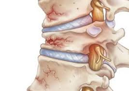
types of compression fxs
step defect
wedge deformity
linear zone of impaction
displaced endplates
loss of IVD height
paraspinal edema
abdominal ileus
describe displaced endplates
anterior shearing of the IVD may avulse the bony rim of the endplate or displace it ant
How will displaced endplates appear
greater ant/post diameter of the vertebral body at the involved endplate (lateral view)
describe loss of IVD height
herniation of the disc will cause a decrease in the potential space and possibly a misalignment of adjacent vertebrae
describe paraspinal edema
soft tissue edema or hematoma, often associated with compression fx
describe abdominal ileus
disturbance to the visceral autonomic nerves/ganglia leading to excessive amounts of gas in the small or large bowel
What does abdominal ileus indicate
severe underlying trauma and great likelihood of fx
describe step defect
step off at the anterior portion due to the endplate no longer being connected
What patho is this
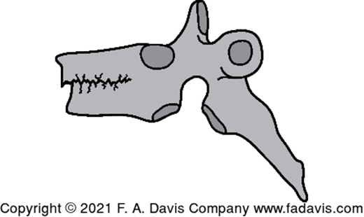
step defect
What patho is this

step defect with endplate disruption
What patho is this
wedge deformity
describe wedge deformity
anteriorly, trapezoidal collapse
how much loss of height indicates significant damage
30%
How is fx stability determined
number of columns involves (ant/middle/post)
hyperflexion injury affect on ant column
compression fx
hyperflexion injury affect on middle column
tear PLL
hyperflexion injury affect on post column
tear posterior lig complex
hyperflexion + rotation injury affect on ant column
endplate fx
disc rupture
hyperflexion + rotation injury affect on middle column
tear PLL
hyperflexion + rotation injury affect on post column
tear PLC
dislocation/fx facet
hyperflexion + shear injury affect on ant column
fx of vert body or disc
hyperflexion + shear injury affect on middle column
tear PLL and body fx
hyperflexion + shear injury affect on post column
fx pedicles and PLC tear
seatbelt injury affect on ant column
horizontal body fx
seatbelt injury affect on middle column
horizontal body fx
seatbelt injury affect on post column
fissuring of lamina, pedicles, and t.p.
describe a seatbelt injury
hyperflexion over fixed restraint
What injury is this
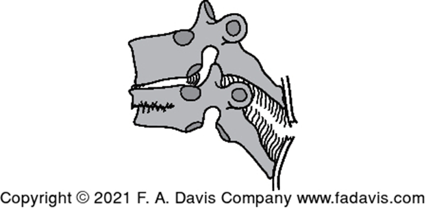
hyperflexion
What injury is this
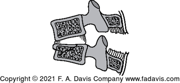
hyperflexion+ rotation
What injury is this
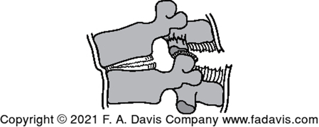
hyperflexion + shear
What injury is this

seatbelt
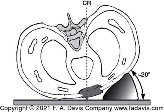
standard imaging for sternum
posterior oblique
lateral
standard imaging for ribs
PA
AP
anterior oblique
posterior oblique
What ribs can be seen during inspiration
1-10
What ribs can be seen during expiration
8-12
Where is the CR for sternal imaging
through SC joint, body of sternum, or the clavicle
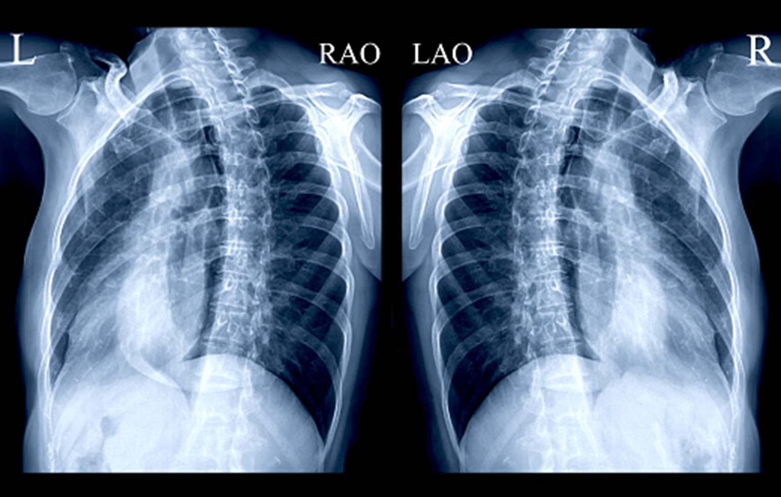
What view is this
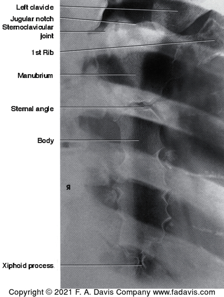
sternal posterior oblique
What view is this
sternal posterior oblique
Which ribs are analyzed with a AP image
posterior ribs
Which ribs are analyzed with a PA image
anterior
difference between ant and post ribs
Ant: angulated/shadows
post: horizontal
What view is this
lateral rib
What occurs if their are 3 or more consecutive rib fxs
flail chest
chest imaging options
x-ray
nuclear scanning
MRI
CT
US
angiography
indications for chest imaging
evaluation of s/s
eval for line placement
pneumothorax (PTX) screen
symptoms indicative of chest imaging
pain
SOB/cough
hemoptysis
fever
chest pain
sings indicative of chest imaging
trauma
hypoxemia
What is the most common chest imaging
CXR
routine projections of CXR
PA
lateral
What side is a lateral CXR taken to see the heart
L
Why is CXR typically taken first
determine pulm v cardiac
identifies patho to begin tx
narrows differential dx/further testing needed
What CXR view exaggerates the size of the heart? Why?
AP because the heart is further from the image receptor
steps for CXR interpretation
Airway
Bone and soft tissue
Cardiac silhouette
Diaphragm & Pleura
Effusion
Fields, fissures, foreign bodies
Gastric bubble and great vessels
Hila and mediastatum
Inspiration
J - all that jazz
What is the normal ratio for thoracic assessment for cardiomegaly
1 heat: 2 thorax