Psyc 100 lec 3 NEUROSCIENCE
1/69
There's no tags or description
Looks like no tags are added yet.
Name | Mastery | Learn | Test | Matching | Spaced | Call with Kai |
|---|
No analytics yet
Send a link to your students to track their progress
70 Terms
list 5 different techniques for studying the brain
-examine AUTOPSY tussue
-TEST PATIENTS w BRAIN DAMAGE
-record electrical activity from brain (EEG Electroencephalography)
-animal studies
-TMS (Transcranial Magnetic Stimulation) (delivery of electromagnetic pulse)
what does EEG stand for and what does it do
Electro-ence-phalography
-record electrical activity from brain
list 6 neuroimaging techniques and briefly explain them
-MRI (magnetic renosance imaging) -clear images, best for detecting SOFT TISSUE INJURIES
-fMRI (functional magnetic renosance imaging) - detects oxygen use (measure brain activity
-CAT or CT (computerized axial tomography) - clear 2D X-ray images. can be combined to create 3D images
-PET (positron emission tomography) - radioactive chemicals INJECTED into bloodstream →enters brain (detect radioactive material flowing thru brain)
NEWEST: -DTI (diffusion tensor imaging) - detect diffusion of H2O to generate contrast in MR images (MAPS WHITE MATTER IN BRAIN)
-MEG (magneto-ence-phalography) -records magnetic field caused by electric currents in brain
Nervous system is consisted of 3 main things:
Central nervous system (CNS) - brain & spinal cord (CONTROL ALL MENTAL/PHYSICAL processes)
Peripheral nervous system (PNS) - nerves that carry info to and from CNS
Neurons (cells that carry info between parts of body & nervous system)
3 Types of Neurons
Afferent neurons (SENSORY)(carry signal PNS to CNS)
Efferent neurons (MOTOR)(carry signal CNS to PNS, muscles, & glands)
Interneurons (relay cells between diff neuron types (ex. found in spinal cord between motor and sensory neurons)
remember: afferent start with A, closer to C. so brings to CNS. efferent cause signal to EXIT CNS into PNS. also think: AHH when touch hot pot, which is sensory, and go to brain CNS to tell u to lift hand off stove. motor comes from brain and go to muscles/nerves to move
2 parts of PERIPHERAL nervous system
SOMATIC nervous system (all nerves that a) gather sensory info from body n deliver it to spinal cord/brain b) send movement info from CNS to muscles of body/neck/head c) are under voluntary control (goal-directed))
AUTONOMIC nervous system (sympathetic & parasympathetic nervous system (involuntary basic life functions like heartbeat/stress response)
remember: PNS: somatic (voluntary) and autonomic (involuntary, sympathetic and parasympathetic)
2 systems within Autonomic Nervous System
SYMPATHETIC nervous system (activated under STRESS) (fight or flight)
-pupils dilate, heart rate/respiration increase, glucose released for energy, blood to brain/muscles
PARASYMPATHETIC nervous system (active during REST)
-REVERSES EFFECT of sympathetic nervous system (return to resting state)
think: sympathetic (stress) and parasympathetic (undo stress and go back to rest). both from autonomic system in PNS (peripheral)
Now the central nervous system, what 2 things it consist of?
brain and spinal cord
the spine extends down from what? and whats main function?
-spine extend down from BASE of BRAIN
-MEDIATE sensory and motor info (act as bridge or “middle” point between afferent neurons sending sensory info to brain AND efferent neurons sending motor info to PNS)
does the spine facilitate voluntary or involuntary movement
-some if voluntary, but sometimes spine influences behaviour without involving brain at all first
whats one way the spinal cord can operate independently without brain
REFLEXES
-axon of sensory (afferent) neuron send PAIN INFO to spinal cord
-sensory neuron synapses (gap between 2 neurons that allows them to communicate) w/ an interneuron, which connects w/ motor neuron, initiate muscle response (CONTRACT)
-PAIN INFO travel to brain (takes time b/c must cross more synapses)
what exactly does it mean when a spinal cord injured
the NERVES in the spinal cord are injured
what are the 2 kinds of spinal cord injuries (location on spinal cord determind what area of body paralyzed)
Quadriplegic (quad-rip-legic): injury CLOSER TO BRAIN (paralyzed everywehere below head/neck
Paraplegic (Para-plegic): injury FURTHER DOWN SPINE (paralyzed lower limbs)
true or false: spinal cord can still function after spinal cord injury
TRUE SOMETIMES, but subsequent inflamation can permanently damage spinal cord
what kind of spinal injury can cause PERMANENT damage
subsequent inflammation
list 3 general ways of treating spinal cord injuries
-boost electrical signals between brain & lower body
-help paralyzed person regain autonomy (freedom)
-stem cell treatments create enviro where injured cells recover (still experimental)
now whats the second part of central nervous system
brain
what part of brain is closest to spinal cord
hindbrain (responsible for basic functions) ex. breathing
what are the 4 sections of hindbrain
medulla
pons
cerebellum
reticular formation (neuron network)
whats 3 sections of brain
forebrain, midbrain, hindbrain
what does the medulla regulate? what part of brain its in
-medulla regulate sneeze, heartbeat, breathe, cough
-hindbrain (close to spinal cord, near bottom of brain)
what is the bridge between the medulla and other brain areas
Pons
Pons function? what part of brain
-hindbrain
-Pons: important for breathe (like medulla), swallow, sleep/dream, EYE movements, facial sensation/expression
Cerebellum function and what part of brain
-hindbrain
-Cerebellum important for MOTOR coordination and learning movement like learn play piano
what does reticular formation regulate. what part of brain
-hindbrain
-regulate SLEEP/WAKE CYCLE (involved in wakefulness, arousal, mood)
-remember: medulla and pons breathe, but medulla also heartbeat and nose stuff (sneeze/cough)
-pons BRIDGE for medulla. also swallow/eye move/face expression
-pons: sleep/dream but reticular formation: sleep CYCLE
-cerebellum: motor learning
whats main structure in midbrain? what is midbrain important for
-substantia nigra
-important in FLUIDITY of movement
6 parts of the forebrain?
Thalamus
Hypothalamus
Pituitary gland
Limbic system (2 parts)
Basal ganglia
Cerebral cortex
function of thalamus vs hypothalamus
thalamus: “RELAY STATION” for incoming SENSORY info
hypothalamus: important for MOTIVATION, drives, CONTROLS ENDOCRINE SYSTEM
pituitary gland function. what part of brain
-forebrain
-regulates hormones
2 parts of limbic system? function of systm, and each part
LIMBIC SYSTEM: regulates motivation, emotion, learning/memory
Amygdala (process FEAR)
Hippocampus (learning/memory)
function of Basal ganglia? what part of brain. and what does it include
-forebrain
-COGNITIVE flexibility and VOLUNTARY MOVEMENT control
-includes nucleus accumbens (nerve cell cluster) important for MOTIVATION/REWARD LEARNING
whats the name for the cluster of cells thats important for motivation and reward learning? what part of brain
nucleus accumbens
-in basal ganglia in forebrain
whats the structure in forebrain responsible for complex functions, and makes humans more distinct from other animals
cerebral cortex
what are some of the complex functions the cerebral cortex is responsible for
consciousness, language, thought
3 major functional areas of cerebral cortex and what they register
Sensory Cortex - register SENSORY neurons (TOUCH)
Motor Cortex - register MOTOR neurons (MUSCLES)
Association Cortex - register complex functions (high-order sensory processing, think, plan)
4 major lobes of cerebral cortex and location
Occipital lobe (back of brain)
Temporal lobe (sides)
Parietal lobe (behind frontal)
Frontal lobe (front of brain) also includes prefrontal lobe
Occipital lobe location and function
-back of brain
-VISION
Temporal lobe location and function
-sides of brain
-AUDITORY (sound, language production/comprehension)
-recognize complex visual stimuli (faces)
what are the two areas of cerebral cortex for speech/language? what lobe are these in
-Broca’s area (in frontal lobe) - speech production (damage means trouble speaking)
-Wernicke’s area (in temporal (side) lobe) - language comprehension (can speak but trouble understanding, may seem less smart)
Parietal lobe function and location
-behind frontal lobe (between front and back)
-SENSORY INTEGRATION:
-primary somatosensory area (process tactile(touch) info)
-high-order visual processing
frontal lobe function and location
-large cortical region in front (larger in humans than in other animals)
-involved in temporal planning, social relationships, movement
-voluntary movement
-includes prefrontal lobe: HIGHER ORDER THINKING: memory, morality, mood, planning
if someone had a stick thru skull, and recovered physically, but experienced a change in personality, what may have been damaged
prefrontal cortex (part of frontal lobe in cerebral cortex)
what is parallel processing
communication in/between lobes of brain allow us to perform complex functions at same time (how we can do more than one action at a time)
4 basic parts of nervous system
-neuron (nerve cell)
-sensory neuron
-motor neuron
-interneuron (communicate w/ sensory/motor neurons and other interneurons)
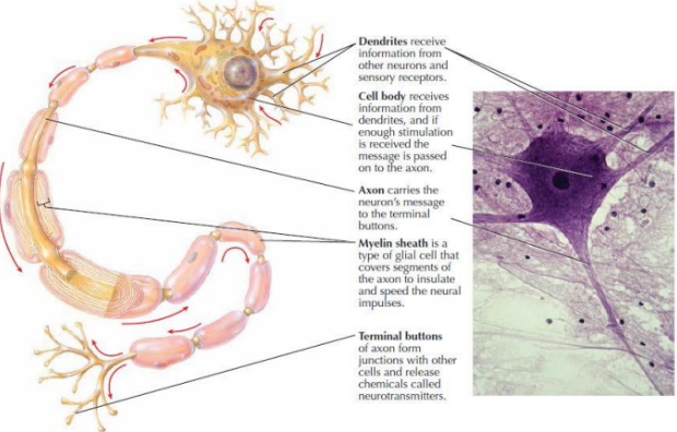
draw out the neuron structure and label dendrites, cell body, axon, myelin sheath, terminal buttons. briefly say function of each
dendrite: RECIEVE INFO (from neurons/receptors)
cell body: RECIEVE INFO FROM DENDRITE (passed to axon)
axon: CARRY MESSAGE to terminal buttons
myelin sheath: glial cell that covers segments of axons to INSULATE/SPEED the neural impulses
terminal buttons (of axon): FORM JUNCTIONS w/ other cells and RELEASE NEUROTRANSMITTERS
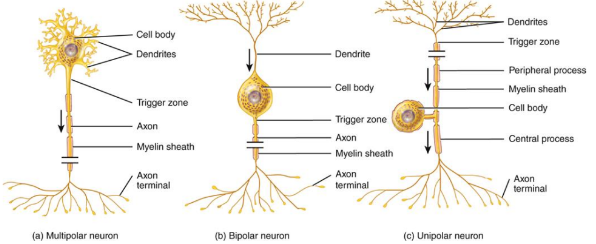
3 different neuron shapes? draw it out
MULTIpolar neuron
BIpolar neuron
UNIpolar neuron
2 major types of nervous system cells
neurons and glia
3 types of glia?
Astroglia (creates barrier between blood/brain, influence neuron communication, heals brain damage)
Oligodendroglia (oligo-dendro-glia): provides MYELIN to speed neuron transmission
Microglia: CLEAN up dead cells/prevent brain INFECTION
simply describe how neurons work
-neurons have negative charges & chemical messengers
-when triggered, neuron “fires”, sending electrical cue to release chemicals that trigger adjacent neuron
whats resting vs action potential in how neurons work
resting potential: at rest, neurons negatively charged inside (compared to outside)
action potential (electrical signal go down axon): when neuron fires, ion channels (pores in neuron) OPEN to let charged ions flow in/out neuron.
-NEURON BECOMES MORE POSITIVE ON INSIDE (relative to outside)
-this shift in charge triggers axon terminals to release neurotransmitters
describe the all-or-none principle for neurons
-either a neuron is stimulated enough to start an action potential or not
-right after neuron fires, cant fire again (refractory period)
-the strength of a nerve impulse is related to frequency, not degree of stimulation
what is the strength of a nervous impulse related to
frequency or amount of nerve cells firing
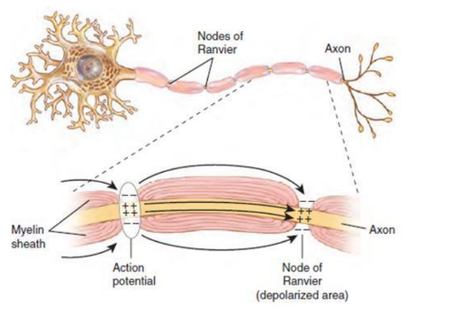
nodes of ranvier
regions of BARE AXON between areas wrapped in myelin
-action potentials travel fown myelinated axons by jumping from node to node
action potential triggers the relase of what into what?
-release of neurotransmitters into synapse (space between neurons)
neurotransmitters are contained in?
synaptic vesicles
what takes up neurotransmitters
neurotransmitter receptors (proteins in membran that recognize specific molecules) in nearby neuron
what are postsynaptic potentials
-ELECTRICAL EVENTS in postsynaptic neurons
-occur when neurotransmitter binds to receptor
-when receptor activated, ions flow thru receptor into neural
what event occurs when a neurotransmitter binds to one of its receptors
postsynaptic potentials (when receptor activated, + or - ions can flow thru recpetor into neuron)
2 types of neurotransmitter receptors and how they affect neuron and likelihood of action potential
-Excitatory postynaptic potentials (EPSPs) depolarize neuron, increase likelihood of action potential
-Inhibitory postsynaptic potentials (IPSPs) hyperpolarize neuron, decrease likelihood of action potential
list some common neurotransmitters and drugs associated with each (drug change availability/responsiveness of receptor for neurotransmitter)
Glutamate - ketamine
GABA - Vallium/ambien
Acetylcholine - nicotine
Dopamine - cocaine, heroin, mathamphetamine
Serotonin - Ecstasy
Norepinephrine - Adderall
what are the few layers of protection between brain and skin
cranium (beneath skin) and cranial meninges (dura mater, arachnoid mater, pia mater) below cranium and above brain.
2 types of brain injury and examples
-Traumatic brain injury (concussion, spinal cord)
-Acquired brain injury (infection, toxin, tumour, disease, stroke)
what type of brain injury is a stroke
acquired
what do brain injury treatments typically focus on
restore blood flow, reduce swelling, treat infections
neuroplasticity is the brains ability to
make NEW neural connections or REORGANIZE after injury
whats the corpus callosum. what does it connect
connects the two brain hemispheres
-dense bundle of neural fibres (axons) allow for communication of info from one side of brain to other
what conencts the 2 brain hemispheres? what allows for communcation between both sides
-corpus callosum connects
-AXONS (dense bundle of neural fibres) allow for communication between sides
what is performed to treat severe epilepsy
corpus callosotomy
what might happen to visual field if corpus callosum is severed? recall what corpus callosum is
corpus callosum connects both hemispheres of brain, so if severed, some visual info only processed by one side of brain
4 important observations by darwin:
animals change over time
aspects of species that seem different outside had structural similarities underneath
selective breedings leads to appearance changes
not all animals born will survive to maturity and able to reproduce